• No results found

Ependymal cells of the mouse brain express urate transporter 1 (URAT1)

N/A
N/A
Protected

Academic year: 2020

Share "Ependymal cells of the mouse brain express urate transporter 1 (URAT1)"

Copied!
7
0
0

Loading.... (view fulltext now)

Full text

Figure

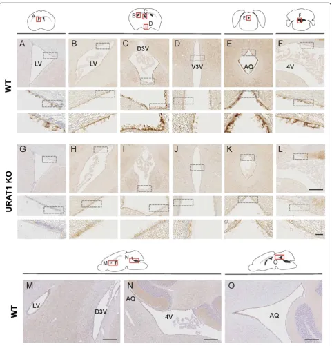
Figure 1 Distribution of URAT1 in mouse brain. Paraffin sections from nine-week-old WT (A–F, coronal section; M–O, sagittal section) andURAT1 KO (G–L, coronal section) mice were stained with a polyclonal antibody to URAT1 (anti-URAT1 antibody) and counters
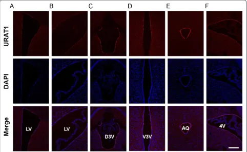
Figure 2 Expression levels of URAT1 differ between ventricles. (A–F) Frozen coronal sections from nine-week-old WT mice were stainedwith anti-URAT1 (red) and DAPI (blue) and imaged by confocal microscopy
Figure 3 URAT1 localizes at the apical surface of ependymal cells. Frozen coronal sections from nine-week-old WT and URAT1 KO micewere stained with anti-acetylated-α-tubulin antibody (green), anti-URAT1 antibody (red) and DAPI (blue)

References

Related documents