INTRODUCTION
Doxorubicin (DOX) is one of the most commonly used broad-spectrum chemotherapeutic drugs; however, its
clinical application is limited because of its dose-dependent cardiotoxicity, which may lead to the development of irre-versible cardiomyopathy and/or heart
failure (1). The mechanism of DOX’s cardiotoxicity is complex and may in-volve oxidative (2,3), nitrosative and ni-trative stress (4,5), mitochondrial dys-function/ toxicity (1,6–8), dysregulation of various metabolic (9) and lipid sig-naling pathways (10–12), activation of various stress kinases and cell death mechanisms (both apoptotic and necrotic) (13), triggering of secondary inflammation and remodeling (14), eventually culminating in cardiac dys-function and heart failure (1,15).
Cannabidiol (CBD) is the most abun-dant nonpsychoactive constituent of
mar-Cardiomyopathy by Modulating Mitochondrial Function and
Biogenesis
Enkui Hao,
1,2Partha Mukhopadhyay,
1Zongxian Cao,
1Katalin Erdélyi,
1Eileen Holovac,
1Lucas Liaudet,
3Wen-Shin Lee,
1,4György Haskó,
5Raphael Mechoulam,
6and Pál Pacher
11
Laboratory of Physiologic Studies, National Institute on Alcohol Abuse and Alcoholism, National Institutes of Health, Bethesda, Maryland, United States of America; 2Department of Cardiology, Shandong Provincial Qianfoshan Hospital, Shandong University, Jinan, China; 3Department of Intensive Care Medicine, BH 08-621 University Hospital Medical Center, Lausanne, Switzerland; 4
Division of General Medicine, Department of Medicine, Taipei Veterans General Hospital, National Yang-Ming University School of Medicine, Taipei, Taiwan; 5Departments of Surgery, Rutgers New Jersey Medical School, Newark, New Jersey, United States of America; and 6Department for Medicinal Chemistry and Natural Products, Faculty of Medicine, Hebrew University of Jerusalem, Ein Kerem, Jerusalem, Israel
Doxorubicin (DOX) is a widely used, potent chemotherapeutic agent; however, its clinical application is limited because of its dose-dependent cardiotoxicity. DOX’s cardiotoxicity involves increased oxidative/nitrative stress, impaired mitochondrial function in cardiomyocytes/endothelial cells and cell death. Cannabidiol (CBD) is a nonpsychotropic constituent of marijuana, which is well tolerated in humans, with antioxidant, antiinflammatory and recently discovered antitumor properties. We aimed to explore the effects of CBD in a well-established mouse model of DOX-induced cardiomyopathy. DOX-induced cardiomyopathy was char-acterized by increased myocardial injury (elevated serum creatine kinase and lactate dehydrogenase levels), myocardial ox-idative and nitrative stress (decreased total glutathione content and glutathione peroxidase 1 activity, increased lipid peroxida-tion, 3-nitrotyrosine formation and expression of inducible nitric oxide synthase mRNA), myocardial cell death (apoptotic and poly[ADP]-ribose polymerase 1 [PARP]-dependent) and cardiac dysfunction (decline in ejection fraction and left ventricular frac-tional shortening). DOX also impaired myocardial mitochondrial biogenesis (decreased mitochondrial copy number, mRNA ex-pression of peroxisome proliferator-activated receptor γcoactivator 1-alpha, peroxisome proliferator-activated receptor alpha, estrogen-related receptor alpha), reduced mitochondrial function (attenuated complex I and II activities) and decreased myo-cardial expression of uncoupling protein 2 and 3 and medium-chain acyl-CoA dehydrogenase mRNA. Treatment with CBD mark-edly improved DOX-induced cardiac dysfunction, oxidative/nitrative stress and cell death. CBD also enhanced the DOX-induced impaired cardiac mitochondrial function and biogenesis. These data suggest that CBD may represent a novel cardioprotective strategy against DOX-induced cardiotoxicity, and the above-described effects on mitochondrial function and biogenesis may contribute to its beneficial properties described in numerous other models of tissue injury.
Online address: http://www.molmed.org doi: 10.2119/molmed.2014.00261
Address correspondence toPál Pacher, Section on Oxidative Stress Tissue Injury, Labora-tory of Physiological Studies, National Institutes of Health/NIAAA, 5625 Fishers Lane, MSC-9413, Bethesda, Maryland 20892-MSC-9413, USA. Phone: 301-443-4830; Fax: 301-480-0257; E-mail: [email protected].
ijuana (Cannabis Sativa), which was con-sidered initially to be biologically inactive (16,17). It has a negligible effect on con-ventional cannabinoid 1 and 2 receptors (CB1/2) in vivo(16,17) and is very safe in humans (18). A pioneering study by J Ax-elrod and D Wink in 1998 (19) demon-strated that CBD was a potent neuropro-tective antioxidant. They found that it was more protective against glutamate-induced neurotoxicity than either ascor-bate or α-tocopherol, suggesting that it has potential therapeutic utility in neu-rodegenerative disorders associated with oxidative stress (19). Later, large numbers of preclinical studies also confirmed po-tent antioxidant and antiinflammatory ef-fects of CBD in preclinical models of coli-tis, ischemic reperfusion injury, various neurodegenerative and cardiovascular disorders, diabetes and diabetic compli-cations (20), among others (17,21). An oromucosal spray containing 50% CBD (Sativex) is approved in the United King-dom, Canada and various other Euro-pean countries to alleviate pain and spas-ticity associated with multiple sclerosis (22), and CBD recently received orphan drug approval by FDA for the treatment of refractory childhood epilepsy.
In this study, we aimed to explore the effects of CBD in a well-established, clinically relevant mouse model of DOX-induced cardiomyopathy (4,5,23), particularly focusing on oxidative and nitrative stress and mitochondrial dysfunction/ biogenesis. Our results may also provide a novel mechanistic insight on the protective effects of CBD in various models of tissue injury and a promising tool for the prevention of devastating cardiovascular complica-tions of DOX chemotherapy.
MATERIALS AND METHODS
Animals/Drugs
All protocols involving the use of ani-mals were approved by the Institutional Animal Care and Use Committees and were performed in line with the Guide-lines for the Care and Use of Laboratory Ani-malsadopted by the National Institutes of
Health (NIH) (24). Male C57BL/6J mice weighing 22–30 g were acutely adminis-tered with single high dose (20 mg/kg) of DOX (Sigma-Aldrich, St. Louis, MO, USA) intraperitoneally (IP) freshly dis-solved in physiological saline. CBD ob-tained from Tocris (Ellisville, MO, USA) or isolated as described earlier (25) was dissolved in vehicle solution (one drop of Tween-80 in 3 mL 2.5% DMSO in saline). Mice were treated with CBD 10 mg/kg IP, or vehicle started 1.5 h before the DOX injection and once every day. Mice were subjected to hemodynamic measure-ments at the end of study (5 d). Hearts were excised and snap frozen in liquid nitrogen for biochemical measurements or fixed for histological evaluation as de-scribed previously (10,11).
Hemodynamic Measurements in Mice Echocardiography was performed by using the VisualSonics Vevo0770 system (VisualSonics Inc., Toronto, ON,Canada), which is equipped with a 30 MHz me-chanical scan probe which can obtain high resolution 2-dimensional images. Animals were anesthetized with isoflu-rane (2%) inhalation anesthesia. B mode images were obtained in the plane con-taining aortic and mitral valves. M mode images were obtained from the paraster-nal short-axis view at the level of papil-lary muscles. LV end-diastolic diameters, ejection fraction and fractional shortening were calculated by using Vevo Analysis software as described previously. The in-vestigators performing echocardiography were blinded to the treatment status.
Serum Creatine Kinase (CK) and Lactate Dehydrogenase (LDH) Levels
The blood samples were collected and the serum was removed immediately for CK and LDH measurements using a clin-ical chemistry analyzer system (VetTest 8008, IDEXX Laboratories, Westbrook, ME, USA).
Myocardial 3-Nitrotyrosine (NT) Determination
Myocardial 3-nitrotyrosine content, a marker of nitrative stress (26), was
deter-mined by nitrotyrosine enzyme-linked immunosorbent assay (ELISA) according to the protocol supplied with the kit (Cell Biolabs, San Diego, CA, USA) as previously described (11,12).
Determination of Myocardial Caspase 3/7 and Poly(ADP-Ribose) Polymerase (PARP) Activities
Caspase 3/7 activity in the heart ho-mogenates was determined using the flu-orimetric-based Apo-ONE homogenous assay kit (Promega, Madison, WI, USA) as described (11,12). Myocardial PARP activity was assayed using a colorimetric kit according to manufacturer’s protocol (Trevigen, Gaithersburg, MD, USA) as described (11,12).
Myocardial DNA Fragmentation ELISA The quantitative determinations of cytoplasmic histone-associated-DNA-fragmentation (mono and oligonucleo-somes) due to in vivocell death were mea-sured using ELISA kit (Roche Diagnostics GmbH, Indianapolis, IN, USA) (12).
Determination of Myocardial Glutathione (GSH) Content
Myocardial GSH content was deter-mined using kits from Trevigen accord-ing to the manufacturer’s protocols, as previously reported (12).
Histological Examination of Heart Sections
Heart samples were fixed in 4% buffered formalin. After embedding and cutting 5 μm slices, all sections were stained with nitrotyrosine (Cayman Chemical, Ann Arbor, MI, USA) accord-ing to the manufacturer’s protocol and/or as described (20). Nitrotyrosine-stained sections were counterNitrotyrosine-stained with hematoxylin. The specific staining was visualized and images were ac-quired using BX-41 microscope and U-TV1 × 2 camera (Olympus, Tokyo, Japan) with 200 × fold magnification.
(Invitro-gen [Thermo Fisher Scientific Inc., Waltham, MA, USA]) according to manu-facturer’s instructions. The isolated RNA was treated with RNase-free DNaseI (Ambion [Thermo Fisher Scientific]) to re-move traces of genomic DNA contamina-tion. Each RNA sample (1 μg) was mixed with 2 μL of 5× Genomic DNA Elimina-tion Buffer to remove any residual ge-nomic DNA as supplied with the kit and made up to a final volume of 10 μL. RT cocktail (prepared according to the manu-facturer’s instruction) was added to RNA containing Genomic DNA Elimination Mixture and incubated at 42° C for 15 min, followed by heating at 95° C for 5 min. Each 20 μL of cDNA Synthesis Re-action received an addition of 91 μL of sterile water. Real-time PCR was per-formed using Oxidative Stress and Anti-oxidant Defense RT Profiler PCR array or specific primer sets (SAB Bioscience, Fred-erick, MD, USA). Relative quantification was calculated using the comparative CT method. The normalizer used for each cDNA sample was housekeeping gene β-actinas described (11,12,20,27).
PARP Activity
Myocardial PARP activity was assayed using a colorimetric kit according to the manufacturer’s protocol (Trevigen) as described (12).
Myocardial 4-Hydroxy-2-Nonenal (4-HNE) Content
Lipid peroxides are unstable indicators of oxidative stress in cells that decom-pose to form more complex and reactive compounds such as 4-HNE, which has been shown to be capable of binding to proteins and forming stable HNE adducts. HNE in myocardial tissues was determined using a kit (Cell Biolabs). In brief, BSA or Myocardial tissue extracts were adsorbed onto a 96-well plate for overnight at 4° C. HNE adducts present in the sample or standard were probed with anti-HNE antibody, followed by an HRP-conjugated secondary antibody. The HNE protein adduct content in an unknown sample was determined by comparing with a standard curve (12).
Myocardial Glutathione Peroxidase Assay
Glutathione peroxidase enzyme activ-ity was assayed using a SpectraMax spectrophotometer (Trevigen) according to the manufacturer’s instructions.
Determination of Myocardial Mitochondrial Complex Activity
Microplate assay kits were obtained from MitoSciences (Eugene, OR, USA) and used to determine the activity of mi-tochondrial complex I, complex II and complex IV according to the manufac-turer’s instructions. The complex en-zymes were immunocaptured in the mi-croplate and activities were determined in Spectramax M3 (Molecular Devices [Thermo Fisher Scientific]). Complex tivities were expressed as percentage ac-tivity compared with the heart samples of the vehicle-treated mice similar to that described previously for liver (27).
Mitochondrial DNA Content Mitochondrial DNA (mtDNA) copy/content determination was per-formed as described previously (27).
Analyses of Data
Results are expressed as means ± SEM. Statistical significance among groups
was determined by paired student ttest or ANOVA followed by Tukey post hoc test for multiple comparisons using GraphPad Prism 6 software (San Diego, CA, USA). Probability values of P< 0.05 were considered significant.
RESULTS
CBD Attenuates DOX-Induced Cardiac Injury and Cardiac Dysfunction
First we investigated if CBD attenuates the DOX-induced increased serum LDH and CK levels (markers of tissue/ cardiac injury) and cardiac dysfunction (evalu-ated by measurement of ejection fraction [EF] and fractional shortening [FS] using echocardiography). DOX-markedly in-creased serum levels of LDH and CK (Figure 1A) and reduced EF and FS (Fig-ures 1B, C). The DOX- induced tissue in-jury and cardiac dysfunction were mark-edly attenuated by CBD treatment (Figures 1A, B).
CBD Attenuates DOX-Induced Myocardial Oxidative Stress
Next we explored the effects of CBD on myocardial oxidative stress. DOX in-duced marked increase in myocardial HNE content (Figure 2A), reduced total
glutathione content (Figure 2B), attenu-ated glutathione peroxidase (Figure 2C) and mitochondrial complex I and II (but not IV) activities (Figures 2D, E) (the unchanged complex IV activity is not shown). DOX also induced a marked in-crease in the myocardial expression of reactive oxygen species generation NADPH oxidase isoform NOX2 (Fig-ure 2F). All markers of DOX- induced oxidative stress were attenuated by CBD treatment (see Figures 2A–F).
CBD Attenuates DOX-Induced Myocardial Nitrative Stress
We also explored effects of CBD on DOX-induced increased nitrative stress. DOX markedly increased myocardial in-ducible nitric oxide synthase (iNOS)
ex-pression (Figure 3A) and 3-nitrotyrosine formation (Figures 3B, 4). CBD largely at-tenuated the DOX-induced increased iNOSexpression and nitrotyrosine for-mation (Figures 3, 4).
CBD Enhances DOX-Induced Impaired Mitochondrial Biogenesis
Since DOX-induced cardiotoxicity is known to be associated with mitochon-drial injury and dysfunction (see also Figure 2), we further explored the ef-fects of CBD on mitochondrial copy number and various markers of mito-chondrial biogenesis. DOX decreased the myocardial mitochondrial copy number and attenuated expression of mRNA of various markers of mito-chondrial biogenesis such as
peroxi-some proliferator-activated receptor γ coactivator 1-α(PGC1A), peroxisome proliferator-activated receptor α (PPARA), estrogen-related receptor α (ERRA), uncoupling protein 2 and 3 (UCP2, UCP3) and medium-chain acyl-CoA dehydrogenase (MCAD) (Figures 5 A–G). CBD largely pre-vented the above-mentioned changes (see Figures 5A–G), suggesting that it enhanced mitochondrial biogenesis in damaged hearts.
CBD Attenuates DOX-Induced Myocardial Cell Death and Matrix Metalloproteinases 2 and 9 (MMP2 and MMP9 ) Activation
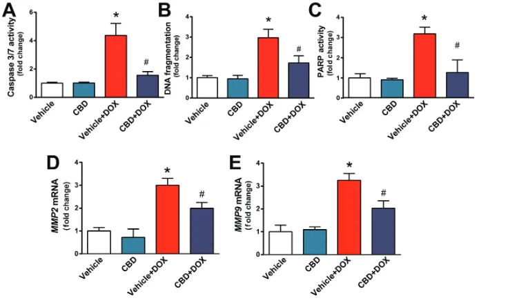
Cell death of cardiomyocytes and en-dothelial cells, as well as activation of proteolytic MMP enzymes, is a conse-quence of increased oxidative and nitrative stress and mitochondrial in-jury. We found that DOX-induced
Figure 2.CBD treatment attenuates DOX-induced myocardial oxidative stress and mito-chondrial dysfunction. The effect of DOX and CBD treatment on myocardial (A) HNE adducts, (B) total glutathione content, (C) GPX activity, (D,E) mitochondrial complex I and II activities and (F) NOX2 mRNA expression. *P < 0.05 versus vehicle group; #P < 0.05 versus DOX group; n = 4–6/group.
Figure 3.CBD treatment attenuates DOX-induced myocardial iNOS expression and 3-nitrotyrosine accumulation. The effect of DOX and CBD treatment on myocardial (A) iNOS mRNA and (B) protein 3-nitrotyrosine content measured by ELISA *P < 0.05 ver-sus vehicle group; #P < 0.05 versus DOX
marked increases in myocardial apo -ptotic and PARP-dependent cell death in the myocardium and in mRNA ex-pression of MMP2and MMP9, which all were largely attenuated by CBD treatment (Figures 6A–E).
CBD Attenuates DOX-Induced Myocardial Inflammation
Myocardial injury is known to trig-ger secondary proinflammatory re-sponse. We found that DOX increased myocardial expression of mRNAs of tumor necrosis factor-α(TNFA), interleukin-1β(IL1B) and monocyte chemoattractant protein-1 (MCP-1) (Figures 7A–C). These changes were at-tenuated by CBD treatment (see Fig-ures 7A–C).
DISCUSSION
DOX-induced cardiomyopathy/ heart failure is a serious complication of chem-otherapy with very limited therapeutic options, mostly including supportive treatment or heart transplantation (1). The currently available preventive strate-gies are not satisfactory and there is an urgent need for development of novel approaches to prevent this devastating complication of DOX chemotherapy. CBD is a nonpsychoactive constituent of marijuana with potent antioxidant and antiinflammatory effects in preclinical disease models which are independent from classical G-protein coupled cannabinoid 1 and 2 receptors (16,17).
Herein, we show that CBD exerts protective effects against DOX-induced
cardiotoxicity and cardiac dysfunction by (i) attenuating oxidative and nitra-tive stress, (ii) improving mitochondrial function, (iii) enhancing mitochondrial biogenesis, (iv) decreasing cell death and expression of MMPs and (v) de-creasing myocardial inflammation.
Consistent with numerous previous reports (1–8) we found that DOX treat-ment markedly increased oxidative stress and impaired antioxidant defense in the heart. DOX also decreased myo-cardial mitochondrial complex I and II activities, as well as the activity of the glutathione peroxidase (GPX). This is consistent with studies suggesting a key role for mitochondrial dysfunction and reactive oxygen species generation in triggering the deleterious cascade of the DOX-induced cardiotoxicity (4,5,28,29). In addition to mitochondr-ial, NADPH oxidase- dependent ROS generation (particularly NOX2 dependent) also may contribute to DOX-induced cardiotoxicity (4,30). We also found that DOX increased myocar-dial expression of iNOS and enhanced 3-nitrotyrosine (3-NT) formation. 3-NT is a marker of peroxynitrite formation, or, more broadly, of nitrative stress (26). Peroxynitrite formed from the diffusion limited reaction of superoxide anion and NO recently was implicated as the key downstream effector of promoting DOX-induced cardiotoxicity (26). Perox-ynitrite may induce lipid peroxidation, nitration of key contractile proteins and/or sarco/endoplasmic reticulum Ca2+pump, trigger activation of mito-gen-activated protein kinases promot-ing apoptotic cell death and impair mi-tochondrial function favoring increased reactive oxygen species generation, eventually culminating in cardiac dys-function (26). In addition, peroxynitrite also can diffuse to the nucleus, induc-ing DNA breaks and consequent over-activation of the nuclear enzyme PARP-1, which, in turn, leads to energetic crisis and cell necrosis (26). Indeed, both neu-tralization of peroxynitrite by peroxyni-trite decomposition catalysts or inhibi-tion of iNOS or PARP is protective
against DOX-induced cardiotoxicity in murine models of DOX-induced car-diomyopathies. (4,5,13). DOX via in-creased generation of reactive oxygen and nitrogen species also leads to acti-vation of MMP enzymes (31), which in turn promotes pathological remodeling (4,14,32,33).
CBD attenuated DOX-induced myo-cardial lipid peroxidation and the de-cline in antioxidant glutathione level
and in the activity of glutathione perox-idase, which could be the consequence of attenuated formation or enhanced in-activation of reactive oxygen and nitro-gen species. CBD also decreased the DOX- induced increased iNOS expres-sion in the myocardium, most likely contributing to the attenuation of DOX-induced myocardial nitrotyrosine for-mation as a consequence of decreased NO availability for peroxynitrite
forma-tion from NO and superoxide anion. In agreement with this, CBD also attenu-ated nuclear factor-κB (NF-κB) and/or iNOS expression in a model of liver is-chemia-reperfusion injury (34),
cisplatin-induced nephropathy (35), and in diabetic cardiomyopathy (20,36). In these models, CBD also attenuated ox-idative stress by attenuating mitochon-drial dysfunction and reactive oxygen species generation or by decreasing the expression of various reactive oxygen species generating NADPH oxidase iso-forms (20,35,36). CBD also attenuated the DOX-induced decreased myocardial mitochondrial complex I activity, in agreement with its effect in a liver is-chemia-reperfusion injury model (34). Furthermore, CBD also reduced the DOX-induced decline in myocardial mi-tochondrial copy number and enhanced mitochondrial biogenesis in injured hearts. Consistent with previous stud-ies, DOX also induced both apoptotic and PARP-dependent cell death in hearts (5,12,23,37), which could be largely attenuated by CBD. CBD also attenuated the DOX-induced increased myocardial inflammation. These results are in agreement with the attenuation of proinflammatory response by CBD in diabetic hearts (20) or in human car-diomyocytes (20) or coronary artery en-dothelial cells (36) exposed to high glu-cose, likewise with beneficial effects of CBD in rodent models of myocardial in-farction, primary diabetes and diabetic complications (17,38–40).
So far, over 50 metabolites of CBD were identified in the urine, showing considerable variations between differ-ent species (including humans), how-ever their biological activity was not evaluated in detail (41,42). Substantial evidence from in vitroor in vivostudies suggests that CBD by itself exerts po-tent antiinflammatory and antioxidant effects, independently from cannabi-noid receptors (for example in liver ischemia/ reperfusion injury model CBD exerted comparable antiinflamma-tory effects in both cannabinoid 2 re-ceptor knockout mice and their
type littermates). Nevertheless, the po-tential protective effect of certain CBD metabolites in vivocannot be excluded either.
Another issue which deserves some discussion is whether the mentioned antioxidant and antiinflam-matory effects of CBD would interfere with the antitumor activity of DOX. The answer is that it is not likely to do so. For example, mitochondrial-targeted antioxidants appear to have synergistic effects with various chemotherapeutic agents in tumor cell killing (43). Activa-tion of MMPs may be involved in tumor progression and metastasis for-mation, which is inhibited by CBD, likewise the NF-κB signaling. Further-more, CBD by itself has been reported to have antitumor effects in a large va-riety of cancer cell lines, as well as in some explanted tumor models (44–46), which would rather predict a synergis-tic effect with antineoplassynergis-tic drugs. In fact, Insys Therapeutics just received FDA orphan drug designation for CBD as a potential treatment for glioblas-toma multiforme in humans.
CONCLUSION
In summary, our results indicate that CBD may represent a novel promising approach for the prevention of induced cardiomyopathy/heart failure by attenuating oxidative/nitrative stress, mitochondrial dysfunction, cell death and inflammation, and by pro-moting mitochondrial biogenesis. These results, coupled with the excellent safety of CBD in humans, its protective effects against cisplatin-induced nephropathy, its recently reported antineoplastic properties in various ma-lignancies and its orphan drug ap-proval for glioblastoma multiforme and childhood epilepsy, are particularly en-couraging from a therapeutic point of view.
ACKNOWLEDGMENTS
This study was supported by the Intra-mural Research Program of National In-stitutes of Health/NIAAA. P Pacher is grateful to George Kunos, Scientific Di-rector of NIAAA, for continuous sup-port, and dedicates this study to collabo-rator and friend, Itai Bab. The study also
was supported by the Intramural Re-search Program of the National Institute on Alcohol Abuse and Alcoholism (to P Pacher).
DISCLOSURES
The authors declare that they have no competing interests as defined by Molec-ular Medicine, or other interests that might be perceived to influence the re-sults and discussion reported in this paper.
Figure 6.CBD treatment attenuates DOX-induced myocardial cell death and MMP2 and MMP9 mRNA expression. Effect of DOX and CBD treatment on myocardial (A) cas-pase 3/7 activity, (B) chromatin fragmentation and (C) PARP activation. (D,E) Expression of MMP2 and MMP9 mRNA. *P < 0.05 versus vehicle group; #P < 0.05 versus DOX group;
n = 4–6/group.
Figure 7.CBD attenuates DOX-induced proinflammatory response in the heart. Real-time PCR shows significant increase of (A) TNFA, (B) IL1B, (C) MCP-1 mRNA level. Pretreatment with CBD significantly attenuates DOX-induced increased proin-flammatory chemokine levels. *P < 0.05 versus vehicle group; #P < 0.05 versus DOX
REFERENCES
1. Carvalho FS, et al. (2014) Doxorubicin-induced car-diotoxicity: from bioenergetic failure and cell death to cardiomyopathy. Med. Res. Rev. 34:106–35. 2. Rajagopalan S, Politi PM, Sinha BK, Myers CE.
(1988) Adriamycin-induced free radical forma-tion in the perfused rat heart: implicaforma-tions for cardiotoxicity. Cancer Res. 48:4766–9.
3. Sterba M, et al. (2013) Oxidative stress, redox sig-naling, and metal chelation in anthracycline car-diotoxicity and pharmacological cardioprotec-tion. Antioxid. Redox. Signal. 18:899–929. 4. Pacher P, et al. (2003) Potent metalloporphyrin
peroxynitrite decomposition catalyst protects against the development of doxorubicin-induced cardiac dysfunction. Circulation. 107:896–904. 5. Mukhopadhyay P, et al. (2009) Role of
superox-ide, nitric oxsuperox-ide, and peroxynitrite in doxoru-bicin-induced cell death in vivo and in vitro. Am.
J. Physiol. Heart Circ. Physiol. 296:H1466–83. 6. Doroshow JH, Davies KJ. (1986) Redox cycling of
anthracyclines by cardiac mitochondria. II. For-mation of superoxide anion, hydrogen peroxide, and hydroxyl radical. J. Biol. Chem. 261:3068–74. 7. Berthiaume JM, Wallace KB. (2007) Adriamycin-induced oxidative mitochondrial cardiotoxicity.
Cell Biol. Toxicol. 23:15–25.
8. Ichikawa Y, et al. (2014) Cardiotoxicity of doxoru-bicin is mediated through mitochondrial iron ac-cumulation. J. Clin. Invest. 124:617–30.
9. Tokarska-Schlattner M, Zaugg M, Zuppinger C, Wallimann T, Schlattner U. (2006) New insights into doxorubicin-induced cardiotoxicity: The crit-ical role of cellular energetics. J. Mol. Cell Cardiol. 41:389–405.
10. Mukhopadhyay P, et al. (2007) Pharmacological inhibition of CB1 cannabinoid receptor protects against doxorubicin-induced cardiotoxicity. J. Am.
Coll. Cardiol. 50:528–36.
11. Mukhopadhyay P, et al. (2010) CB1 cannabinoid receptors promote oxidative stress and cell death in murine models of doxorubicin-induced car-diomyopathy and in human cardiomyocytes.
Cardiovasc. Res. 85:773–84.
12. Mukhopadhyay P, et al. (2011) Fatty acid amide hydrolase is a key regulator of endocannabinoid-induced myocardial tissue injury. Free Radic. Biol.
Med. 50:179–95.
13. Pacher P, et al. (2002) Activation of ribose) polymerase contributes to development of doxorubicin-induced heart failure. J. Pharma-col. Exp. Ther. 300:862–7.
14. Bai P, et al. (2004) Matrix metalloproteinase acti-vation is an early event in doxorubicin-induced cardiotoxicity. Oncol. Rep. 11:505–8.
15. Azuma J, et al. (1981) Adriamycin cardiotoxicity: possible pathogenic mechanisms. J. Mol. Cell.
Cardiol. 13:381–397.
16. Pacher P, Batkai S, Kunos G. (2006) The endo-cannabinoid system as an emerging target of pharmacotherapy. Pharmacol. Rev. 58:389–462. 17. Izzo AA, Borrelli F, Capasso R, Di Marzo V,
Mechoulam R. (2009) Non-psychotropic plant
cannabinoids: new therapeutic opportunities from an ancient herb. Trends Pharmacol. Sci. 30:515–27. 18. Cunha JM, et al. (1980) Chronic administration of
cannabidiol to healthy volunteers and epileptic patients. Pharmacology. 21:175–185.
19. Hampson AJ, Grimaldi M, Axelrod J, Wink D. (1998) Cannabidiol and (-)Delta9-tetrahydro-cannabinol are neuroprotective antioxidants.
Proc. Natl. Acad. Sci. U. S. A. 95:8268–73. 20. Rajesh M, et al. (2010) Cannabidiol attenuates
cardiac dysfunction, oxidative stress, fibrosis, and inflammatory and cell death signaling path-ways in diabetic cardiomyopathy. J. Am. Coll.
Cardiol. 56:2115–25.
21. Stanley CP, Hind WH, O’Sullivan SE. (2013) Is the cardiovascular system a therapeutic target for cannabidiol? Br. J. Clin. Pharmacol. 75:313–22. 22. Pacher P, Kunos G. (2013) Modulating the
endo-cannabinoid system in human health and dis-ease—successes and failures. FEBS J. 280:1918–43. 23. Mukhopadhyay P, et al. (2007) Pharmacological
inhibition of CB1 cannabinoid receptor protects against doxorubicin-induced cardiotoxicity. J. Am.
Coll. Cardiol. 50:528–36.
24. Committee for the Update of the Guide for the Care and Use of Laboratory Animals, Institute for Laboratory Animal Research, Division on Earth and Life Studies, National Research Coun-cil of the National Academies. (2011) Guide for the Care and Use of Laboratory Animals. 8th edition. Washington (DC): National Academies Press. 25. Gaoni Y, Mechoulam R. (1971) The isolation and
structure of delta-1-tetrahydrocannabinol and other neutral cannabinoids from hashish. J. Am.
Chem. Soc. 93:217–24.
26. Pacher P, Beckman JS, Liaudet L. (2007) Nitric oxide and peroxynitrite in health and disease.
Physiol. Rev. 87:315–424.
27. Mukhopadhyay P, et al. (2014) Poly (ADP-ribose) polymerase-1 is a key mediator of liver inflam-mation and fibrosis. Hepatology. 59:1998–2009. 28. Davies KJ, Doroshow JH. (1986) Redox cycling of
anthracyclines by cardiac mitochondria. I. An-thracycline radical formation by NADH dehy-drogenase. J. Biol. Chem. 261:3060–7.
29. Chandran K, et al. (2009) Doxorubicin inactivates myocardial cytochrome c oxidase in rats: cardio-protection by Mito-Q. Biophys. J. 96:1388–98. 30. Zhao Y, et al. (2010) Nox2 NADPH oxidase
pro-motes pathologic cardiac remodeling associated with Doxorubicin chemotherapy. Cancer Res. 70:9287–97.
31. Pacher P, Schulz R, Liaudet L, Szabo C. (2005) Ni-trosative stress and pharmacological modulation of heart failure. Trends Pharmacol. Sci. 26:302–10. 32. Kizaki K, et al. (2006) Enhanced gene expression
of myocardial matrix metalloproteinases 2 and 9 after acute treatment with doxorubicin in mice.
Pharmacol. Res. 53:341–6.
33. Spallarossa P, et al. (2006) Matrix metalloproteinase-2 and -9 are induced differently by doxorubicin in H9c2 cells: The role of MAP kinases and NAD(P)H oxidase. Cardiovasc. Res. 69:736–45.
34. Mukhopadhyay P, et al. (2011) Cannabidiol pro-tects against hepatic ischemia/reperfusion injury by attenuating inflammatory signaling and re-sponse, oxidative/nitrative stress, and cell death.
Free Radic. Biol. Med. 50:1368–81.
35. Pan H, et al. (2009) Cannabidiol attenuates cis-platin-induced nephrotoxicity by decreasing ox-idative/nitrosative stress, inflammation, and cell death. J. Pharmacol. Exp. Ther. 328:708–14. 36. Rajesh M, et al. (2007) Cannabidiol attenuates
high glucose-induced endothelial cell inflamma-tory response and barrier disruption. Am. J. Phys-iol. Heart Circ. Physiol. 293: H610–9.
37. Pacher P, et al. (2006) Beneficial effects of a novel ultrapotent poly(ADP-ribose) polymerase inhibitor in murine models of heart failure. Int. J. Mol. Med. 17:369–75.
38. Weiss L, et al. (2006) Cannabidiol lowers inci-dence of diabetes in non-obese diabetic mice. Au-toimmunity. 39:143–51.
39. Weiss L, et al. (2008) Cannabidiol arrests onset of autoimmune diabetes in NOD mice. Neurophar-macology. 54:244–9.
40. Horvath B, Mukhopadhyay P, Hasko G, Pacher P. (2012) The endocannabinoid system and plant-derived cannabinoids in diabetes and diabetic complications. Am. J. Pathol. 180:432–42. 41. Martin BR, Harvey DJ, Paton WD. (1977)
Bio-transformation of cannabidiol in mice. Identifica-tion of new acid metabolites. Drug Metab. Dispos. 5:259–67.
42. Harvey DJ, Samara E, Mechoulam R. (1991) Com-parative metabolism of cannabidiol in dog, rat and man. Pharmacol. Biochem. Behav. 40:523–32. 43. Mukhopadhyay P, et al. (2012)
Mitochondrial-tar-geted antioxidants represent a promising ap-proach for prevention of cisplatin-induced nephropathy. Free Radic. Biol. Med. 52:497–506. 44. Solinas M, et al. (2013) Cannabidiol, a
non-psy-choactive cannabinoid compound, inhibits prolif-eration and invasion in U87-MG and T98G glioma cells through a multitarget effect. PloS One8:e76918.
45. Pacher P. (2013) Towards the use of non-psy-choactive cannabinoids for prostate cancer. Br. J.
Pharmacol. 168:76–8.
46. Massi P, Solinas M, Cinquina V, Parolaro D. (2013) Cannabidiol as potential anticancer drug.
Br. J. Clin. Pharmacol. 75:303–12.