ORIGINAL ARTICLE
Induced membrane technique using
enriched bone grafts for treatment
of posttraumatic segmental long bone defects
F. Piacentini
*, M. J. Ceglia, L. Bettini, S. Bianco, R. Buzzi and D. A. Campanacci
Abstract
Background: Reconstruction of posttraumatic bone defects represents a difficult challenge. The induced membrane technique is an effective two-stage procedure for bone defect reconstruction. To overcome the problems of autolo-gous bone grafting, different graft substitutes have been investigated. The aim of the present study is to evaluate our clinical experience in reconstruction of critical posttraumatic bone defects using an induced membrane technique based on a combination of autologous graft and allograft (cancellous bone) enriched with platelet-rich plasma (PRP) and bone marrow concentrate aspirate (BMCA).
Materials and methods: Between 2009 and 2014, we reconstructed 18 posttraumatic bone defects in 16 patients. Their average length was 6.4 cm (range 1.6–13.2 cm). The defect location was the femur in nine cases (50%), the tibia in eight (44%) cases, and the humerus in one (6%) case. In all cases, we used a combination of autologous and cancel-lous allograft graft enriched with PRP and BMCA. Bone fixation was achieved using intramedullary nailing in 2 cases (11%), plating in 15 cases (66%), and external fixation in 1 case (6%).
Results: Both clinical and radiographic union were achieved in 13 (72%) cases (13 patients). Five (28%) cases (four patients) developed nonunion. Nonunion was observed in two of eight (25%) tibial defects and in three (33%) of nine femoral defects (ns). Three of 4 (75%) double defects had delayed union, whereas 2 of 14 (14%) single defects did not heal (p= 0.016). The average length of the 13 defects that united was 6 cm (range 1.6–11.8 cm), while the length of the 5 defects that did not unite was 10.3 cm (range 6–13.2 cm) (p= 0.009).
Conclusions: In this series using an induced membrane technique based on a combination of autograft and allo-graft enriched with BMCA and PRP, the healing rate was lower than in other series where autologous bone allo-graft alone was employed. Nonunion was more frequent in longer and double defects. Further research aimed at developing effective alternative options to autogenous cancellous bone graft is desirable.
Level of evidence: III
Keywords: Bone defect, Reconstruction, Induced membrane technique
© The Author(s) 2019. This article is distributed under the terms of the Creative Commons Attribution 4.0 International License (http://creat iveco mmons .org/licen ses/by/4.0/), which permits unrestricted use, distribution, and reproduction in any medium, provided you give appropriate credit to the original author(s) and the source, provide a link to the Creative Commons license, and indicate if changes were made.
Introduction
A bone defect (BD) can result as a consequence of an open fracture, debridement of infection, or oncologic resection. Reconstruction of bone defects represents a challenge that can be managed using several tech-niques, including autologous bone grafting, distraction
osteogenesis, the induced membrane technique, and vascularized bone grafts. Each technique suffers from its own difficulties and complications [1].
To date, autologous cancellous bone graft has rep-resented the gold standard [2] for posttraumatic bone defect reconstruction, although it is limited in quantity and may result in donor-site morbidity [3, 4].
The induced membrane technique developed by Masquelet allows use of autologous grafts for man-agement of large defects up to 25 cm [5, 6]. It is a
Open Access
*Correspondence: fedepiace@live.it
two-stage procedure: the first stage includes debride-ment, positioning of a cement spacer into the bone defect, bone stabilization, and soft tissue reconstruc-tion; the second stage includes sharp dissection of the membrane, cement removal, and autologous grafting of the defect. The induced membrane behaves as a new periosteum which preserves dead space, contains the graft, promotes vascularization, releases growth fac-tors, and prevents graft resorption [7, 8]. Encourag-ing clinical results for this technique were reported by its originators [9] and confirmed by other authors [10, 11].
To overcome the problems of autologous bone graft-ing, namely limited graft availability and donor-site morbidity, alternative graft sources and different graft substitutes have been investigated [12, 13]. It is com-monly accepted that major factors involved in bone regeneration are osteogenic cell populations, the oste-oinductive stimulus, and the osteoconductive matrix [14]. Use of a scaffold enriched by stem cells and growth factors has been proposed as an alternative to autologous bone grafting [15, 16].
The aim of this study is to evaluate our clinical expe-rience in reconstruction of critical posttraumatic bone defects using an induced membrane technique based on a combination of autologous and homologous can-cellous graft enhanced with bone marrow concen-trated aspirate (BMCA) and platelet-rich plasma (PRP) (Table 1).
Materials and methods
Between 2009 and 2014, we reconstructed 18 posttrau-matic bone defects in 16 patients, including 12 (75%) males and 4 (25%) females with average age of 45 years (range 23–66 years). In all cases, the bone defect resulted from an open fracture and surgical debridement. The initial open fracture was classified as Gustilo–Ander-son grade II in six cases, grade IIIA in five cases, and grade IIIB in seven cases.
Fracture was due to traffic injury in 13 (81%) cases, gunshot wound in 2 (12%) cases, and attempted suicide in 1 (6%) case.
All defects were complete. Their average length meas-ured on postdebridement radiographs with correction for 15% magnification was 6.4 cm (range 1.6–13.2 cm). The defect location was the femur in nine cases (50%), the tibia in eight (44%) cases, and the humerus in one (6%) case. Two (12%) patients were affected by a double defect, involving the distal femur bilaterally in one case and distal femur and distal tibia in the other.
Four (22%) defects involved the diaphyseal region and 14 (78%) the metadiaphyseal region with distal involve-ment in 11 (61%) defects and proximal involveinvolve-ment in 3 (17%). Associated fractures were present in 13 (81%) patients. Fractures underwent irrigation and debride-ment within 8 h from trauma.
Initial fracture stabilization was achieved with exter-nal fixation in 12 (67%) cases, intramedullary nailing in 3 (17%) cases, and plating in 3 (17%) cases. The bone defect was filled with an antibiotic-loaded cement spacer. A commercially available gentamycin-loaded cement (Copal, Heraeus, Germany) was utilized. Two grams of vancomycin powder per package of cement was added. Cement overstuffing was avoided to facilitate soft tissue closure. Care was taken to place cement in and around bone stumps.
Soft tissue treatment included primary closure in 10 cases. The remaining eight cases were covered with either a vacuum-assisted closure (VAC, KCI Medical SRL, Assago, Italy) or an incision drape [17]. The deci-sion-making criteria were excessive soft tissue tension for primary closure and availability of the VAC system. After 48 h, patients underwent a second-look proce-dure. Either delayed primary closure was performed (11 cases, 61%) or soft tissue reconstruction was scheduled (7 tibial fractures, 39%). The choice of the procedure was based on the dimensions of the soft tissue defect and skin viability. Soft tissue reconstruction implied
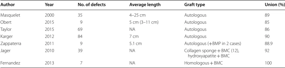
Table 1 English literature case series results of long bone defect reconstruction by induced membrane technique
Author Year No. of defects Average length Graft type Union (%)
Masquelet 2000 35 4–25 cm Autologous 89 Obert 2015 9 5 cm (3–11 cm) Autologous 85
Taylor 2015 69 NA Autologous 86
Karger 2012 84 7 cm Autologous 90
Zappaterra 2011 9 5.1 cm Autologous (+BMP in 2 cases) 88.9 Jager 2010 39 NA Collagen sponge + BMC (12),
rotational flap of the soleus muscle in four cases, pro-peller rotational flaps in two cases, and free anterolat-eral thigh flap in one case. The average time interval between trauma and soft tissue reconstruction was 8.5 days (range 6–12 days). The second stage of the pro-cedure was performed after an average of 3.2 months from the injury; this interval was shorter than 2 months in 5 fractures (28%), between 2 and 4 months in 11 fractures (61%), and longer than 4 months in 2 frac-tures (11%). The procedure included a skin incision over the previous one, sharp dissection of the mem-brane, removal of cement spacer, and opening of med-ullary canals. Intraoperative cultural tests were drawn routinely, but only one (6%) proximal femur defect was positive for Staphylococcus aureus.
In all cases, we utilized a combination of autologous graft and cancellous allograft. Autologous graft was vested from the ipsilateral anterior iliac crest. After har-vesting, it was not manipulated any more. Fresh-frozen cancellous bone allograft, provided by the local bone bank, was thawed in hot saline solution and added to bulk up the defect. BMCA and PRP were employed to enrich the graft. Both were prepared according to the technique recommended by Regen Lab SA (Le Mont-sur-Lausanne, Switzerland). BMCA requires 20 cc medul-lary blood aspirated from the contralateral anterior iliac crest. Five cc medullary blood is drawn each time, redi-recting the needle in various directions within the bone to avoid aspiration of venous blood. The blood is then inserted into dedicated tubes and centrifuged for 8 min at 3400 rpm to isolate the mesenchymal cell component.
The procedure for PRP requires 14 cc peripheral venous blood from an antebrachial vein. This drawing is centrifuged for separation of PRP. BMCA and PRP are subsequently added to the graft with addition of calcium gluconate to ensure gelification of the preparation to pro-mote adhesion of mesenchymal cells and platelets to the graft. Ten minutes later, the graft is ready to be placed into the defect. Accurate suturing of the membrane over the graft is mandatory; usually, suction drains are not recommended.
Definitive bone fixation was achieved using intramed-ullary nailing in 2 cases (11%), plating in 15 cases (66%), and external fixation in 1 case (6%).
Patients were evaluated clinically and radiographi-cally with average follow-up of 37 months (range 25–92 months). They were investigated for residual pain, joint range of motion, walking ability, deformity, and graft integration.
The radiological criterion for long bone healing was at least two healed corticals.
Statistical analysis was carried out using the chi-square and Student t-test, with p value < 0.05 considered significant.
Results
Both clinical and radiographic union were achieved in 13 (72%) cases in 13 patients (Fig. 1). Five (28%) cases in four patients showed delayed union. Nonunion was observed in two of eight (25%) tibial defects and in three of nine (33%) femoral defects (ns). The single humeral defect united uneventfully.
Two patients were affected by a double defect, involv-ing the distal femur bilaterally in one case and distal femur and distal tibia in the other case. Three of 4 (75%) double defects showed delayed union, whereas 2 of 14 (14%) single defects did not heal (p = 0.016).
The average length of the 13 defects that united was 6 cm (range 1.6–11.8 cm), and the length of the 5 defects that did not unite was 10.3 cm (range 6–13.2 cm) (p= 0.009). The average age of the five patients who suf-fered nonunion was 51.5 years (range 28–66 years). The average age of the 11 patients bearing defects that united was 41.6 years (ns).
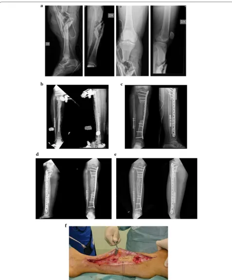
healed after 5 months. The third case of nonunion occurred in a 60-year-old male affected by multiple fractures including a grade IIIB open fracture of the left distal tibia which was stabilized with an EF and covered with an anterolateral thigh flap. Four months later, the bone defect was grafted and definitive fixation with an anterolateral plate undertaken. Eleven months later, radiographic evaluation demonstrated nonunion with slight varus deformity. The patient underwent autolo-gous bone grafting and repeated fixation with con-solidation of the fracture. The last case occurred in a 50-year-old man with a grade IIIB open fracture of the right tibia. The fracture was stabilized with an external fixator, and an anterolateral thigh flap was used for soft tissue reconstruction. Five months later, the 12.3-cm defect was filled with grafts and stabilized with a 4.5-mm medial plate. After 2 years, radiographic signs of incomplete healing with initial valgus deformity were
evident. The plate was removed, and no macroscopic signs of osteointegration of the graft were observed (Fig. 2). The anterolateral plate was substituted, filling the defect with an autologous bone graft. At last follow-up (42 months), consolidation was achieved.
Bone union occurred without loss of reduction in 12 defects stabilized with internal fixation, while a tibial defect, stabilized with external fixation, healed with 5° deformity in the coronal plane and 10° in the sagittal.
Thirteen patients with 13 healed defects that united were also clinically evaluated. Five (38%) defects united with average shortening of 2 cm (range 1.5–3 cm). Short-ening was compensated with a corresponding lift in the patient’s shoe.
The knee range of motion for patients affected by fem-oral defects averaged 108° (range 90–125°) with average extension lag inferior to 5°.
For patients affected by tibial defects, the average ankle range of motion was 10° in dorsiflexion (range 0–20°), 32° in plantar flexion (range 10–55°), with 50% loss of pro-nosupination as compared with the unaffected ankle. Five (38%) patients were free from pain, 7 (54%) complained of occasional pain, and 1 (7%) patient managed pain with daily consumption of antiinflammatory drugs.
Twelve patients (13 fractures) were satisfied with the final outcome, while four patient (5 fractures) were not satisfied.
Discussion
We reconstructed 18 critically sized posttraumatic long bone defects using an induced membrane technique based on an autologous cancellous graft augmented with fresh-frozen bone chips, BMCA, and PRP. This combina-tion represents a modificacombina-tion of the technique described by Masquelet, where autologous cancellous graft is rec-ommended. The cited author admits augmentation with allograft chips as long as the allograft/autologous graft ratio does not exceed 1:3 [7]. In our series, the allograft/ autologous graft ratio was 1:1 or higher in all cases. Graft enrichment with BMCA and PRP was supposed to increase the osteogenic and osteoinductive potential.
Bone union occurred in 13 (72%) out of 18 cases, while nonunion occurred in 5 (28%) cases. Our success rate remains inferior to the series reported by the origi-nators of the technique, where autologous bone graft was employed [6, 18]. Such series report a union rate of 85–89%. Three out of four (75%) double defects showed delayed union, while single bone defects showed delayed union in 2 of 14 cases (14%) (p = 0.045). The higher nonunion rate of double defects may be explained by the greater allograft/autologous graft ratio of the bone graft. A single anterior iliac crest was used to harvest the autologous graft in single as well as double defects. Furthermore, the average length of the double defects was 9.5 cm, while it was 6 cm for the single defects (p < 0.0001). The larger size of the double defects and the higher allograft/autologous graft ratio may have con-tributed to the higher incidence of nonunion for these defects.
The nonunion rate of the femur (three out of nine cases; 33%) was greater than that for the tibia (two of eight cases; 25%), although this difference did not reach statistical significance. All three femoral nonunions occurred at the distal femur. It may be considered that reconstruction of a complete bone defect of the distal femur with a lateral locking plate may be prone to varus instability, delayed union, and fixation failure. Stability
could be increased by adding a medial cortical bone strut or medial plate [19]. The average length of defects with delayed union was 10.7 cm (range 8–13.2 cm), while in those defects that united the average length was 6 cm (range 1.6–11.8 cm) (p = 0.009). These results suggest that larger defects suffer from a higher risk of nonunion.
We did not observe any stress fractures with this tech-nique, as reported by other authors [6]. This may proba-bly be due to the fixation device, which was not removed so far. For definitive osteosynthesis, we used a nail in 2 cases, a plate in 12 cases, and an external fixator in only 1 case. Stress fracture could eventually be an issue in case of hardware removal.
Management of posttraumatic bone defects using the induced membrane technique offers several advantages. Early application of a cement spacer prevents soft tissues from entering the bone defect and increases local deliv-ery of antibiotics. It is usually possible to perform inter-nal fixation with a plate or nails, which are more tolerable for the patient and do not interfere with soft tissue care.
An issue of concern is the amount of cancellous auto-graft available from each crest. It has been calculated that reconstruction of a diaphyseal tibial or femoral defect requires an average of 7 cm3 of autologous graft per cen-timeter of defect. If the defect is located in the meta-physeal region, the average amount required increases to 12 cm3 [20]. Anterior and posterior iliac crests pro-vide an average of 21 and 36 cc of graft, respectively [3]. Although the posterior iliac crest provides a larger graft volume, its harvesting requires prone positioning of the patient. In this series, all operations were carried out with the patient in supine position, thus we preferred to har-vest the graft from the anterior iliac crest. A diaphyseal bone defect greater than 5 cm in length requires harvest-ing from two or more iliac crests. The rate of complica-tions after iliac crest harvesting varies from 0.7 to 19.4% [21].
technique carried out by Stafford suggested RIA to be a valid alternative to iliac graft. On the other hand, Karger pointed out the issue of irregular ossification of the RIA graft when used alone. It is therefore advis-able to use RIA graft as an augmentation of autologous graft [18].
BMCA is another option for enhancing bone healing, and can be used with autologous bone graft or with a bone graft substitute. In an experimental study [15], use of hydroxyapatite, BMCA, and PRP demonstrated the same healing potential as autologous graft. Use of hydroxyapatite and PRP or use of hydroxyapatite alone led to inferior results. In a clinical study, bone mar-row concentrate was used in combination with either a collagen sponge (12 patients) or bovine hydroxyapa-tite (27 patients) to treat 39 bone defects [23]. At radi-ographic follow-up, all patients demonstrated new bone formation, but two patients underwent revision surgery due to insufficient bone healing. Complete bone healing was achieved after 17.3 weeks in the hydroxyapatite group, compared with 22.4 weeks in the collagen group. The authors concluded that BMCA combined with hydroxyapatite can reduce bone heal-ing time when compared with BMCA with collagen sponge [16].
Platelet-rich plasma (PRP) is a technique for concen-trating platelets and their growth factors to optimize bone healing. Both in vitro and in vivo experimental studies suggest the efficacy of PRP to stimulate a bone healing response, but the best approach for its clinical use remains unclear. The best method for delivery of growth factors in active form has still not been defined, and additional confounding factors include the small clinical series available and the large number of com-mercially available preparation kits.
Bone morphogenetic proteins are osteoinductive agents that are Food and Drug Administration (FDA) approved for spinal fusion procedures. Previous enthu-siasm in literature for off-label use of BMPs in segmen-tal bone defects has diminished more recently, mainly due to rising concerns regarding their adverse effects. In a recent study [24] carried out on 118 patients who underwent long bone nonunion surgery either with autogenous graft alone (50 patients) or with autogenous graft in combination with rhBMP-2, the authors were unable to demonstrate the desired synergistic effect of rhBMP-2.
In conclusion, reconstruction of long bone defects using the induced membrane technique appears to be a reasonable option. Autogenous cancellous bone graft still remains the gold standard. In our series, the use of a combined allograft/autogenous graft with added BMCA and PRP led to poorer results than described in literature
with exclusive use of autogenous cancellous graft. Fur-ther research aimed to develop alternative options to autogenous cancellous bone graft is desirable to provide adequate amounts of graft, shorten operating times, and avoid donor-site morbidity.
Authors’ contributions
FP, LB, SB carried out the controls over the patients. FP, SB, DAC and RB car-ried out the interventions. MJC participated in the manuscript drafting. The authors declare that they have no competing interests. The authors have no financial interests to declare in relation to the content of this article. All authors read and approved the final manuscript.
Competing interests
The authors declare that they have no competing interests. The authors have no financial interests to declare in relation to the content of this article.
Ethics approval and consent to participate
All procedures performed in studies involving human participants were in accordance with the ethical standards of the institutional and/or national research committee and with the 1964 Helsinki Declaration and its later amendments or comparable ethical standards. Informed consent was obtained from all individual participants included in the study.
Funding
No funding was used for this study.
Publisher’s Note
Springer Nature remains neutral with regard to jurisdictional claims in pub-lished maps and institutional affiliations.
Received: 24 July 2017 Accepted: 4 July 2018
References
1. Mauffrey C, Barlow BT, Smith W (2015) Management of segmental bone defects. J Am Acad Orthop Surg 23(3):143–153
2. Calori GM, Mazza E, Colombo M, Ripamonti C (2011) The use of bone-graft substitutes in large bone defects: any specific needs? Injury 42(Suppl. 2):S56–S63
3. Dawson J, Kiner D, Gardner W, Swafford R, Nowotarski PJ (2014) The reamer-irrigator-aspirator as a device for harvesting bone graft compared with iliac crest bone graft: union rates and complications. J Orthop Trauma 28(10):584–590
4. Schmidmaier G, Herrmann S, Green J et al (2006) Quantitative assessment of growth factors in reaming aspirate, iliac crest, and platelet preparation. Bone 39(5):1156–1163. https ://doi.org/10.1016/j.bone.2006.05.023
5. Masquelet AC (2003) Muscle reconstruction in reconstructive surgery: soft tissue repair and long bone reconstruction. Langenbecks Arch Surg 388(5):344–346
6. Masquelet AC, Fitoussi F, Begue T, Muller GP (2000) Reconstruction of the long bones by the induced membrane and spongy autograft. Ann Chir Plast Esthétique 45(3):346–353
7. Masquelet AC, Begue T (2010) The concept of induced membrane for reconstruction of long bone defects. Orthop Clin N Am 41(1):27–37 8. Taylor BC, French BG, Fowler TT, Russell J, Poka A (2012) Induced
mem-brane technique for reconstruction to manage bone loss. J Am Acad Orthop Surg 20(3):142–150
9. Masquelet A-C, Benko PE, Mathevon H, Hannouche D, Obert L (2012) Harvest of cortico-cancellous intramedullary femoral bone graft using the Reamer-Irrigator-Aspirator (RIA). Orthop Traumatol Surg Res 98(2):227–232
11. Apard T, Bigorre N, Cronier P, Duteille F, Bizot P, Massin P (2010) Two-stage reconstruction of post-traumatic segmental tibia bone loss with nailing. Orthop Traumatol Surg Res 96(5):549–553
12. Vadalà G, Di Martino A, Russo F et al (2016) Autologous bone mar-row concentrate combined with platelet-rich plasma enhance bone allograft potential to induce spinal fusion. J Biol Regul Homeost Agents 30(4):165–172
13. Hernigou P, Dubory A, Roubineau F et al (2017) Allografts supercharged with bone-marrow-derived mesenchymal stem cells possess equivalent osteogenic capacity to that of autograft: a study with long-term follow-ups of human biopsies. Int Orthop 41(1):127–132
14. Giannoudis PV, Einhorn TA, Marsh D (2007) Fracture healing: the diamond concept. Injury 38(4 Suppl):S3–S6
15. Hakimi M, Grassmann J-P, Betsch M et al (2014) The composite of bone marrow concentrate and PRP as an alternative to autologous bone graft-ing. PLoS ONE 9(6):e100143
16. Fernandez-Bances I, Perez-Basterrechea M, Perez-Lopez S et al (2013) Repair of long-bone pseudoarthrosis with autologous bone marrow mononuclear cells combined with allogenic bone graft. Cytotherapy 15(5):571–577
17. Henry SL, Ostermann PA, Seligson D (1993) The antibiotic bead pouch technique. The management of severe compound fractures. Clin Orthop Relat Res 295:54–62
18. Karger C, Kishi T, Schneider L, Fitoussi F, Masquelet A-C (2012) Treatment of posttraumatic bone defects by the induced membrane technique. Orthop Traumatol Surg Res 98(1):97–102
19. Ehlinger M, Ducrot G, Adam P, Bonnomet F (2013) Distal femur fractures. Surgical techniques and a review of the literature. Orthop Traumatol Surg Res 99(3):353–360
20. Abdollahi K, Kumar PJ, Shepherd L, Patzakis MJ (1999) Estimation of defect volume in segmental defects of the tibia and femur. J Trauma 46(3):413–416
21. Dimitriou R, Mataliotakis GI, Angoules AG, Kanakaris NK, Giannoudis PV (2011) Complications following autologous bone graft harvesting from the iliac crest and using the RIA: a systematic review. Injury 42(Suppl 2):S3–15
22. Nauth A, Lane J, Watson JT, Giannoudis P (2015) Bone graft substitution and augmentation. J Orthop Trauma 29(Suppl 1):S34–S38
23. Jäger M, Herten M, Fochtmann U et al (2011) Bridging the gap: bone marrow aspiration concentrate reduces autologous bone grafting in osseous defects. J Orthop Res 29(2):173–180