O R I G I N A L R E S E A R C H
Open Access
Evaluation of the etiology of persistent iritis
after cataract surgery
Kristin Neatrour
*, Allison McAlpine, Timothy Brooks Owens, Rupal H. Trivedi and Lynn J. Poole Perry
Abstract
Background:The purpose of this study was to evaluate patients with persistent iritis after cataract surgery to determine its incidence and risk factors. Adjusting the management of patients at risk could allow for a more predictable post-operative course and outcome. A retrospective chart review was performed of patients who had post-operative iritis longer than 1 month after cataract surgery during a 2-year period at Storm Eye Institute at the Medical University of South Carolina (MUSC) in Charleston, South Carolina. Patient demographics and various pre-operative, intra-pre-operative, and post-operative factors were analyzed for trends.
Results:Thirty-nine patients (49 eyes) met the inclusion criteria, and this group was compared to a control cohort of 40 patients (66 eyes) who did not have persistent iritis after cataract surgery. The overall incidence of post-operative iritis was 1.75%. In all patients with post-post-operative iritis lasting greater than 1 month, African American race and pupil expansion device use were statistically significant factors. After excluding patients with a history of ocular inflammation or known inflammatory or autoimmune diagnosis (1.20% incidence), there were still a significantly higher proportion of African Americans compared to the control group. When patients with post-operative iritis of less than 6 months in duration were additionally excluded, the incidence was 0.32%, and history of diabetes was statistically significant in addition to race.
Conclusions:Risk factors for persistent iritis after cataract surgery include being diabetic, of African American racial background, and pupil expansion device use. These patients can be better informed of the higher risk of prolonged inflammation in their post-operative course, and peri-operative management can be tailored accordingly.
Keywords:Post-operative uveitis, Post-operative management, Iritis, Inflammation, Risk factors, Cataract surgery complications, Pseudophakia, Epidemiology, Incidence
Background
Cataract surgery techniques have evolved significantly in recent years. Modern surgical advances and medications have led to improved peri-operative management of in-flammation and better outcomes, even with challenging uveitic eyes [1]. Routine cataract surgery results in the release of inflammatory mediators, leading to some de-gree of post-operative inflammation in all patients. This post-operative inflammation is usually easily controlled with topical steroid tapers and typically resolves by 1 month after surgery.
In the presence of certain risk factors, such as a his-tory of uveitis, this post-operative inflammahis-tory period may be prolonged for weeks or months, punctuated by
periods of “rebound” inflammation when topical steroid drops are tapered. Studies have shown that the recur-rence rate of uveitis following cataract surgery is as high as 51% [2]. A study performed at Vanderbilt University found a significant association between postsurgical uve-itis and intra-operative complications, as well as worse visual acuity outcomes. The median duration of
inflam-mation was 10 months [3]. In a review of rheumatoid
arthritis patients undergoing cataract surgery, elevated pre-operative rheumatoid factor (RF) serum titers were associated with 1+ aqueous cell 1 month after surgery. Interestingly, analysis of patients with this level of post-operative cell and low poperative RF titers re-vealed that 75% of these patients had diabetes [4].
In the absence of a known predisposing etiology such as a history of uveitis or surgical complications, however, some patients still experience a prolonged course of * Correspondence:[email protected]
Storm Eye Institute, Medical University of South Carolina, 167 Ashley Avenue, Charleston, SC 29425, USA
inflammation after cataract surgery. The unique charac-teristics shared by this subset of patients have not been previously studied.
The purpose of this study was to evaluate patients who developed prolonged post-operative iritis, but who had no known risk factors, in order to determine the in-cidence and possible underlying etiologies or predispos-ing factors. To investigate this, a 2-year retrospective chart review was performed of patients at Storm Eye In-stitute at the Medical University of South Carolina (MUSC) who developed post-operative iritis persisting longer than 1 month after cataract surgery. Patient demographics and various pre-operative, intra-operative, and post-operative factors were evaluated for trends. These results were then compared to a cohort of pa-tients who did not have persistent post-operative iritis to evaluate for statistically unique characteristics.
Methods
The MUSC Institutional Review Board approved this retrospective study, and the research followed the tenets of the Declaration of Helsinki. The data requests were submitted through the Services, Pricing, and Application for Research Centers (SPARC) at MUSC, and this process complied with the Health Insurance Portability and Accountability Act. A list of medical record num-bers was provided for all patients in the 2-year time period (from November 2013 to September 2015) whose charts had International Classification of Diseases (ICD) codes for both “pseudophakia” (ICD-10 Z96.1) and “ iri-docyclitis”(ICD-10 H20–H20.9). These charts were then
analyzed for pre-operative, intra-operative, and
post-operative characteristics as discussed below. To de-termine the incidence of persistent post-operative iritis, another SPARC data query produced the number of total procedures performed in the 2-year time period that were billed with a Current Procedural Terminology (CPT) code of 66982 or 66984. The incidence was calcu-lated strictly based on surgery date occurring within the study period, whereas the subsequent analyses included the cohort of patients with clinic visits for prolonged post-operative iritis in that time period in order to study a larger sample size.
Pre-operative characteristics included basic demo-graphics of age, gender, and race. It was noted if this was the first or second eye undergoing cataract surgery, as well as the course of the other eye, if applicable. If pa-tients had a history of ocular inflammation, then the ophthalmic diagnosis, underlying systemic inflammatory diagnosis with supporting lab or imaging studies, base-line systemic or topical anti-inflammatory medications, and pre-operative anti-inflammatory prophylaxis regi-men were recorded. No prophylaxis was prescribed un-less there was a uveitic history, in which we routinely
prescribe an oral prednisone taper at the time of surgery. All ophthalmic diagnoses, prior ocular surgeries, prior ocular trauma, and other medical diagnoses were also included. For patients with glaucoma, medical and surgi-cal treatments were listed. Diagnosis of diabetes was re-ported along with duration, peri-operative hemoglobin A1c (HbA1c), status of diabetic retinopathy (DR)
includ-ing macular edema, and if a recent intravitreal injection was administered, based on information available in the chart. The type of cataract (senile, uveitic, or traumatic) and grade (for nuclear, cortical, and posterior subcapsu-lar classifications) were noted.
Intra-operatively, the attending surgeon’s name was doc-umented in the operative note, but the resident’s level of participation in the case was typically not included. There were five attending cataract surgeons at MUSC during this time period. It was noted if femtosecond laser-assisted cataract surgery (FLACS) was performed and if intracam-eral antibiotics were administered, if known. Any docu-mented findings outside of the standard operative note template were recorded, including the use of a pupil ex-pansion device, intra-operative floppy iris syndrome (IFIS), posterior capsule violation, anterior vitrectomy, and intra-ocular lens (IOL) suture fixation and the use of triamcino-lone, and if the cataract surgery was combined with any other procedures, such as a trabeculectomy or minimally invasive glaucoma surgery (MIGS). The type of IOL (e.g., SN60WF) and implanted location (posterior chamber, sul-cus, or anterior chamber) were also recorded.
Post-operative topical drops used at our institution
in-clude a month of a topical non-steroidal
anti-inflammatory drug (typically ketorolac four times daily) and a 4-week taper of prednisolone (four times per day for 1 week followed by a weekly tapering off the drop). Post-operative findings included the duration of persistent iritis (in months) as well as the extent of an-terior chamber cell and flare (evaluated with slit lamp examination according to the Standardization of Uveitis
Nomenclature system) [5]. Patient symptomatology was
noted if specified in the record. Response to topical ther-apy was categorized as rapid or indolent and persistent, and any topical and/or systemic adjunctive treatment was listed. Any documentation indicating good or poor
compliance was included. Lastly, post-operative
best-corrected visual acuity (BCVA) and maximum in-traocular pressure (IOP) were recorded.
Patient and peri-operative characteristics were ana-lyzed with descriptive statistic calculations. Continuous variables were analyzed with a two-tailedt test and cat-egorical variables were evaluated with the Fisher exact test. Visual acuity based on the Snellen chart was con-verted into logarithm of the minimum angle of
reso-lution (logMAR) units. A p value of < 0.05 was
considered statistically significant for the analysis.
Results
During the 2-year time period, 2169 cataract surgeries were performed at MUSC. Of these cases, 38 eyes had post-operative iritis lasting longer than 1 month (1.75%). Eliminating eyes with a prior history of ocular inflamma-tion or pre-existing systemic inflammatory or auto-immune diagnosis led to an incidence of 1.20% (26 eyes). After excluding the eyes with both post-operative iritis less than 6 months in duration and a prior history of inflammation, the incidence was 0.32% (7 eyes).
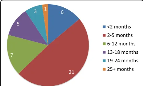
Thirty-nine patients (49 eyes) were seen in the clinic for the evaluation and management of prolonged post-operative iritis during the study time period. Fig-ure 1 shows the relative distribution of patients catego-rized by the duration of post-operative iritis. Six [6] patients were excluded from this subset analysis because 3 patients were lost to follow-up and 3 patients had on-going iritis at the time of data collection (n= 43). The average duration in this subset was 7.2 (± 7.7) months, and the median was 4 months.
The analysis of all patients comparing the cases of pro-longed post-operative inflammation with the control cases showed that African American race and the use of an intra-operative pupil expansion device were statisti-cally significant. When patients with prior ocular inflam-mation or pre-existing diagnosis were excluded, African American race remained statistically significant. After additionally excluding patients who had iritis for less
than 6 months post-operatively, history of diabetes be-came significant, in addition to African American race.
Table 1 summarizes the patient demographic and
pre-operative/intra-operative characteristics.
The analysis of uveitis patients showed that 13 patients had a history of prior ocular inflammation. The types of uveitis cases reported were as follows: panuveitis (1); ir-itis (4); anterior and intermediate uveir-itis (2); episclerir-itis (2); and uveitis, not further specified (5). Four patients were taking prednisone or a disease-modifying antirheu-matic drug at baseline. Four of the cataracts were de-scribed as uveitic. Intra-operatively, pupil expansion devices were used in 6 cases (5 iris hooks and 1 malyu-gin ring). No cases were complicated by a posterior cap-sular tear or anterior vitrectomy. The average duration of iritis amongst these patients was 5.8 (± 5.0) months, and there were 7 cases of iritis lasting greater than 6 months. Post-operatively, about 50% of patients reported iritis symptoms (6 patients/8 cases) including pain, photophobia, and/or blurry vision. Additionally, 50% showed a rapid improvement with additional treatment.
Table 2 provides a detailed summary of findings
amongst diabetic patients in the iritis and control groups.
There were 19 patients diagnosed with glaucoma or as
a glaucoma suspect receiving treatment. Table3
summa-rizes the treatment differences between the iritis and control groups. There were no statistical differences be-tween the two groups.
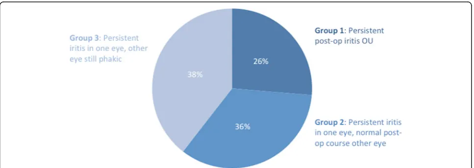
The patients with prolonged iritis were separated into groups according to whether one or both eyes were af-fected (see Fig.2). Group 1 (10 patients) had persistent post-operative iritis in both eyes. Group 2 (14 patients) had both eyes operated, but only one eye had persistent iritis, which was evenly divided between the first or sec-ond eye being affected. Group 3 (15 patients) had per-sistent iritis in the one eye, but were still phakic in the other eye. All of the patients in group 1 were females, whereas the majority of patients in group 2 were males. Group 2 was compared to group 1 to assess the charac-teristics of cases where both eyes underwent cataract surgery and the one eye versus both eyes was affected. In group 2, 2 patients had iritis only in the eye where a pupil expansion device was used compared to the fellow eye where it was not used. Similarly, the patient with prior ocular trauma had prolonged iritis only in the af-fected eye. This analysis suggests that these factors may have a correlation with persistent iritis in this subset of patients, but the sample size is too small to derive statis-tical significance. History of diabetes was not statisstatis-tically significant between groups 1 and 2.
Assessment of type and grade of the cataracts revealed that all patients in the control group had senile cata-racts, whereas 4 patients in the persistent iritis group Fig. 1Number of eyes versus the length of iritis. Legend: Fig.1
had uveitic cataracts (the remainder were senile). Twenty-nine of the 66 eyes (43.9%) in the control group had grade 3 or greater cataracts versus 15 of the 49 eyes (30.6%) in the persistent iritis group.
Intra-operative characteristics of both groups were an-alyzed. Femtosecond laser-assisted surgery was used for 23 cases in the control group and 2 cases in the iritis
group. Table 4 summarizes the type of pupil expansion
device used. One patient in the iritis group had IFIS re-quiring anterior vitrectomy (no cases of IFIS in the con-trol group). That was also the only case where a sulcus IOL was placed (versus posterior chamber IOL in all
other patients in both groups). Nine of the surgeries in the control group were combined with other procedures (e.g., trabeculectomy) versus only 3 of the surgeries in patients who developed persistent iritis. In the persistent iritis group, intracameral antibiotics were given in 85.7% of cases in the persistent iritis group and 95.4% of cases in the control group.
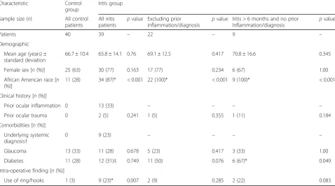
Post-operatively, average BCVA in the control group was 0.16 (± 0.17) logMAR, and the average BCVA in the persistent iritis group was 0.26 (± 0.43) logMAR. Aver-age maximum IOP was 20 in the control group and 16 in the iritis group. Further analysis of the persistent iritis Table 1Comparison of characteristics between patients with persistent post-operative iritis versus control group with associatedp values
Characteristic Control
group
Iritis group
Sample size (n) All control
patients
All iritis
patients p
value Excluding prior
inflammation/diagnosis p
value Iritis > 6 months and no prior
Inflammation/diagnosis p
value
Patients 40 39 – 22 – 9 –
Demographic
Mean age (years) ± standard deviation
66.7 ± 10.4 65.8 ± 14.1 0.76 69.1 ± 12.5 0.417 70.8 ± 16.6 0.345
Female sex [n(%)] 25 (63) 30 (77) 0.163 17 (77) 0.234 6 (67) 1.00
African American race [n (%)]
11 (28) 34 (87)* < 0.001 22 (100)* < 0.001 9 (100)* < 0.001
Clinical history [n(%)]
Prior ocular inflammation 0 13 (33) – – – –
Prior ocular trauma 0 2 (5) 0.241 1 (5) 0.355 1 (11) 0.184
Comorbidities [n(%)]
Underlying systemic diagnosis†
0 9 (23) – – – –
Glaucoma 13 (33) 11 (28) 0.678 5 (23) 0.417 3 (33) 1.00
Diabetes 11 (28) 12 (31)‡ 0.749 11 (50) 0.076 6 (67)* 0.049
Intra-operative finding [n(%)]
Use of ring/hooks 1 (3) 9 (23)* 0.007 2 (9) 0.285 2 (22) 0.083
*Significant at the 0.05 probability level
†The diagnoses were as follows: ulcerative colitis (1), sarcoidosis (4), multiple sclerosis (2), rheumatoid arthritis (1), ankylosing spondylitis (1), and HSV (1) ‡Of the 12 patients in this group, 0 had a recent intravitreal injection, and the peri-operative HbA1cranged from 5.9–7.8%. Four of these patients had
documented DR without cystoid macular edema (CME). One patient with diabetes was noted to have CME not due to DR. Two other patients were found to have pre-existing CME without the diagnosis of diabetes
Table 2Comparison of diabetic history and demographics between the iritis and control groups
Iritis patients Control patients
Diabetic patients 12 11 (16 cases)
+ Diabetic retinopathy 4 3
+ Diabetic macular edema 1 0
+ Recent intravitreal injection 0 0
Female sex 9 6
African American race 11 6
Average HbA1c 6.42% (5/12 known) 6.83% (10/16 known)
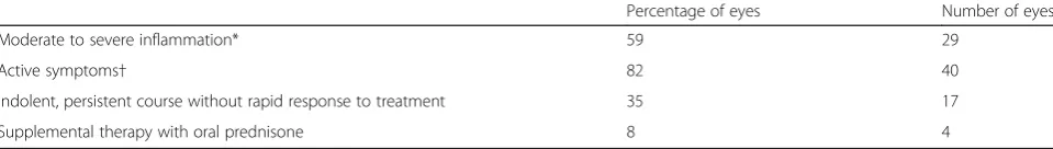
group is summarized in Table 5. Compliance ratings were assigned based on attendance at scheduled follow-up visits and attending comments on adherence to treatment regimens. Two patients (2 eyes) were noted to have poor compliance.
Discussion
This is the first report of risk factors associated with prolonged post-operative iritis in patients who under-went uncomplicated cataract surgery by phacoemulsifi-cation and who did not have a history of uveitis or other
underlying inflammatory disorders. Prolonged
post-operative iritis represents a significant burden amongst uveitis cases seen in the southeastern part of the USA. A study of uveitis diagnoses at the University of Virginia over a 30-year period revealed that 10% of all cases of uveitis were “post-procedural,” with 48.7% of those cases (approximately 5% of the total burden) asso-ciated with cataract extraction and IOL implantation [6]. In our study, there was a significant association between a diagnosis of diabetes and an extended duration of
post-operative iritis lasting longer than 6 months. Afri-can AmeriAfri-can race was also signifiAfri-cantly correlated with prolonged post-operative iritis when predisposing factors such as known prior uveitis episode(s) or an underlying inflammatory disease diagnosis were excluded.
The association of diabetes with prolonged post-operative inflammation after cataract surgery is perhaps not surpris-ing in light of the link between inflammation and diabetes [7]. In recent years, the theory that obesity leads to chronic systemic inflammation by activation of immune cells with the release of inflammatory mediators, and thereby in-creased the risk of metabolic disorders such as diabetes, has become well established [8]. Several studies have confirmed higher levels of flare correlating with inflammatory markers in the aqueous fluid in patients who are diabetic compared to non-diabetic patients [9,10]. Higher levels of flare are as-sociated with a breakdown of the blood-aqueous barrier in diabetic patients and may persist for many months after cataract surgery [11]. It may be theorized that persistent hyperglycemia and a chronic systemic pro-inflammatory state predispose diabetic patients to a prolonged course of Table 3Treatment comparison of glaucoma or glaucoma suspect patients
Treatment All patients Iritis patients Control patients
Medical
Alpha agonist 6 3 3
Carbonic anhydrase inhibitor (topical) 8 4 4
Beta blocker 11 5 6
Prostaglandin analogue 10 5 5
Direct cholinergic agonist 1 0 1
Carbonic anhydrase inhibitor (oral) 1 1 0
Laser
Selective laser trabeculoplasty 5 2 3
Laser peripheral iridotomy 3 2 1
post-operative iritis refractory to standard 1-month topical steroid treatment tapers.
There have been several studies that have examined the differences in aqueous markers in diabetic and non-diabetic patients after cataract surgery. Miric et al. studied lens oxidative stress markers in patients after se-nile cataract extraction. Serum and lens levels of xanthine oxidase were higher in the diabetic than the non-diabetic group, and they positively correlated with HbA1c concentration, suggesting that the diabetic state
could possibly contribute to earlier cataract formation [12]. Additionally, Mitrovic et al. found significantly al-tered levels of inflammatory mediators in the aqueous humor of diabetic patients following cataract surgery, leading to a predisposition for corneal edema [13]. While the exact mechanism behind increased inflammation in diabetic eyes is unknown, there are probably many
po-tential pathways involved. Prolonged inflammation
within the eye may not follow the same patterns as
in-flammation elsewhere in the body due to its
immune-privileged state.
African American race remained a statistically signifi-cant factor in all comparisons. This may be attributed to the fact that the African American race is an independ-ent risk factor for inflammation and oxidative stress, positing that the inflammatory response following cata-ract surgery may be more robust in these patients [14].
Unsurprisingly, intra-operative pupil expansion device use correlated with prolonged post-operative iritis, likely due to iris trauma during insertion and manipulation of pupil expansion devices leading to disruption of the
blood-aqueous barrier. The density of the cataract or history of previous ocular trauma did not appear to have an increased risk of prolonged post-operative inflamma-tion in this study.
The incidence of persistent iritis in our study was 0.32%, which is similar to that previously reported by Patel et al. [3]. The incidence was 0.24% in Patel’s study with the exclusion criteria similar to those of our own study: duration of iritis less than 6 months, prior uveitis or underlying systemic disease, penetrating trauma, en-dophthalmitis, retained lens material, neovascular glau-coma, and prior intraocular surgery.
This study is limited by sample size as well as the ex-pected limitations of any retrospective data collection. The results were confined to information available in the electronic medical record and were subject to variability in provider documentation (e.g., various exam documen-tation styles, grading scales, codes), which can introduce bias. The incidence rate may be underestimated because the charts for the CPT data query were not all reviewed, so some cases of prolonged post-operative inflammation may have been missed. Certain parameters such as pa-tient symptomatology and compliance with treatment regimen could not be evaluated unless they were deliber-ately reported in the charts. Multivariate analyses could not be run due to the relatively small sample size, which was partially limited by the ability to perform a compre-hensive chart review since some information did not transfer or was not easily searchable during or pre-dating the conversion to electronic medical records. Despite these limitations, this report provides an ex-tremely important first step in the evaluation of risk fac-tors for prolonged post-operative iritis, which have not been previously reported in the literature.
Overall, in both groups, the post-operative refractive outcomes were excellent. Long-term refractive outcomes after cataract surgery are dependent on many factors, in-cluding pre-existing ocular morbidity, intra-operative complications, and development of post-operative se-quelae such as cystoid macular edema or retinal detach-ment. While a prolonged post-operative inflammatory course may not necessarily lead to a worse visual acuity
outcome, it can be associated with delayed
post-operative visual recovery and with patient
Table 4Pupil expansion devices used for cases in the iritis and control groups and comparison of device choice based on inflammation history
Malyugin ring Iris hooks
Total number of cases 5 5
Iritis group 4 5
Control group 1 0
Cases with prior ocular inflammation 1 5
Cases with known inflammatory disease 1* 3†
*Ulcerative colitis and ankylosing spondylitis (1) †Multiple sclerosis (2) and sarcoidosis (1)
Table 5Post-operative symptomatology, exam findings, and treatment course in the prolonged iritis group
Percentage of eyes Number of eyes
Moderate to severe inflammation* 59 29
Active symptoms† 82 40
Indolent, persistent course without rapid response to treatment 35 17
Supplemental therapy with oral prednisone 8 4
*Defined as having at least 2+ cell or moderate/3+ flare
discomfort. Supplementary peri-operative medications can improve control of post-operative outcomes in pa-tients who are at high risk pre-operatively for having prolonged or rebound post-op iritis.
Conclusions
This analysis showed that diabetes was a significant in-dependent risk factor for the development of chronic ir-itis lasting greater than 6 months in patients without a history of inflammation. Additionally, African American race was significantly correlated with post-operative iritis lasting greater than 1 month. We propose that altering the peri-operative treatment regimen for patients who are diabetic and/or African American could shorten and improve their post-operative recovery. Further investiga-tion with a large-scale prospective trial would allow for a more powerful statistical analysis and may help to ex-plain how these factors may contribute to post-operative inflammation.
Abbreviations
BCVA:Best-corrected visual acuity; DR: Diabetic retinopathy; FLACS: Femtosecond laser-assisted cataract surgery; HbA1c: Glycated
hemoglobin; hemoglobin A1c; ICD: International Classification of Diseases;
IFIS: Intra-operative floppy iris syndrome; IOL: Intraocular lens; IOP: Intraocular pressure; MIGS: Minimally invasive glaucoma surgery; MUSC: Medical University of South Carolina; RF: Rheumatoid factor; SPARC: Services, Pricing, and Application for Research Centers
Acknowledgements
Not applicable.
Funding
Not applicable.
Availability of data and materials
The datasets used and analyzed during the current study are available from the corresponding author upon reasonable request.
Declarations
The authors declare that this manuscript is original, has not been published before and is not currently being considered for publication elsewhere. There are no known conflicts of interest associated with this publication nor has there been financial support for this work that could have influenced its outcome. The manuscript has been read and approved by all named authors and there are no other persons who satisfied the criteria for authorship but are not listed.
Authors’contributions
KN led and coordinated the study concept and design and had major roles in data acquisition and analysis as well as in all the stages of the manuscript preparation. AM and TO collected and analyzed the data and contributed to the drafts of the manuscript. RT participated in the project concept and design, data interpretation and statistical analysis, and critical supervision of the manuscript. LP supervised the study with significant contributions to the design methodology, data analysis, and manuscript revision and preparation. All authors read and approved the final manuscript.
Ethics approval and consent to participate
The MUSC Institutional Review Board approved this retrospective study and the research followed the tenets of the Declaration of Helsinski (IRB Study #Pro00048917). The data requests were submitted through Services, Pricing, and Application for Research Centers (SPARC) at MUSC, and this process complied with the Health Insurance Portability and Accountability Act.
Consent for publication
Not applicable.
Competing interests
The authors declare that they have no competing interests.
Publisher’s Note
Springer Nature remains neutral with regard to jurisdictional claims in published maps and institutional affiliations.
Received: 13 August 2018 Accepted: 4 February 2019
References
1. Taravati P, Lam DL, Leveque T, Van Gelder RN (2012) Postcataract surgical inflammation. Curr Opin Ophthalmol 23(1):12–18
2. Agrawal R, Murthy S, Ganesh SK, Phaik CS, Sangwan V, Biswas J (2012) Cataract surgery in uveitis. Int J Inflamm 2012:548453
3. Patel C, Kim SJ, Chomsky A, Saboori M (2013) Incidence and risk factors for chronic uveitis following cataract surgery. Ocul Immunol Inflamm 21(2):130–134 4. Matsuo T, Fujiwara M, Matsuo N (1995) Inflammation after cataract
extraction and intraocular lens implantation in patients with rheumatoid arthritis. Br J Ophthalmol 79(6):549–553
5. Jabs DA, Nussenblatt RB, Rosenbaum JT (2005) Standardization of uveitis nomenclature for reporting clinical data. Results of the First International Workshop. Am J Ophthalmol 140(3):509–516
6. Bajwa A, Osmanzada D, Osmanzada S, Khan I, Patrie J, Xin W et al (2015) Epidemiology of uveitis in the mid-Atlantic United States. Clin Ophthalmol (Auckland, NZ) 9:889–901
7. Wellen KE, Hotamisligil GS (2005) Inflammation, stress, and diabetes. J Clin Invest 115(5):1111–1119
8. Hotamisligil G (2017) Inflammation, metainflammation and immunometabolic disorders. Nature 542:177–185
9. Takamura Y, Tomomatsu T, Arimura S, Tomomatsu Y, Matsumura T, Takihara Y et al (2013) Anterior capsule contraction and flare intensity in the early stages after cataract surgery in eyes with diabetic retinopathy. J Cataract Refract Surg 39(5):716–721
10. Ino-ue M, Azumi A, Shirabe H, Tsukahara Y, Yamamoto M (1994) Laser flare intensity in diabetics: correlation with retinopathy and aqueous protein concentration. Br J Ophthalmol 78(9):694–697
11. Liu Y, Luo L, He M, Liu X (2004) Disorders of the blood-aqueous barrier after phacoemulsification in diabetic patients. Eye (London, England) 18(9):900–904 12. Miric DJ, Kisic BB, Zoric LD, Mitic RV, Miric BM, Dragojevic IM (2013)
Xanthine oxidase and lens oxidative stress markers in diabetic and senile cataract patients. J Diabetes Complicat 27(2):171–176
13. Mitrovic S, Kelava T, Sucur A, Grcevic D (2016) Levels of selected aqueous humor mediators (IL-10, IL-17, CCL2, VEGF, FasL) in diabetic cataract. Ocul Immunol Inflamm 24(2):159–166