R E S E A R C H
Open Access
Double regularization medical CT image
blind restoration reconstruction based on
proximal alternating direction method of
multipliers
Jingyu Zhang
1, Yunshan Sun
2*, Yuan Zhang
1and Jianfu Teng
1Abstract
To solve the problem of CT image degradation, a double regularization CT image blind restoration
reconstruction method was proposed. The objective function including both a clear image and point spread function was established. To avoid the over-smoothing phenomenon and protect the detail, the objective function includes two constraint regularization terms. They are total variation (TV) and wavelet sparsity respectively. The objective function was solved by the alternating direction multiplier method (ADMM), and the optimal solution was obtained. Firstly, the CT image blind restoration reconstruction was decomposed into two sub-problems: reconstructed image estimation and point spread function estimation. Furthermore, each sub-problem can be solved by the proximal alternating direction method of multipliers. Finally, the CT image blind restoration reconstruction was realized. The experimental results show that the proposed algorithm takes into account the degradation effect of projection data, and the proposed algorithm is superior to other existing algorithms in the subjective visual effect. At the same time, in the aspect of objective evaluation, the proposed algorithm improves the objective image quality metrics such as peak signal-to-noise ratio (PSNR), structural similarity index metric (SSIM), and universal image quality index (UIQI).
Keywords:Blind restoration, Image reconstruction, CT image, Proximal alternating direction method of multipliers
1 Introduction
Computer tomography (CT) is that X-ray beam is used to scan some certain thickness parts of the body. X-ray through the detection layer is received by the detector. CT reflects the degree of organ and tissue absorption of X-ray. It is conducive to find small lesions in any part of the body. CT can greatly improve the early detection rate and patho-logical diagnosis rate. CT detection has become one of the most important diagnostic basis in the field of medical image diagnosis. However, in the imaging and transmission process, CT image will be affected by many factors, such as electronic radiation detection effect of defocus blur, detector number and spacing, frame geometry,
reconstruction algorithm and other factors. A point of hu-man tissue can be mapped to hu-many points on CT image. In other words, each point on CT image is a mixture of many points of human tissue. And its correspondence is generally referred to as a point spread function, that is, the transmission characteristics of the whole system. Image restoration can be achieved by measuring the point spread function of CT system to eliminate the effect of degrad-ation [1, 2].
Medical CT image blind restoration algorithm use only CT degradation image of the CT system to estimate the point spread function. Then, the purpose of image restor-ation is achieved and the measurement of the point spread function is avoided. Jiang et al. [3] firstly proposed the con-cept of medical CT blind restoration. They used the ex-pectation maximization strategy to estimate point spread function and image restoration. And, they gave the * Correspondence:[email protected]
2School of Information Engineering, Tianjin University of Commerce, Tianjin,
China
Full list of author information is available at the end of the article
assumption that the degradation function of CT system has Gaussian distribution. Multiple sets of bone interfaces and the iterative strategy [4–6] were utilized to estimate the point spread function. Blind equalization algorithm was applied to the two-dimensional CT blind image restor-ation [7–9]. Combining with the dimension reduction the-ory and orthogonal transform, without any information of degradation, only the features of degraded images were uti-lized to estimate the clear image. In addition, there are spe-cial uses of image restoration technology to eliminate specific CT artifacts [10–13]. In general, in the process of CT imaging, there is always the degradation. CT image degradation or artifact was eliminated by the use of special-ized techniques, but it is bound to increase the complexity of the algorithm. If we can consider the influence of the projection data degradation and overcome it, it will im-prove the quality of the reconstructed image.
Total variational (TV) is a very important optimization constraint in CT reconstruction and was widely studied and applied. Candes et al. [14] proposed a compressive sensing theory. This theory proved that sparse signals can be accurately restored by data far less than the sampling theorem, which greatly facilitates the research and application of TV constraints in CT reconstruction, especially sparse projection data CT reconstruction [15–18]. Hu et al. [19] proposed a reconstruction method that the pre-scan CT images or analytic recon-struction images were used as the iterative initial condi-tions to improve the speed of TV constraint reconstruction. In the whole variation method, the as-sumption that the image segmentation smoothing will cause the small edge of the CT image and the texture of the texture are blurred, resulting in the “smooth” phenomenon, there is also the problem of slow recon-struction. In order to solve the above problems, the re-searchers also combine some sparse constraints with the TV method, or directly use sparse constraints. Wang et al. [20] studied a TV-based optimization model for Micro-CT reconstruction based on an up-sampling of the reconstruction grid with original detector and X-ray dose. Using an extension of the gra-dient projection method, an alternating minimization algorithm is employed to solve the corresponding en-ergy function. Lu-zhen et al. [21] proposed a hybrid compression-aware reconstruction method based on the total variation and non-aliasing contourlet transform. The method uses non-aliasing sontourlet transform as a sparse representation of CT image. The objection function is solved by splitting the Bregman algorithm. In a word, it is helpful to improve the effect of image reconstruction by adding proper regularization term. Chen et al. [22] pro-posed image domain artifacts used in low-dose CT image suppression dictionary learning method. The establish-ment of sparse representation of the discriminant
dictionary filters a part of the artifacts and noise. And then, the general dictionary learning was used to further reduce noise and residual artifacts.
Alternating direction multiplier method (ADMM) is a simple and effective method for solving separable convex programming problems, especially on solving large-scale problems. In the ADMM algorithm, the objective function of the original problem is divided into several sub-problems which are easy to find local solutions, and the global solution of the original problem is obtained. It is also applied in the reconstruction of the compression percep-tion. Ramani et al. [23] used the method of variable separ-ation to separate the shift and shift invariant in the statistical data model and established two equivalent con-straint problems. Then, the objection function is solved by the alternating direction multiplier methods. Sawatzky et al. [24] established a multi-channel framework for the re-construction of medical CT images and optimized the so-lution by the alternating direction multiplier method.
At present, CT image reconstruction algorithm often does not consider the influence of CT system point spread function, but this assumption is unreasonable. CT image is often affected by the system point spread function in the imaging process. The point spread func-tion results in image degradafunc-tion [25]. In this paper, a new blind reconstruction model of medical CT image is established. Considering the influence of sampling data by degradation factors in image reconstruction, the pro-posed method avoids the elimination of point spread function alone. So it is beneficial to improve the quality of reconstructed image, reduce the complexity of the al-gorithm, and improve the ability of real-time processing.
2 Method
2.1 Proximal alternating direction method of multipliers for solving unconstrained optimization problems
Suppose an unconstrained optimization problem as follows:
min
x∈ℜn fð Þ þx gðGxÞ ð1Þ
where f:ℜn→(−∞, +∞) and g:ℜm→(−∞, +∞) are closed convex function and G∈ℜm×n is a transform-ation matrix.
ADMM is an effective method to solve formula (1), which combines the advantages of multiplier method and alternating minimization method. This method in-troduces dummy variables u and d. And then, an itera-tive solution is obtained by the idea of alternating minimization, that is,
xkþ1←arg min
x fð Þ þx
μ
2kGx−uk−dkk
2
2 ð2Þ
ukþ1←arg min
u gð Þ þu
μ
2kGxkþ1−u−dkk
2
2 ð3Þ
dkþ1←dk−ðGxkþ1−ukþ1Þ ð4Þ
Formulas (2)–(4) are iterated to obtain the optimal so-lution formula (1). The feasibility and convergence of ADMM has been demonstrated.
To improve the application of ADMM in the image processing problem, here considering a generic uncon-strained optimization problem:
min x∈ℜn fð Þ þx
X
i¼1 J
gi Hð Þix
ð5Þ
where gi(⋅) is a closed convex function. Hð Þj∈ℜmjn is a random matrix, andm=m1+m2+⋯+mJ. DefinitionG = [H(1),H(2),⋯,H(J)]∗∈ℜm×n, ∗ represents a conjugate transposition of matrix or a vector.
In image reconstruction, assumef(x) = 0, formula (2) be-comes a quadratic optimization problem in this case. The parametersμin formula (2) affect the convergence speed. In the sub-optimization problem, different weight
coefficients are used to improve the ability of the algo-rithm, that is, the quadratic optimization problem becomes
xkþ1←arg min
x ðGx−u−dÞ Υ
Gx−u−d
ð Þ ¼ arg min
x ðGx−ζÞ Υ
Gx−ζ ð Þ
ð6Þ
Here,Υ¼ diag μ1;⋯;μ1
|fflfflfflfflffl{zfflfflfflfflffl} m1 elements
;⋯;μj;⋯;μj |fflfflfflffl{zfflfflfflffl} mjelements
;⋯;μ|fflfflfflfflffl{zfflfflfflfflffl}J;⋯;μJ
mJelements 0
B B @
1 C C A.
The quadratic problem can be done using the gradient method (Gx−ζ)∗Υ(Gx−ζ) to solve, getting
xkþ1←ðGΥGÞ−1GΥζ¼ X
j¼1
J
μj Hð Þj
Hð Þj
" #−1
X
j¼1
J
μj Hð Þj
ζð Þj
ð7Þ
where ζð Þj ¼uð Þkj þd j
ð Þ
k ; ζ j
ð Þ;
uð Þkj with d j
ð Þ
k ðj¼1;⋯;JÞ
are respectively sub-vectors ofζ,uk, anddk. In formula (5), assume
gð Þ ¼u X
j¼1 J
gj uð Þj
ð8Þ
where uð Þj∈ℜmj is a sub-vector of u, u= [(u(1))∗,
⋯, (u(J))∗]∗.
Solving the iterative optimization, formula (8) is decom-posed into J-independent sub-optimization problems. Sub-optimization problem is solved by the proximity op-erator. Different optimization problems require different proximity operators. As can be seen, formula (8) is trans-formed intoJproximity operators to solve, which is
ukþ1←proxg=μðGxkþ1−dkÞ ð9Þ
where prox(⋅) is a proximity operator. The most fre-quently used proximity operators include Moreau prox-imity operator and vector soft approximation operator.
Formula (5) can be optimized by iterating formulas (7), (9), and (4).
2.2 Double regularization CT image blind restoration reconstruction
CT blind image restoration reconstruction model repre-sents as follows:
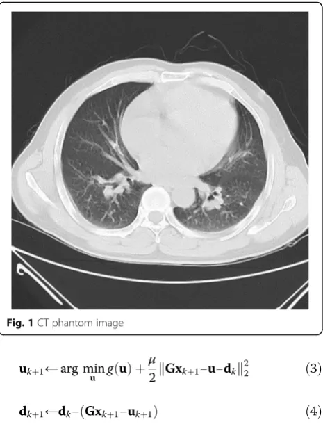
Fig. 1CT phantom image
P¼AHpsfxþn ð10Þ
wherePrepresents the measured projection data of CT.
A represents the sampling matrix of the system. Hpsf represents an unknown image degradation matrix. x
indicates that a clear image. nis the noise vector intro-duced in the measurement.
CT image reconstruction is the process that a clear image is reconstructed in the case of a given projection dataP. In order to ensure reconstruction quality, two or more regularization terms were used to solve unconstrained optimization problem. An iterative formula is utilized to achieve the CT image reconstruction. Then, it is possible to accurately reconstruct X from P=AHpsfx+n. In practical applications, in order to improve the quality of reconstruc-tion, some regularization terms are usually used in image re-construction. In this way, an ill-posed problem is transformed into a well-posed problem. This will guarantee the existence, uniqueness, and stability of the solution. It is helpful to obtain a relatively stable and meaningful solution from the meaning of the solution. Total variation (TV) transformation is a kind of commonly used regularization prior model. For example, reference [15] used TV minimization constraint for cone beam CT reconstruction. However, for low-contrast images, the method sometimes results in loss of information. In order to overcome this shortcoming, a new image reconstruction method is pro-posed in this paper. The method uses two kinds of
regularization terms: the total variation transformation and the sparse transformation. Sparse transform has two advan-tages. First, it is very good to maintain the edge structure. Second, it has a protective effect on low-contrast informa-tion. The total variation transform can effectively suppress noise interference and strip artifacts. The model proposed in this paper can achieve a good balance between artifact sup-pression and detail protection. In CT image reconstruction, the image does not necessarily satisfy the sparsity. Therefore, the good sparse transform can ensure that the image sparse representation has a good sparsity. In other words, when α=Θx, the image representation αis more sparse than the original imagex.Θrepresents the sparse wavelet transform.
The problem of blind restoration of double regularization CT images under incomplete projection data can be achieved by the following unconstrained minimization objective function.
^
x¼arg min
x μ
2P−AHpsfx
2
2þλ1 kΘxk1þλ2
X
i¼1
n
Dix
k k2 ð11Þ
Among them, Diis used to calculate the difference be-tween the horizontal and vertical directions of theithpixel. The first term in formula (11) represents the difference between the projection of the reconstructed projection and the measured projection, that is, the data fidelity item; the second term is the wavelet sparse transformation con-straint term; the third term is the total variation difference constraint term.
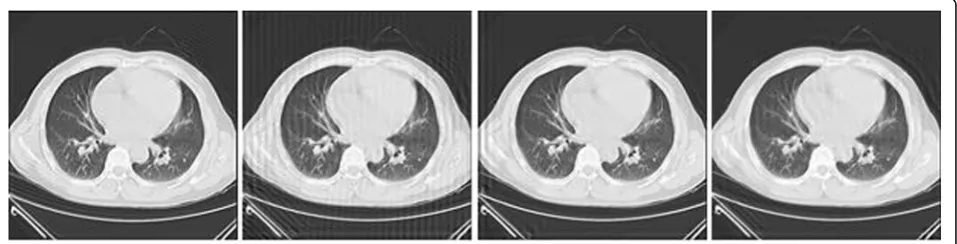
Fig. 3Comparison of reconstructed images with different reconstruction methods (SNR = 20 dB)
2.3 ADMM method for solving double regularized CT image blind rebuild reconstruction
Reconstruction of CT image blind restoration can be di-vided into image restoration reconstruction sub-problem and degraded function estimation sub-problem. To solve one of both variables, the other one can be regarded as a constant amount.
The purpose of CT image blind reconstruction is to solve formula (11). ADMM was used to solve the optimization problem. Formula (11) is converted into formula (5), as follows
g1 uð Þ1
¼P−uð Þ122 ð12Þ
g2 uð Þ2
¼λ1 uð Þ2 1 ð13Þ
gj uð Þj
¼λ2 uð Þj 2; Among them; j¼3;4 ð14Þ
Hð Þ1∈ℜln; Hð Þ1 ¼AHpsf ð15Þ
Hð Þ2∈ℜdn; Hð Þ2 ¼Θ ð16Þ
Hð Þj∈ℜ2n; Hð Þj ¼Dj−2 Among them; j¼3;4 ð17Þ
Formulas (12)–(17) are taken into formula (7) and let
μ3=μ4=μ, then
X
j¼1
J
μj Hð Þj
Hð Þj ¼μ1HpsfAAHpsfþμ2ΘΘþμ X
j¼3 nþ2
Dj−2Dj−2
¼μ1HpsfAAHpsfþμ2ΘΘþμ Dh
Dhþð ÞDvDv
h i
¼μ1HpsfA
AHpsf þμ2ΘΘþμDD
ð18Þ
Among them, (⋅)∗ represents a conjugate transpose
matrix. D¼ D h
Dv
; D1¼Dh calculates the difference of
the image in the horizontal direction, and D2=Dv -calculates the difference in the vertical direction.
HpsfA
AHpsf is a diagonal matrix. αij represents any element of the matrix. ∀αijαji≥0 and x≥0, so the data
P−AHpsfx
2
2 in formula (11) is a positive definite quad-ratic function.
For ease of calculation, a periodic boundary difference operator that can be diagonalized by Fourier transform is
Dh¼FΔhF ð19Þ
Dv¼FΔvF ð20Þ
Among them,Fis a two-dimensional discrete Fourier trans-form,Fis a unitary matrix,Δh, andΔvare diagonal matrix.
Thus, formula (21) can be rewritten as
X
j¼1 4
μj Hð Þj
Hð Þj ¼μ
1FHpsfAAHpsfþμ2ΘΘþμ
X
j¼3 4
Dj−2Dj−2
¼μ1HpsfAAHpsfþμ2ΘΘþμ Dh
Dhþð ÞDvDv
h i
¼μ1HpsfA
AHpsfþμ2ΘΘþμFΔhFFΔhFþFΔvFFΔvF
¼μ1HpsfAAHpsfþμ2ΘΘþμFΔhΔhFþFΔvΔvF
ð21Þ
The quadratic problem in the algorithm is solved by the gradient method
xkþ1←K μ1HpsfA
uð Þ1
k þd
1
ð Þ
k
þμ2Θ u 2
ð Þ
k þd
2
ð Þ
k
þμX j¼3 4
Dj−2 u
j
ð Þ
k þd j
ð Þ
k
" #
ð22Þ
where,K¼ μ1HpsfAAHpsfþμ2ΘΘþμ P j¼3
4 Dj−2Dj−2
−1
.
For optimization problems in different forms, different proximity operators are required. For the uð Þk1þ1 , the essence is to solve a quadratic programming problem, you can use the gradient method to get
uð Þk1þ1 ¼proxg1=μ1 AHpsfxkþ1−d 1 ð Þ
k
¼ arg min
u
1
μ1
y−u
k k2
2þ
1
2 u− AHpsfxkþ1−d
1 ð Þ k 2 2 ¼ 2
2þμ1 yþ
μ1
2 AHpsfxkþ1−d
1 ð Þ k 0 @ 1 A
ð23Þ
In order to solveℓ1norm, the soft threshold function as a proximity operator is usually used, which is
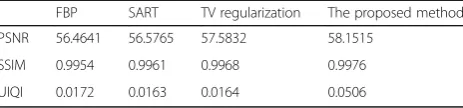
Table 1Comparison of image evaluation parameters of
different reconstruction algorithms (SNR = 20 dB)
FBP SART TV regularization The proposed method
PSNR 56.4641 56.5765 57.5832 58.1515
SSIM 0.9954 0.9961 0.9968 0.9976
UIQI 0.0172 0.0163 0.0164 0.0506
Table 2Comparison of image evaluation parameters of
different reconstruction algorithms (SNR = 40 dB)
FBP SART TV regularization The proposed method
PSNR 58.6194 59.9776 60.0533 61.6325
SSIM 0.9982 0.9991 0.9992 0.9996
UIQI 0.0152 0.0570 0.0832 0.1565
Table 3Comparison of image evaluation parameters of
different reconstruction algorithms (head CT)
FBP SART TV regularization The proposed method
PSNR 58.6891 58.8119 58.9634 61.8236
SSIM 0.9966 0.9967 0.9983 0.9993
softðv;τÞ ¼ signð Þv ⊗maxfj jv−τ;0g ð24Þ
Among them, ⊗ represents the multiplication of the corresponding element.
uð Þk2þ1 ¼proxg2=μ1 Θxkþ1−d
2 ð Þ
k
¼ arg min
u
λ1 μ2
u
k k1þ1
2 u− Θxkþ1−d
2 ð Þ
k
2
2
¼soft Θxkþ1−d2
ð Þ
k ;
λ1 μ2
0 @
1 A
ð25Þ
Among them, soft(⋅) is a soft threshold function.
uð Þkjþ1 ¼proxgj=μ D
j−2xkþ1−dkð Þj
¼ arg min
u
λ2 μk ku 2þ
1 2 u− D
j−2xkþ1−dj ð Þ
k
2
2
¼vector−soft Dj−2xkþ1−dj ð Þ
k ;
λ2 μ
0 @
1
A j¼3;4
ð26Þ
Among them, vector‐soft(⋅) is vector soft threshold function. vector−softðv;λÞ ¼k kvv2 max k kv 2−λ;0
. The iterative relationship of the introduced intermedi-ate variabledis as follows:
dð Þk1þ1¼d 1 ð Þ
k − RFpxkþ1−u 1 ð Þ
kþ1
ð27Þ
dð Þk2þ1¼d 2 ð Þ
k − Θxkþ1−uð Þk2þ1
ð28Þ
dð Þkjþ1¼d j
ð Þ
k − D
j−2xkþ1−uð Þkjþ1
j¼3;4 ð29Þ
The optimal solution of formula (11) can be obtained by iterative algorithm. In other words, we obtain an esti-mate of the reconstructed image.
For solving the point spread function sub-problems, we always see x as a constant to solve Hpsf. In order to ensure the convergence, we add the total variance para-digm of each element in Hpsf as a constraint term,
∇hpsf
1¼
P ffiffiffiffiffiffiffiffiffiffiffiffiffiffiffiffiffiffiffiffiffiffiffiffiffiffi h2psf;xþh2psf;y q
. Among them, ∑ repre-sents the superposition of all the elements.
^
Hpsf ¼ arg min Hpsf
μ
2P−AHpsfx
2
2þγ ∇hpsf1 ð30Þ
As before, the optimal solution of formula (30) can be obtained by ADMM method too.
3 Results and discussion 3.1 Results
To verify the effectiveness of the proposed algorithm, hu-man lung CT images were used for the first simulation ex-periments, and phantom image is 512 × 512 pixels, as shown in Fig. 1. In this paper, the fan beam source is
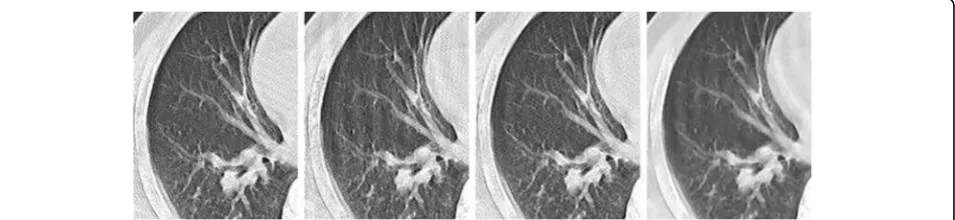
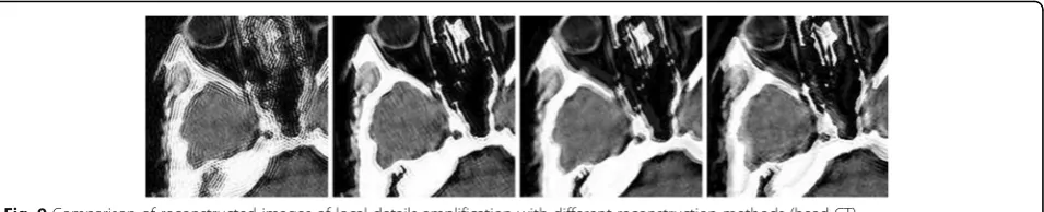
Fig. 5Comparison of reconstructed images of local details amplification with different reconstruction methods (SNR = 20 dB)
simulated and the center of the image is coincident with the center of rotation. The projection data was obtained by the uniform sampling in the range of 0 ~ 360. Each projection data was used by FBP, SART, TV regularization, and the al-gorithm proposed for image reconstruction. All the recon-struction methods are simulated at the same degradation condition. In the experiment, the degradation process of CT image is approximated by a Gauss point spread function, the degraded image is generated by the Gauss filter ofσ= 1, and the Gauss white noise with the noise power of σ2n is superimposed. Signal-to-noise ratio (SNR) is defined as SNR ¼10 log10 k kx 22=σ2n
. The experimental analysis is carried out respectively under the condition of SNR of 20 and 40 dB. The projection data under under different signal-to-noise ratios 20dB and 40dB is shown in Fig. 2. The selected sparse wavelet transform is db20 wavelet, and the decom-position level is 2. The iterative algorithm termination con-dition is kxkþ1−xkk22=kxkþ1k22≤210−6. The maximum number of iteration is set to 50. On the other hand, in the sub-problem of point spread function estimation, the algo-rithm termination condition is‖h(l+ 1)−h(l)‖/‖h(l)‖≤10−3. The maximum number of iteration is set to 100.
The experiments were performed in a computer Intel(R) i7-4770 @3. 60 GHz with 16 G RAM with MATLAB 2016a programming. The experimental parameters are as follows:
μ= 1, λ1= 10−3μ, λ2= 10−4μ, γ= 10−3μ. For ADMM pa-rameters, we used heuristic rules, which lead to a good per-formance of the algorithm. μ1 and μ2 are chosen to be proportional toλ2specificallyμ1= 10λ2andμ2= 100λ2.
From the reconstruction effect pictures that can be seen, FBP algorithm is seriously affected by the noise with the serious artifacts and poor effect. SART algo-rithm is better than FBP algoalgo-rithm, but the artifacts are still serious; although there is no obvious streak artifact in TV regularization algorithm, the image is too smooth and the detail is lost. The proposed algorithm in this paper is the best in terms of clarity, contrast, and detail retention. In order to see more clearly the details of the retention effect, a part of Figs. 3 and 4 was taken to enlarge and contrast in this paper, as shown in Figs. 5 and 6. As can be seen from the figure, the method can effectively suppress the noise, preserve the image details, clearly see the small blood vessels, and have good recon-struction effect. The artifacts of FBP and SART algo-rithm are serious, and the TV algoalgo-rithm does not have obvious artifacts, but it is too smooth and it lost too many details.
To evaluate the restored images, three metrics, i.e., peak signal-to-noise ratio (PSNR), universal image qual-ity index (UIQI) [26], and structural similarqual-ity index metric (SSIM) [27], are employed. Among these metrics, the ideal value of PSNR is +∞, and the ideal values of both UIQI and SSIM are 1. These three metrics can only be used in the simulated experiments because they require the existence of a reference image. The above four metrics tabulated for each experiment are the aver-age values over 10 times repetitions. The results are shown in Tables 1 and 2.
3.2 Discussion
The experimental results show that the structural simi-larity of image restoration method based on double regularization is increased, and the image quality is im-proved. The proposed method can obtain better visual effect and improve the PSNR. Compared with other
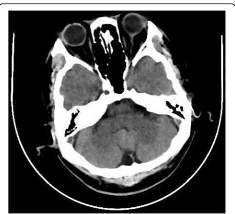
Fig. 7Head CT phantom image
methods, the proposed algorithm has stronger ability to recover details. It can be seen from Tables 1 and 2 that the new algorithm improves the objective image quality metrics such as the PSNR, structural similarity, and UIQI value. At the same time, under different parame-ters, the new algorithm has a better effect of restoration and reconstruction, showing that the algorithm has better robustness.
In the second simulation, a head CT image is chosen to verify the algorithm performance as shown in Fig. 7. The image size contains 512 × 512 pixels.
For comparison purposes, the projection data is denoised by Hanning filter. In the case of limited-angle projection, the number of projection angle was set 180. The limited-angle projection was reconstructed by algorithms of by FBP, SART, TV regularization, and the proposed algorithm.
The reconstructed images are shown in Fig. 8. To view the texture more clearly, a part of an image is amplified, as shown in Fig. 9. From the reconstruction figures that can be seen, the reconstructed images by FBP and SART have some artifacts in the case of limited-angle projection. The reconstructed image by TV regularization algorithm does not have obvious stripe-shape artifacts, and the images are too smooth. The proposed algorithm has the best effects such as sharpness, contrast, and detail preservation.
To quantitatively evaluate the effectiveness of the proposed algorithm, the reconstructed images shown in Fig. 8 and the ideal phantom shown in Fig. 7 are com-pared using SSIM, PSNR, and UIQI, as shown in Table 3.
From Table 3, we can see that PSNR and UIQI are im-proved compared with the other algorithms. SSIM of the proposed algorithm is much close to 1. That means the method proposed can get an image that is highly similar to the ideal phantom image.
4 Conclusions
An algorithm for double regularization medical CT image blind restoration reconstruction based on prox-imal alternating direction method of multipliers is pro-posed. The proposed method combines the total variation and wavelet domain sparsity regularization constraint for image restoration and reconstruction. To
solve the large-scale optimization problem in image res-toration, an approximate alternating direction multiplier method is used. The experimental results show that, compared with the current image reconstruction algo-rithm, the proposed algorithm has a better subjective visual effect. In the aspect of objective evaluation, the al-gorithm proposed in this paper improves the objective image quality metrics such as the peak signal-to-noise ratio, structural similarity index metric, and universal image quality index. At the same time, multi-regularization constraint reduces the sensitivity of regularization parameter in a certain range, which is beneficial to the maintenance of the complex edge pen-alty terms, and preserves the image edges and details.
Acknowledgements
The authors thank the editor and anonymous reviewers for their helpful comments and valuable suggestions.
Funding
This work was supported by Tianjin Research Program of Application Foundation and Advanced Technology (Nos.16JCYBJC28800 and 13JCYBJC15600), Tianjin Excellent Postdoctoral International Training Program, Tianjin Commerce University Young and Middle-aged Teacher International Training Program.
Availability of data and materials We can provide the data.
Authors’contributions
JZ and JT conceived and designed the study. YS and YZ performed the experiments. JZ wrote the paper .JZ, JT, and YS reviewed and edited the manuscript. All authors read and approved the final manuscript.
Competing interests
The authors declare that they have no competing interests.
Publisher’s Note
Springer Nature remains neutral with regard to jurisdictional claims in published maps and institutional affiliations.
Author details
1School of Electrical and Information Engineering, Tianjin University, Tianjin,
China.2School of Information Engineering, Tianjin University of Commerce, Tianjin, China.
Received: 14 July 2017 Accepted: 12 October 2017
References
1. Z Liang, B Kun, H Kuidong, et al., Multi-pinhole imaging measurement and assessment method for PSF of cone-beam CT system. Chin. J. Sci. Instrum.
33(9), 2061–2066 (2012)
2. A Kayugawa, M Ohkubo, S Wada, Accurate determination of CT point-spread-function with high precision. J. Appl. Clin. Med. Phys.14(4), 216–226 (2013) 3. M Jiang, G Wang, MW Skinner, et al., Blind deblurring of spiral CT images.
IEEE Trans. Med. Imaging22(7), 837–845 (2003)
4. JY Xu, K Taguchi, BMW Tsui, Statistical projection completion in X-ray CT using consistency conditions. IEEE Trans. Med. Imaging29(8), 1528–1540 (2010) 5. A Pakdel, JG Mainprize, N Robert, et al., Model-based PSF and MTF
estimation and validation from skeletal clinical CT images. Med. Phys.41(1), 8 (2014) Article ID: 011906
6. T Taniguchi, G Akamatsu, Y Kasahara, et al., Improvement in PET/CT image quality in overweight patients with PSF and TOF. Ann. Nucl. Med.29(1), 71–77 (2015)
7. YS Sun, LY Zhang, J Zhang, et al., Neural network blind equalization algorithm applied in medical CT image restoration. Math. Probl. Eng. ,2013(28) Article ID:743546 (2013)
8. YS Sun, LY Zhang, HY Zhang, et al., Multichannel blind CT image restoration via variable splitting and alternating direction method. Method Trans. Tianjin Univ.21(6), 524–532 (2015)
9. YS Sun, LY Zhang, HY Zhang, Complex system blind equalization algorithm for equivalent multichannel CT image restoration. J. Med. Imaging Health Informatics6, 555–561 (2016)
10. GP Chang, TT Chang, TS Pan, et al., Joint correction of respiratory motion artifact and partial volume effect in lung/thoracic PET/CT imaging. Med. Phys.37(12), 6221–6232 (2010)
11. JH Lewis, R Li, X Jia, et al., Mitigation of motion artifacts in CBCT of lung tumors based on tracked tumor motion during CBCT acquisition. Phys. Med. Biol.56(17), 5485–5502 (2011)
12. XH Meng, YC Chen, DQ Zheng, Correcting respiratory motion blur in PET images based on phantom and deconvolution algorithm. Chin. J. Biomed. Eng.32(6), 655–662 (2013)
13. C Mory, V Auvray, B Zhang, et al., Removing streak artifacts from ECG-gated reconstructions using deconvolution. J. Xray. Sci. Technol.22(2), 253–270 (2014) 14. EJ Candès, JK Romberg, T Tao, Stable signal recovery from incomplete and
inaccurate measurements. Commun. Pure Appl. Math.59(8), 1207–1223 (2006) 15. EY Sidky, CM Kao, X Pan, Accurate image reconstruction from few-views
and limited-angle data in divergent-beam CT. J. Xray. Sci. Technol.14(2), 119–139 (2009)
16. E Sidky, X Pan, Image reconstruction in circular cone-beam computed tomography by constrained, total-variation minimization. Phys. Med. Biol.
53(17), 4777–4807 (2008)
17. LA Vese, Expectation maximization and total variation-based model for computed tomography reconstruction from undersampled data//SPIE medical imaging. Int. Soc. Opt. Photonics,7961(3),165-177 (2011) 18. J Chen, J Cong, LA Vese, et al., A hybrid architecture for compressive sensing
3-D CT reconstruction. IEEE J. Emerg. Top. Circuits Syst.2(3), 616–625 (2012) 19. Z Hu, H Zheng, Improved total variation minimization method for few-view
computed tomography image reconstruction. Biomed. Eng. Online13(1), 1–10 (2014)
20. LY Wang, ZH Wei, SH Luo, et al., Image super reconstruction for micro-CT based on compressed sensing. J. Image Graph.17(4), 487–493 (2012) 21. L-z Deng, P Feng, M-y Chen, P He, Q-s Vo, B Wei, A CT reconstruction algorithm based on non-aliasing contourlet transform and compressive sensing. Comput. Math. Methods Med.11, 9 (2014) Article ID: 753615 22. Y Chen, LY Shi, QJ Feng, J Yang, HZ Shu, LM Luo, JL Coatrieux, WF Chen,
Artifact suppressed dictionary learning for low-dose CT image processing. IEEE Trans. Med. Imaging33(12), 2271–2292 (2014)
23. S Ramani, JA Fessler, A splitting-based iterative algorithm for accelerated statistical X-ray CT reconstruction. IEEE Trans. Med. Imaging31(3), 677–688 (2012) 24. A Sawatzky, QF Xu, CO Schirra, et al., Proximal ADMM for multi-channel
image reconstruction in spectral X-ray CT. IEEE Trans. Med. Imaging33(8), 1657–1688 (2014)
25. Y Hu, L Xie, L Luo, et al., L0 constrained sparse reconstruction for multi-slice helical CT reconstruction. Phys. Med. Biol.56(4), 1173–1189 (2011) 26. AYA Yusra, CS Der, Comparison of image quality assessment: PSNR, HVS,
SSIM, UIQI. Int. J. Sci. Engineering3(1), 1–5 (2012)