REVIEW
Mitochondria, cholesterol and cancer
cell metabolism
Vicent Ribas
1,2, Carmen García‑Ruiz
1,2,3and José C. Fernández‑Checa
1,2,3*Abstract
Given the role of mitochondria in oxygen consumption, metabolism and cell death regulation, alterations in mito‑ chondrial function or dysregulation of cell death pathways contribute to the genesis and progression of cancer. Cancer cells exhibit an array of metabolic transformations induced by mutations leading to gain‑of‑function of oncogenes and loss‑of‑function of tumor suppressor genes that include increased glucose consumption, reduced mitochondrial respiration, increased reactive oxygen species generation and cell death resistance, all of which ensure cancer progression. Cholesterol metabolism is disturbed in cancer cells and supports uncontrolled cell growth. In par‑ ticular, the accumulation of cholesterol in mitochondria emerges as a molecular component that orchestrates some of these metabolic alterations in cancer cells by impairing mitochondrial function. As a consequence, mitochondrial cholesterol loading in cancer cells may contribute, in part, to the Warburg effect stimulating aerobic glycolysis to meet the energetic demand of proliferating cells, while protecting cancer cells against mitochondrial apoptosis due to changes in mitochondrial membrane dynamics. Further understanding the complexity in the metabolic alterations of cancer cells, mediated largely through alterations in mitochondrial function, may pave the way to identify more efficient strategies for cancer treatment involving the use of small molecules targeting mitochondria, cholesterol homeostasis/trafficking and specific metabolic pathways.
Keywords: Mitochondria, Cholesterol, Tumor metabolism, Warburg effect, Reactive oxygen species, Apoptosis
© 2016 The Author(s). This article is distributed under the terms of the Creative Commons Attribution 4.0 International License (http://creativecommons.org/licenses/by/4.0/), which permits unrestricted use, distribution, and reproduction in any medium, provided you give appropriate credit to the original author(s) and the source, provide a link to the Creative Commons license, and indicate if changes were made.
Introduction
Cancer cells exhibit critical metabolic transforma-tions induced by mutatransforma-tions leading to gain-of-function of oncogenes and loss-of-function of tumor suppres-sor genes that result in cell deregulation associated with increased cellular stress. Hanahan and Weinberg identified the six conceptual hallmarks of human can-cer: (1) self-sufficient growth signaling, (2) evasion of growth suppressors, (3) cell death resistance, (4) replica-tive immortalization, (5) angiogenesis and (6) invasion/ metastasis [1]. Other common characteristics of cancer cells include enhanced anabolism, avoidance of immune destruction and altered autophagy [2, 3]. Of these charac-teristic features of cancer cells, mitochondria are directly involved in a number of them. Indeed, mitochondria are
critical mediators of apoptosis and the source of reactive oxygen species (ROS) generation and energy production. Consequently, altered mitochondrial function of cancer cells underlies several phenotypes, including: (1) resist-ance to apoptosis; (2) increased biosynthetic anabolism to support uncontrolled growth and proliferation; (3) increased ROS generation that activates metastatic pro-teases, tumor-promoting inflammation, genetic instabil-ity and DNA mutagenesis; (4) decreased mitochondrial oxidative phosphorylation (OXPHOS), increased aerobic glycolysis and decrease of pH in the extracellular milieu. Furthermore, due to its role as a hub in several signal-ing pathways [4], mitochondria are central for key met-abolic alterations of cancer cells, some of which will be described below.
Experimental evidence indicates that high cell prolif-eration [5, 6] and tumor growth [7, 8] are closely asso-ciated with enhanced cholesterol requirement. Some types of cancers, such as hepatocellular carcinoma (HCC), are dependent on cholesterol for growth [9],
Open Access
*Correspondence: Checa229@yahoo.com
2 Liver Unit‑Hospital Clínic, Centre Esther Koplowitz, IDIBAPS, CIBEREHD, Planta Cuarta, C/Rosselló 149, Barcelona 08036, Spain
and observational studies show a protective association between the use of statins and the risk of developing liver cancer [10], although this trend has been also observed in other cancer types, such as prostate and gastrointestinal cancers [11]. In line with this, genome-scale metabolic models of hepatocellular carcinoma found that among 101 metabolites relevant to HCC development, 30 % of them are related to cholesterol biosynthesis [12]. This protective effect of statins has been attributed to the inhi-bition of the mevalonate pathway (see below), prevent-ing the posttranslational modification of the oncogenes MYC, RAS and RHO [11, 13, 14]. Moreover, analyses of the Cancer Genome Atlas (TCGA) database revealed a correlation between increased activity of the cholesterol synthesis pathway and decreased survival in patients with sarcoma, acute myeloid leukemia and melanoma [15,
16], supporting the concept that cholesterol promotes carcinogenesis. In this regard, cholesterol trafficking to mitochondria has been reported in tumor cells [17, 18] and may account for the recognized mitochondrial dys-function and contribute to chemotherapy and apoptosis resistance and metabolic reprogramming of cancer cells, which will be discussed in the following sections.
Mitochondria in cell life and death Life‑sustaining functions
Mitochondria are complex organelles, which differ from the often-held view of isolated, small rounded double-membrane structures. They constitute a dynamic net-work that continuously undergoes fusion and fission controlled by specific mechanisms [19], and have inter-actions with other cell structures such as cytoskeleton and endoplasmic reticulum (ER) [20, 21]. Mitochondria contain multiple copies of their own maternally-inher-ited mitochondrial DNA (mtDNA), with an epigenetic complexity not completely understood [22]. Mitochon-drial DNA is a circular molecule of approximately 16.5 kilobases present from hundreds to thousands of copies per cell, which encodes 13 polypeptides of the OXPHOS and respiratory chain, as well as 2 ribosomal RNAs and 22 transfer RNAs necessary for translation of polypep-tides inside mitochondria. Most mitochondrial proteins (approximately 1500) are encoded by nuclear DNA, translated in the cytosol and imported into the mito-chondria through specific translocator complexes (TIM and TOM) of the mitochondrial inner (MIM) and outer membranes (MOM), respectively. In addition, a disulfide relay molecular device consisting of MIA40 and aug-menter of liver regeneration (ALR) are responsible for the import of nuclear encoded sulfur Fe/S cluster proteins to the mitochondrial intermembrane space that are essen-tial for mitochondrial function [23, 24]. Recent data have shown that ALR links mitochondrial function to HCC
development [25, 26]. Indeed, mitochondrial proteome has significant cell-type differences, allowing mitochon-dria to serve in a highly adaptive fashion to the cellular specific functional requirements [27].
Mitochondria are the power plants of the cell, provid-ing the energy for countless cellular functions through OXPHOS. OXPHOS is coordinated by a cascade of redox reactions organized in five protein complexes embed-ded in the MIM, known as the electron transport chain (ETC), which transfers electrons to oxygen [28, 29]. The fall in electron potential energy through the ETC is used to pump protons out of the mitochondrial matrix to the intermembrane space, generating an electrochemical gradient known as the mitochondrial transmembrane potential (Δψm), which induces a proton motive force used by complex V to regenerate ATP from ADP. Moreo-ver, many additional mitochondrial processes, especially those related to transport of solutes across the MIM [30] are dependent on the electrochemical driving force of the Δψm. Additional metabolic pathways that are located within mitochondria comprise the tricarboxylic acid cycle (TCA or Krebs cycle), β-oxidation of fatty acids, steroidogenesis, metabolism of amino acids, formation of Fe/S clusters, heme biosynthesis as well as reactions involved in lipogenesis, gluconeogenesis, ketogenesis and ammonium detoxification (urea cycle) [31].
Physiologically under aerobic conditions, cells degrade glucose via glycolysis to pyruvate, which is imported into mitochondria. Pyruvate enters the TCA cycle in the form of acetyl-CoA that along with oxaloacetate gener-ates citrate, in a reaction catalyzed by citrate synthase. Citrate is processed in the TCA cycle to generate reduc-ing equivalents that feed the ETC and generate energy with the consumption of oxygen. However, in conditions where macromolecular biosynthesis is active, citrate may be exported to cytosol where is converted to acetyl-CoA by ATP citrate lyase (ACLY), which is used for lipogen-esis. Besides their role in metabolism, mitochondria are involved in calcium homeostasis, innate immunity, inte-gration of signaling pathways and autophagy [32, 33]. Moreover, in response to metabolic and genetic stress mitochondria and nucleus engage in bidirectional signal-ing pathways, which modulate cell function [4, 34, 35].
superoxide [40], which is produced in the mitochondrial matrix and undergoes dismutation to hydrogen perox-ide (H2O2) [40], a reaction catalyzed by mitochondrial superoxide dismutases (SOD2). Hydrogen peroxide is further inactivated by the mitochondrial glutathione per-oxidase (mGSH/GPX) and peroxiredoxin/thioredoxin (Prx/Trx) antioxidant systems [41]. Both systems use the reducing equivalents of NADPH to regenerate the mito-chondrial oxidized glutathione (mGSSG) and Trx back to the reduced forms. The Prx/Trx system is thought to be responsible for scavenging hydrogen peroxide at nanomolar concentrations, while mGSH/GPX system is important for buffering high ROS levels [42, 43]. How-ever, both systems are mutually regulated, as selective depletion of mGSH results in decreased levels of Trx2 and Prx3 [44], highlighting the central role of mGSH in maintaining an adequate hydrogen peroxide homeosta-sis. Due to its more stable and diffusible nature, hydro-gen peroxide acts as a second messenger because of its reactions with specific oxidation-prone protein cysteinyl residues [45], which confers properties to hydrogen per-oxide as a mitochondrial signaling molecule [4]. In line with this, mitochondrial hydrogen peroxide bursts have self-sustained circadian oscillations, acting as a redox intracellular pacemaker [46].
Death promoting pathways
Besides their fundamental role in energy generation, mitochondria also play a strategic role in the regulation of several forms of cell death, including apoptosis (both caspase-dependent and independent), necrosis and pro-grammed necrosis [47]. The central mediators of apopto-sis include a group of cysteine proteases named caspases, which become activated by a proteolytic processing cas-cade in response to pro-apoptotic signals. The series of events leading to apoptosis have been categorized in two modes, the extrinsic and intrinsic apoptotic pathways. The extrinsic pathway involves extracellular ligand bind-ing to a transmembrane death receptor, such as TNF receptor or FAS receptor, followed by recruitment of cytosolic adaptor proteins and activation of an initiator caspase (usually caspase-8), which stimulates an effector caspase (such as caspase-3). Conversely, the intrinsic (or mitochondrial) pathway involves the destabilization of the MOM and the release of mitochondrial proteins that activate effector caspases. The BCL-2 family of proteins regulates this pathway with opposing pro-apoptotic effec-tor functions (BAX, BAK), apoptotic BH3-only pro-teins (BAD, BIM, BID, BIK, Noxa, PUMA, HRK, BMF) and anti-apoptotic functions (BCL-2, BCL-xL, MCL-1, A1, BCL-B, BCL-w) [48]. Activation of the intrinsic path-way of apoptosis by a number of stimuli and stresses, trig-gers the binding and activation of pro-apoptotic proteins
BAX or BAK to the MOM leading to the MOM per-meabilization (MOMP) without disruption of the inner membrane and the subsequent release of proteins from the mitochondrial intermembrane space (IMS), such as cytochrome c [49, 50]. Although active BAX or BAK are required to induce MOMP, the underlying mechanism is controversial [51]. While the model of pro-apoptotic activation or neutralization by anti-apoptotic members are still incompletely known, recent findings have shown that BCL-2 ovarian killer (BOK), which displays a high sequence similarity to BAX and BAK, engages the mito-chondrial apoptotic pathway independently of BAK/ BAX [52]. Although mitochondrial proteins are normally secured in the IMS the rupture of the physical barrier (MOM) constitutes a point-of-no-return in cell death [49, 50]. Pro-apoptotic BH3-only proteins act as stress sentinels that relay the diverse array of apoptotic signals via BAX/BAK activation to induce MOMP. In contrast, anti-apoptotic BCL-2-family proteins prevent MOMP and apoptosis by binding BH3-only proteins, preventing their interaction with BAX/BAK, or by binding activated BAX/BAK [53]. Pro- and anti-apoptotic BCL-2 protein interactions are mediated between BH-3 domains and the BH3 binding cleft in anti-apoptotic BCL-2 proteins.
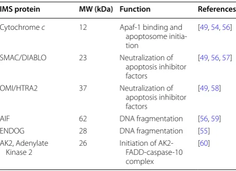
Once released from the mitochondria into the cytosol through MOMP, cytochrome c binds to the adaptor mol-ecule APAF-1, causing it to oligomerise and form a hep-tameric structure called apoptosome [54]. This complex recruits pro-caspase 9, which in turn, activates the execu-tioner caspases-3 and -7, triggering the cascade of events that lead to controlled cell death and fragmentation. In addition to cytochrome c, other IMS proteins (Table 1) are also mobilized and released into the cytosol following MOMP where they promote or counteract caspase acti-vation and hence cell death [55–60].
Table 1 IMS proteins related to apoptosis induction
IMS protein MW (kDa) Function References
Cytochrome c 12 Apaf‑1 binding and apoptosome initia‑ tion
[49, 54, 56]
SMAC/DIABLO 23 Neutralization of apoptosis inhibitor factors
[49, 56, 57]
OMI/HTRA2 37 Neutralization of apoptosis inhibitor factors
[49, 58]
AIF 62 DNA fragmentation [56, 59] ENDOG 28 DNA fragmentation [55] AK2, Adenylate
Kinase 2 26 Initiation of AK2‑FADD‑caspase‑10 complex
For the execution of mitochondrial apoptosis cytochrome c detaches from the MIM and dissoci-ates from the phospholipid cardiolipin, which binds cytochrome c by an electrostatic bond [61]. Cardi-olipin can be oxidized by ROS or by the cardiCardi-olipin– cytochrome c complex [62] resulting in oxidized cardiolipin, which exhibits lower affinity for cytochrome
c than the reduced form, and therefore contributes to cytochrome c detachment from MIM and its release to cytosol. Since mitochondrial ROS are controlled by anti-oxidants [63, 64], mGSH arises as an important modula-tor of apoptotic cell death by indirectly controlling the redox state of cardiolipin [63, 65]. In addition, it has been described that oxidized cardiolipin modulates the bio-physical properties of MOM to allow oligomerized BAX to insert and permeabilize the MOM [63, 65, 66].
Integrin-mediated attachment of normal cells to the extracellular matrix elicits anti-apoptotic and pro-survival signaling. The loss of cell–matrix interaction induces anoikis, a specific form of apoptosis [67]. Cell detachment leads to upregulation and activation of sev-eral BH3-only pro-apoptotic proteins (BID, BIM and BDF) that, in turn, activate BAX and BAK resulting in MOMP and the apoptotic cascade, resulting in cell death [68]. In addition to MOMP, the generation of mitochon-drial ROS in cells undergoing anoikis is required for cell death, as antioxidants treatment suppressed anoikis [69,
70]. Normal cells detached from the matrix undergo dramatic global metabolic changes characterized by decreased mitochondrial respiration and SOD2 induc-tion. Indeed, cells depleted of SOD2 are hypersensitive to cell death by anoikis [71], suggesting the importance of ROS generated in mitochondria in the execution of anoikis.
As opposed to apoptosis, necrosis is a morphologically distinct form of cell death responsible for irreversible tissue destruction due to bioenergetic failure and oxida-tive damage. Permeabilization of the MIM by the mito-chondrial permeability transition (MPT) and secondary rupture of the MOM is a key event of necrosis. MPT is a regulated pore-forming protein complex whose molecular characterization remains elusive [72–74]. Of the MPT components, cyclophillin D is a key constitu-ent, while the role of other putative components, such as voltage-dependent anion channel (VDAC), adenine nucleotide translocase (ANT) and translocator protein (TSPO, also called benzodiazepine receptor, PBR) is con-troversial [49, 75, 76]. Mitochondrial ROS regulate MPT by targeting specific cyclophillin D cysteine residues. Necrosis is characterized by mitochondrial swelling, loss of Δψm, and impaired OXPHOS and ATP generation. The fundamental difference with respect to apoptosis is the rapid loss of cellular membrane potential due to
energy depletion and ion pump/channel failure, leading to swelling and cytolysis. Concomitantly, water influx causes matrix swelling, rupture of MOM and release of apoptogenic proteins sequestered in IMS. These events, however, block apoptotic cell death due to energetic fail-ure, ATP exhaustion and oxidative stress-mediated cas-pase inactivation. Moreover, TNFα has been recently shown to induce a caspase-independent form of pro-grammed cell death, named propro-grammed necrosis or necroptosis [77, 78], involving receptor-interacting ser-ine/threonine-protein kinase 1 (RIPK1) and RIPK3 kinases, which interact with the pseudokinase mixed lin-eage kinase domain-like protein (MLKL). The execution of necroptosis requires mitochondrial ROS generation, which is dependent of MPT and involves cyclophyllin D but it is independent of BAX or BAK [79].
Cholesterol homeostasis and mitochondrial trafficking
to the blockade of isoprenoids (FPP and GGPP) genera-tion. In contrast to statins, the inhibition of SS results in selective cholesterol downregulation without exerting a major effect in the isoprenylation of proteins [82].
As HMGCoAR is the regulatory enzyme in the meva-lonate pathway its feedback and transcriptional control impact in cholesterol and isoprenoids regulation. One mechanism for feedback control involves the rapid degra-dation of HMGCoAR mediated by ER resident proteins, Insigs. Accumulation of sterols in the ER membrane trig-gers binding of the membrane domain of HMGCoAR to a subset of Insigs, which carry a membrane-anchored ubiquitin ligase called GP78 which ubiquitinates HMG-CoAR, marking it for proteasomal degradation [83]. HMGCoAR is regulated at the transcriptional level by the transcription factor SREBP-2, which resides in the ER is an inactive form. When sterols levels are low, SREBP-2 is transported from the ER to the Golgi to undergo a pro-teolytic processing by specific proteases, resulting in the mature form of SREBP-2, which translocates to the nuclei to induce HMGCoAR as well as other targets involved in the regulation of cholesterol homeostasis, including the LDL receptor.
As cholesterol synthesis requires oxygen, which is used for the biotransformation of squalene to cholesterol, an additional mechanism that regulates cholesterol synthe-sis is oxygen availability. Indeed, the bulk for the oxygen requirement centers on the sequential transformation of lanosterol to cholesterol, involving several redox reac-tions. Moreover, hypoxia has been shown to stimulate HMGCoAR degradation through both accumulation of lanosterol and Insigs induction [84]. In contrast to these physiological features, cholesterol synthesis and regula-tion are altered at several levels in cancer cells to meet the unrestricted growth needs [84–87]. Indeed, tumor cells exhibit increased cholesterol levels compared to sur-rounding cells; moreover, cancer tissues display increased upregulation of HMGCoAR, loss of feedback inhibi-tion, decreased expression of cholesterol exporter ATP binding cassette transporter A1 (ABCA1) and increased extracellular cholesterol uptake via LDL receptor [87]. Hence, as briefly described below (“Strategies targeting the mevalonate pathway and cholesterol synthesis in can-cer” section), targeting the mevalonate pathway may be of potential relevance in cancer therapy.
Mitochondrial cholesterol trafficking in cancer
Mitochondria are cholesterol-poor organelles com-pared to other cell bilayers (e.g. plasma membrane). Nevertheless, the limited availability of cholesterol in the MIM plays an important physiological role, includ-ing the synthesis of bile acids in hepatocytes or steroid hormones in specialized tissues through the metabolism
role in carcinogenesis may be related to the regulation of cell death and chemotherapy sensitization, which will be described below.
Role of mitochondria and cholesterol in altered cancer cell metabolism
The oncogenic transformation of cancer cells requires energy metabolism reprogramming in order to support unrestrained growth. The dependence on aerobic gly-colysis despite normal oxygen tension constitutes one of the key metabolic alterations in cancer cells. This event was first described by Otto Warburg in 1930 and has ben coined since then as the Warburg Effect [96–98]. Although the glycolytic phenotype in cancer cells was proposed to be due to defective mitochondrial OXPHOS, many cancer cells exhibit competent OXPHOS activ-ity capable to generate ATP [99]. The dependence on glycolysis is characteristic of many tumors and is widely exploited for clinical tumor imaging using positron emis-sion tomography (PET) with a radiolabeled analog of glucose (18F-fluorodeoxyglucose) [100]. Elevated aerobic glycolysis in cancer cells serves many purposes, ensur-ing ATP generation without reliance on oxygen avail-ability. Moreover, aerobic glycolysis generates bicarbonic and lactic acids, which are released to the extracellu-lar milieu, favoring tumor invasion, angiogenesis and immunosurveillance suppression [101]. Glucose can be diverted to the pentose phosphate pathway to generate nucleotides and NADPH to fuel antioxidant defenses and biosynthetic reactions. Finally cancer cells use interme-diates of glycolytic pathway for biosynthesis of de novo nucleic acids, lipids and amino acids to support their unrestrained growth and proliferation [97, 102, 103]. In line with these changes, a Warburg-like metabolism has been described in many rapidly proliferating embry-onic tissues, supporting the biosynthetic programs of aerobic glycolysis in active proliferating cells [104, 105]. Given that many tumor types rely on oxidative metabo-lism, glucose flux is not necessarily coupled to oxidative glucose metabolism. Oxygen consumption in many can-cer cells is used for mitochondrial oxidation of alternate fuels, such as glutamine [106], suggesting that the fate of glucose for mitochondrial oxidation in cancer cells is probably even lower. Cancer cells undergo a number of metabolic alterations, including the depression of oxida-tive mitochondrial OXPHOS and TCA cycle, which are used for anabolic reactions [107, 108]. Moreover, several transcriptional and posttranslational mechanisms have been proposed to contribute to the metabolic reprogram-ming and dependence on the Warburg effect in cancer cells, involving activation of oncogenes and inactiva-tion of tumor suppressor genes. In this regard, activated oncogenes such as KRAS and MYC along with mutated
tumor suppressors such as TP53 can extensively repro-gram cell metabolism resulting in diversion of carbon skeletons to fuel anabolic reactions for biomass synthesis instead of being completely oxidized through mitochon-drial respiration.
MYC in tumor metabolism reprogramming
MYC is an oncogene that plays a role in cell cycle pro-gression, apoptosis and cellular transformation. In addi-tion, MYC is important for the increased transcription of metabolic enzymes required for anabolism in cancer and fast-growing cells, regulating the conversion of glucose to pyruvate through the activation of important glyco-lytic genes and glucose transporters, while blocking the entry of pyruvate into the TCA cycle via pyruvate dehy-drogenase kinase (PDK1). Interestingly, MYC promotes the metabolic adaptation of tumor cells [109] by activat-ing genes important for mitochondrial biogenesis and function [110, 111]. Moreover, the AMPK-related pro-tein kinase 5 (ARK5), which is involved in maintenance of mitochondrial integrity and bioenergetic homeosta-sis, was identified as a MYC target [112]. This dual role of MYC as a driver of Warburg effect and a promoter of mitochondrial biogenesis underlies the dependence of cancer cells on glutamine oxidation, an essential event for cell survival under conditions with low glucose and oxygen [113]. Moreover, MYC upregulates the glutamine transporters SLC5A1 and SLC7A1, which contribute to glutamine uptake in cancer cells. As MYC induces the flux of 3-phosphoglycerate from glycolysis to the synthe-sis of serine and glycine needed for nucleotide biosynthe-sis, MYC coordinates the synthesis of nucleotides with glutamine metabolism [114]. Indeed, the rate of glutami-nolysis is greater compared to the rate of glycolysis in cells with high MYC expression and are more dependent on mitochondrial oxidative metabolism than cells with low MYC levels.
Tumors are metabolically heterogeneous, exhibiting complex metabolic profiles [115], including the depend-ence on aerobic glycolysis and reliance on OXPHOS [116–119]. For instance, while cancer stem cells are quiescent and exhibit high OXPHOS reliance, they may coexist with other highly cycling cancer cells that rely on glycolysis. The dependence of these cancer stem cells on mitochondrial OXPHOS prompted the use of mitochon-drial OXPHOS inhibitors to selectively target these cells to prevent tumor relapse after cytotoxic treatment [120,
inhibitor of peroxisome proliferator-activated receptor α (PGC1-α) suppressing mitochondrial respiration while activating glycolytic programs [119]. This heterogeneity defines a scenario where therapies targeting specifically highly respiratory or highly glycolytic tumor cells may not be completely effective.
TP53 and tumor metabolism
Reduced expression of the tumor suppressor protein TP53 can also impact metabolic reprogramming in can-cer cells. Defects in P53 function lead to impaired trans-activation of SCO2, a mitochondrial protein required for the correct assembly of the cytochrome c oxidase in the ETC and of TIGAR, an isoform of 6-phospho-fructo-2-kinase, whose expression exerts a tumor sup-pressor function by inhibiting glycolytic flux [122, 123]. Moreover, TP53 activates transcription of glutaminase 2 (GLS2) to promote glutaminolysis to fuel the TCA cycle and facilitate fatty acid oxidation as an alternative source [124]. Collectively, TP53, in addition to its role in orches-trating cell cycle arrest and apoptosis, counteracts the Warburg effect by favoring OXPHOS and minimizing glycolytic metabolism, and therefore its loss-of-function is a requirement for the aerobic glycolysis in most carci-nogenic processes.
Hypoxia‑inducible factor (HIF1α)
Hypoxia is an inherent feature of solid tumor develop-ment that arises due to the disorganized structure and architecture of tumor vasculature resulting in irregular and inefficient oxygen delivery. Hypoxia is considered a negative prognostic factor for response to treatment and survival of cancer patients [125, 126]. Hypoxia-induc-ible factor (HIF) is a key transcription factor activated mainly by hypoxia due to the dependence of HIF-proly hydroxylases (PHD) on oxygen (see below). In addi-tion to hypoxia HIF is also regulated by oxidative stress, inflammation and metabolic stress [127]. HIF1 com-prises a stable β subunit (HIF-1β/Arnt) and a labile α subunit (HIF1α) encompassing three family members, HIF1α, HFI2α and HIF3α (Fig. 1). In normoxia HIF1α is rapidly degraded due to the sequential action of oxygen-dependent PHD and the Von Hippel-Landau E3-ubiq-uitin ligase (pVHL). PHDs primarily function as oxygen sensors so that in normoxia PHDs become activated to hydroxylate HIF1α on two highly conserved proline resi-dues. Hydroxylated HIF1α is then recognized and ubiq-uitinated by the pVHL, marking HIF1α for proteasomal degradation (Fig. 1a). In low oxygen conditions, PHDs are inactivated and therefore HIF1α is stabilized, translocate to the nucleus where heterodimerize with HIF1β/Arnt to form a complex that activates hundreds of genes involved in energy metabolism, autophagy and angiogenesis [128]
(Fig. 1b). Activation of HIF1α promotes the conversion of glucose to pyruvate and lactate by upregulating the tran-scription of glucose transporters (GLUT1), hexokinases (HK1 and HK2), lactate dehydrogenase A (LDHA) as well as the lactate-extruding monocarboxylate transporter 4 (MCT4) [129], supporting the shift to aerobic glyco-lysis. Activated HIF1α increase the transcription of the PDK1, which inhibits PDH, decreasing the conversion of pyruvate to acetyl-CoA, which compromises OXPHOS, therefore linking low oxygen conditions to the depression of mitochondrial function. Moreover, HIF-1 activates transcription of the cytochrome c oxidase subunit 4-2 (COX4-2) and the LON mitochondrial protease, which degrades COX4-1 subunit and allows its substitution by the less efficient COX4-2 subunit [130]. In a scenario with inhibited mitochondrial OXPHOS by genetically down-regulating the master regulator of mitochondrial biogen-esis PGC1α, ROS-mediated HIF1α stabilization is able to rescue cell bioenergetics by activating transcription of glycolytic genes and glycolysis, allowing cancer cells to escape from metabolic stress [131]. In addition, HIF1 α induces the expression of BCL-2/adenovirus E1B 19-kDa interacting protein 3 (BNIP3) and BNIP3-like (BNIP3L), which trigger mitochondrial autophagy, thereby decreas-ing the oxidative metabolism of both fatty acids and glu-cose [132]. Therefore, HIF1α not only counteracts the MYC-mediated suppression of mitochondrial biogenesis by reducing mitochondrial mass and function, but also cooperates with MYC to promote aerobic glycolysis by induction of HK2 and PDK1 [133]. There are three PHDs known in mammals, encoded by three genes (PHD1, PHD2 and PHD3) [134]. Although, PHDs are thought to act as true oxygen sensors due to their requirement of oxygen for hydroxylation of HIF1, they are also depend-ent on iron (Fe2+), ascorbate and on the TCA
interme-diate 2-oxoglutarate (2-OG) as cofactors. Conversely, it has been reported that several TCA intermediates, such as fumarate and succinate competitively inhibit all three PHDs, while citrate and oxaloacetate inhibit factor inhibiting HIF1 (FIH), an asparaginyl hydroxylase which is able to block the transcriptional activity of HIF1α by catalyzing the hydroxylation of an asparagine residue of HIF1α [135]. These effects have important implications as succinate dehydrogenase (SDH) inactivation and isoci-trate dehydrogenase (IDH) neomorphic gain-of-function leading to accumulation of succinate and 2-hydroxyglu-tarate, respectively, contribute to HIF1α stabilization and cancer promotion [136].
Role of ROS in cancer cell biology
a
depending on the context and on the type of species gen-erated [137–140]. ROS are highly reactive molecules with the potential to target and oxidize proteins, lipids and DNA, which derive from different sources and mecha-nisms (Table 2) and from environmental events, such as ultraviolet or ionizing radiation [39, 139, 141].
ROS-induced damage on DNA can lead to enhanced mutation rates, driving the transformation of normal cells into a tumorigenic phenotype. In line with this asso-ciation, moderate intake of antioxidants have shown to reduce the risk of cancer development and slow cancer progression [142–145], leading to the concept that anti-oxidants can prevent ROS-induced damage and there-fore cancer incidence. Moreover, high ROS production in
cancer cells can stabilize survival factors such as HIF1α, which drive tumor initiation and progression [146]. Solid tumor formation, in turn, contributes to hypoxia devel-opment due to the disorganized vasculature, and the limited oxygen supply in solid tumors stimulates mito-chondrial ROS generation and HIF1α stabilization [147,
148]. HIF1α in turn activates ROS generation, estab-lishing a feed-forward loop where HIF1α supports its stability to promote cancer cell survival and malignant progression [141]. However, transformed cells adapt to this oxidative environment by turning on strategies that control the generation of ROS to ensure their role in pro-liferation signaling, while containing the damaging effects of ROS overproduction. An important strategy in this regard is the modulation of mitochondrial ROS genera-tion, which is downregulated in cancer cells by shifting to aerobic glycolysis. This scenario suggests that reduc-ing mitochondrial oxidation not only promotes survival of cancer cells but also increases anabolic metabolism. On the other hand, the pro-apoptotic activity of mito-chondrial inhibitors are reversed by antioxidants [121,
149], lending further support for the association of ROS with tumor prevention [141]. Conversely, large-scale multicenter clinical trials of antioxidant supplementation showed a significant increase in cancer incidence [150–
154]. Quite intriguingly recent preclinical studies con-firmed the pro-tumorigenic and pro-metastatic effects of antioxidant supplementation such as N-acetyl-l-cysteine (NAC), a GSH precursor [155, 156], thus highlighting the relevance of antioxidants in the protection of cancer cells against oxidative damage. Therefore, antioxidant supple-mentation can promote the growth of tumors by rescuing the viability of cells under high oxidative stress.
A key survival strategy of cancer cells is the upregula-tion of antioxidant systems to detoxify the producupregula-tion of ROS. One central factor associated to the resistance of cancer cells is the transcription factor NF-E2-related fac-tor (NRF2). NRF2 is a master regulafac-tor of the antioxidant response and xenobiotic metabolism through the regula-tion of a wide range of antioxidant and detoxificaregula-tion genes [157]. NRF2 is sequestered in the cytoplasm by the Kelch-like ECH-associated protein 1 (KEAP1), which acts as a NRF2 repressor and plays a pivotal role in the regulation of the NRF2 pathway. KEAP1 binds and promotes NRF2 (See figure on previous page.)
Fig. 1 Regulation of HIF1α transcriptional activity. a In normoxia, PHD hydroxylates HIF1α in several proline and asparagine residues, with 2‑OG, ascorbate and Fe3+, acting as cofactors for the reaction. Hydroxyted HIF1α binds with high affinity to the E3 ubiquitin‑ligase pVHL and HIF1α
becomes ubiquitinated, marking it for proteasomal degradation. Through this mechanism, HIF1α is kept at very low concentrations and transcrip‑ tionally inactive. b In low O2 conditions, the activity of PHD is inhibited due to lack of oxygen, resulted in no hydroxylation nor ubiquitination of
HIF1α. These events lead to the HIF1α protein stabilization which can translocate to the nucleus where it forms a complex with HIF1β and recruits CBP/p300 to the promoter of HIF1α target genes, activating the transcription of a vast array of genes responsible of glycolysis, angiogenesis and cell death resistance which are involved in tumor progression
Table 2 Cellular sources of ROS
CIF complex I flavin site, CIQ complex I ubiquinone site, CIIF complex II flavin site
and CIIIQ0 complex IIIQo are sites of the mitochondrial ETC, mGPDH Mitochondrial
glycerol 3-phosphate dehydrogenase, ETFQOR electron-trasferring flavoprotein ubiquinone oxidoreductase, PDH pyruvate dehydrogenase, OGDH 2-oxoglutarate dehydrogenase and BCKDH branched-chain 2-oxoacid dehydrogenase are mitochondrial enzymes capable of generate ROS. Upon stress signaling, cytosolic p66Shc translocates to mitochondria to directly stimulate hydrogen peroxide generation. Nitric oxide synthase (NOS) produces NO.by facilitating the conversion of l-arginine to l-citruline. NADPH oxidase
family of enzymes (NOX) transfer electrons from NADPH to O2 to produce
O2−. Other cellular enzymes incuding xanthine oxidase and cytochrome p450
families also participate in ROS generation in normal biological reactions and in chemicals or xenobiotics detoxification reactions
Source Cellular
compartment Primary radical generated
Complex IF Mitochondria O2−
Complex IQ Mitochondria O2−
Complex IIF Mitochondria O2−
Complex IIQ0 Mitochondria O2−
mGPDH Mitochondria O2−
ETFQOR Mitochondria O2−
PDH Mitochondria O2−
OGDH Mitochondria O2−
BCKDH Mitochondria O2−
P66shc Mitochondria, cytoplasm H2O2
NOS Cytoplasm NO
NOX family Cytoplasm, cell membrane O2−
Xantine oxidase Cytoplasm, peroxisome H2O2
Cytochrome p450 family Endoplasmic reticulum O2−
degradation through the ubiquitin–proteasome path-way. Under oxidative stress or through particular chemi-cal inducers, cysteine residues of KEAP1 are modified and the resulting conformational change leads to the release of NRF2, which is stabilized and translocated to the nucleus, to induce the transcription of a large number of genes [158]. In this regard, NRF2 activators, such as curcumin, butylated hydroxyanisole (BHA) or the synthetic oleane triterpenoids (CDDO), have preventive properties against carcinogenesis [157]. However, given the dual role of ROS on cancer genesis and development, NRF2 activation also provides protection to cancer cells. Therefore, NRF2 is constitutively elevated in many types of cancer cells [159–162] and this increase is associated with a poor prog-nosis in cancer patients [163–165]. A variety of molecular mechanisms contribute to the constitutive expression and/ or stabilization of NRF2 in cancer cells. Loss-of-function by somatic mutations or epigenetic silencing of KEAP1 impairs its binding to NRF2 and abrogates its repres-sive effect [159, 166]. The autophagy protein P62, also named sequestrosome 1, binds and sequesters KEAP1 in autophagosomes, leading to the autophagy-dependent KEAP1 degradation, resulting in increased NRF2 stability and activation of target genes [167–170]. Overexpression of P62 or increased P62 levels due to defects in autophagy leads to persistent activation of NRF2 [171, 172], contrib-uting to carcinogenesis [173]. In addition, activation of oncogenes such as K-RAS, BRAF and MYC stimulates the transcription of NRF2 [174]. There is substantial evi-dence that impaired TCA cycle activates NRF2 [175], in a similar fashion as described for HIF1α. In this case, fuma-rate accumulation can form adducts with KEAP1 on its cysteine residues and provoke NRF2 activation. Physiolog-ical fumarate levels are low due to the activity of fumarate hydratase (FH). However, in cancer cells with loss-of-func-tion of FH, high levels of fumarate are associated with sus-tained NRF2 activation [176, 177]. Nonetheless, activation of NRF2 transcriptional activity leads to the upregulation of antioxidants and detoxifying enzymes that promote not only the survival of cancer cells but also mediate chem-oresistance [178, 179]. Besides these important roles of NRF2 on detoxification, it has also been shown that NRF2 can contribute to other aspects of cancer survival such as the counteraction of cell death by BCL-2 overexpression [180] and altered metabolism by redirecting glucose and glutamine to the production of ribose-5-phosphate for nucleotide synthesis and to the regeneration of NADPH through the activation of the pentose-phosphate pathway [181].
Besides the role of ROS scavenging in cancer progres-sion, this event is also important for cancer metastasis. Hence, it can be postulated that the supplementation of antioxidants would provide an additional advantage
for cancer cells to spread to distant sites by counter-acting their sensitivity to anoikis and oxidative stress. For instance, metastatic cells undergo reversible meta-bolic changes that allow them to counteract oxidative stress [156, 181]. Indeed, it has been recently shown that increased GSH synthesis mediates the metastatic colonization of colorectal cancer cells to the liver [182]. Conversely, other reports showed that inhibition of mito-chondrial oxidative stress prevents metastasis [183, 184] and this apparent paradox might be explained by the dif-ferent targets of antioxidants and their effect in difdif-ferent types of cancer cells [184–186]. Therefore, current anti-oxidant strategies are not clinically effective in cancer therapies, illustrating our limited understanding on the complex role of ROS in tumor initiation, progression and metastasis, which needs to be fully characterized to iden-tify new and more effective therapeutic venues.
Mitochondrial cholesterol in HIF1α regulation
As mentioned above, HIF-1 is the main transcription factor regulating the cellular response to hypoxia and its stabilization is known to promote cell survival and tumor progression. While HIF-1 stabilization is mainly determined through oxygen sensing by PHD and iron availability, PHD activity is also dependent on the cyto-solic levels of 2-OG. Indeed, 2-OG emerges as a potential inhibitor of angiogenesis and cellular transformation by promoting the degradation of HIF1 [187, 188].
HIF-1α activation contributes to the metabolic repro-gramming of cancer cells by impairing mitochondrial phosphorylation and the subsequent stimulation of aerobic glycolysis. Although the physiological levels of mitochondrial cholesterol are low, mitochondrial cho-lesterol accumulation impairs mitochondrial function and the activity of the mitochondrial 2-oxoglutarate car-rier (2-OGC), which exchanges cytosolic GSH by matrix 2-OG. As StARD1 promotes mitochondrial cholesterol accumulation in the inner membrane, StARD1 induc-tion thus contributes to the impairment of OGC car-rier, resulting in the depletion of 2-OG in the cytosol and GSH in the mitochondrial matrix (Fig. 2). As men-tioned above, mGSH is a key mitochondrial antioxidant that controls hydrogen peroxide production [39, 189,
cytosolic 2-OG levels and generation of mitochondrial ROS. As a result, mitochondrial cholesterol loading in cancer cells acts as an additional mechanism governing angiogenesis and novel vessel growth via HIF1α stabiliza-tion, although this molecular link deserves to be further tested and it is currently under investigation. Finally, his-tone lysine demethylases have been recognized as impor-tant players in cancer cell biology by removing methyl moieties from DNA and aberrant expression of these chromatin modifying enzymes is implicated in the course of tumor initiation and progression [192]. Like PHD, his-tone methyl demethylases are also dependent on iron and 2-OG, and therefore mitochondrial cholesterol loading may further modulate cancer progression by the regula-tion of histone lysine demethylases via limitaregula-tion of cyto-solic 2-OG levels, which deserves further investigation.
Role of mitochondria and cholesterol in cancer cell death and chemotherapy resistance
Mitochondria and cell death resistance
Cancer cells have evolved multiple mechanisms to disable programmed cell death to support their survival and pro-liferation. Given that mitochondria are key players in sev-eral pathways of programmed cell death (see above) many strategic battles regulating cell death resistance take place in mitochondria [49]. The most prominent example of
this is the overexpression of pro-survival BCL-2 proteins, a common feature in diverse cancers. The gene encoding BCL-2 was first identified in a chromosomal transloca-tion that resulted in constitutively high levels of BCL-2 in neoplastic B cells [193, 194]. Different mechanisms such as genomic copy number amplification, oncogenic tran-scriptional upregulation or downregulation of microRNA repressors or stabilization of BCL-2 family members con-tribute to the maintenance of high levels of Bcl-2 [195,
196]. On the other hand, due to genomic deletion or pro-moter methylation leading to transcriptional silencing, loss-of-function of several pro-apoptotic proteins such as BAK, BAX and BH3-only family members have been observed in a variety of cancer types. Although BAX and BAK can play redundant roles, recent experimental data argues that in the context of activation of BH3-only pro-tein or anti-apoptotic BCL-2 there is a strict dependence of either BAX or BAK [197, 198]. Although cancer cells are generally resistant to apoptosis, certain stress condi-tions, such as hypoxia and low nutrient availability, lower the threshold for apoptosis susceptibility. Cancer cells often exhibit higher levels of pro-apoptotic BH3-only protein, which is accompanied by higher anti-apoptotic BCL-2 proteins to antagonize apoptosis. This state has been termed as cancer cells “primed for death” [199] and this dependence on anti-apoptotic BCL-2 proteins can be
Fig. 2 Mitochondrial cholesterol‑mediated HIF1α stabilization. Mitochondrial cholesterol loading mediated by StARD1 decreases mitochon‑ drial membrane fluidity which leads to an impaired activity of the OGC, which exchanges mitochondrial 2‑OG by cytosolic GSH. Cytosolic 2‑OG depletion may promote HIF1α stabilization by the impairment of PHD due to their requirement of 2‑OG as a cofactor for HIF1α hydroxylation and subsequent degradation. Moreover, 2‑OGC inhibition results in mGSH depletion, which in turn, limits the detoxification of ROS, in particular, H2O2.
exploited to design more effective pro-apoptotic thera-peutic strategies [200].
Loss–of-function of TP53 is found in more than 50 % of human cancers. In addition to the above-mentioned roles of its inactivation in cancer cell metabolism, TP53 is central in the orchestration of cell death pathways upon cellular stress such as DNA damage by stimulating the transcription of pro-apoptotic proteins (PUMA, BAX), autophagy and cell-cycle arrest. TP53 exerts a vast array of extranuclear functions and therefore the cytoplasmic pool of TP53 cooperates with its nuclear counterpart to activate programmed cell death in response to cer-tain cellular stresses. TP53 is involved in various forms of cell death such as apoptosis, necrosis and necroptosis and is able to mediate both MOMP and MPT opening in response to death stimuli. After stress induced TP53 acti-vation, a small fraction translocates to MOM, resulting in the activation of the intrinsic apoptotic pathway [201]. TP53-mediated MOMP is related to the ability to bind and inactivate anti-apoptotic BCL-2 and BCL-xL, and to transcriptionally induce the expression and activation of pro-apoptotic proteins by direct binding [202]. Moreover, TP53 regulates MPT openings of necrosis/necroptosis via cyclophillin D and dynamin-related protein 1 (DRP1) [203, 204] in response to specific cell death triggers, such as TNFα or oxidative stress. In addition, TP53 inhibits autophagy [205], resulting in impaired mitophagy, con-tributing to the reduced threshold for cell death. Given these protective roles against specific alterations in cell cycle and cell death resistance, many cancer-associated TP53 mutations have been identified. Although most of TP53 mutations has been described as loss-of-function, it has been proposed that some TP53 mutations may have oncogenic capabilities [206].
Although MOMP is considered a point of no return for apoptosis, cancer cells are able to inhibit caspases ensuring survival in certain conditions. This mechanism described in some post-mitotic cells, such as neurons and certain cancer cells, allows the recovery of cancer cells provided that MOMP-inducing stimuli are removed [207–209]. Caspases can be directly inhibited by XIAP or by the neutralization of its inhibitors [200]. In addition, cytochrome c released through MOMP can be targeted for proteasomal degradation thereby avoiding the assem-bly of the apoptosome [209]. Besides caspase inhibition, survival after MOMP requires a pool of intact mitochon-dria in which MOMP has not been triggered [210]. The selective maintenance of cells with intact mitochondria may contribute to carcinogenesis and cancer relapse after cytotoxic therapies due to the increased susceptibil-ity to oncogenic transformation [211]. Moreover, limited mitochondrial permeabilization induced by sub-lethal apoptosis triggers can promote DNA damage, genomic
instability and ultimately carcinogenesis [212, 213]. This mechanism would have two important implications for cancer progression. First, low-level limited apoptosis can drive mutagenesis in surviving cancer cells, serving as a driving force towards malignancy. Second, sub-lethal apoptotic anticancer therapies can increase the tumo-rigenic potential of surviving cancer cells by promot-ing new mutations that favor relapse and chemotherapy resistance.
Mitochondrial cholesterol in cell death and chemotherapy resistance
mitochondrial cholesterol contributes to chemotherapy resistance in HCC by increasing membrane order and resistance of MOM to MOMP. As StARD1 regulates mitochondrial cholesterol trafficking, it is conceivable that this member of the StART family stands as a novel target to regulate cancer cell death and chemotherapy response.
Cancer biology and therapeutics
As described in the previous sections, cancer cells exhibit critical metabolic transformations induced by muta-tions that result in cell cycle deregulation associated with enhanced cellular stress. Adaptation to this stress phenotype is required for cancer cells to survive and involves the participation of genes that regulate metab-olism, bioenergetics, cell death and ROS detoxification
(Fig. 3). In this context, small molecules that selectively kill cancer cells while sparing normal surrounding cells, are the desired approach for the treatment of cancer. To this aim, cancer therapeutics should target the differen-tial features of cancer cells. Here, we briefly summarize the therapeutic strategies that involve mitochondria and their proposed mechanism of action to selectively target transformed cells.
Therapeutics aimed at cancer metabolism and bioenergetics
Cells are addictive to glucose and glutamine and their limitation can cause cell death. This dependence is driven by the activation of MYC and HIF1-α [109] and consequently, targeting pathways regulating glucose/ glutamine metabolism may be of relevance for cancer
a
c
b
d
treatment. The specific GLS1 inhibitor bis -2-(5-pheny-lacetamido-1,3,4-thiadiazol-2-yl)ethyl sulfide (BPTES) inhibits proliferation of lymphoma cells but has no effect on neuroblastoma cells, which express GLS2 [214, 215], implying that the general GLS inhibitor 6-diazo-5-oxo-L-norleucine (DON) may exhibit broader antitumor effects [216, 217]. Inhibitors of glutamate dehydrogenase (GDH) are promising agents to target glutamine addiction of certain cancer cells. For instance, the green tea compo-nent epigallocatechin-3-gallate (EGCG), which inhibits GDH, has been shown to promote apoptosis in several cancers types, resulting in tumor growth inhibition, set-ting the basis for the exploration of its efficacy in phase II clinical trials [214, 218].
The inhibition of aerobic glycolysis in cancer cells is also of potential relevance. While inhibitors such as 2-deoxy-d-glucose and ionidamine, which targets early steps in the glycolysis pathway, exhibit severe toxic side effects [218], inhibition of distal steps in glycolysis are effective. Inhibition of lactate production by inactivation of lactate dehydrogenase (LDH) reduces tumorigenic-ity in several cancer models [219–221]. Additionally, the specific inhibitor of LDHA, FX11, reduces tumor progression in lymphoma in vivo [219]. In some cancers LDHB can replace LDHA, hence limiting the efficacy of the LDHA inhibitors [109, 222]. Inhibition of lactate export from cancer cells results in wide-reaching con-sequences, leading not only to lactate accumulation, alterations in glycolytic intermediates, reduction in glu-cose transport and ATP, NADP and GSH levels but also in mitochondrial damage and cell death [223], suggest-ing that inhibition of lactate transporter MCT1 is a suit-able therapeutic approach. Moreover, the effect of small molecules that block the entry of pyruvate to the mito-chondrial TCA cycle, such as dichloroacetate, a PDK1 inhibitor, may be of potential relevance [109, 218, 224].
Although targeting glycosis may be effective in a spe-cific population of cancer cells exhibiting a highly glyco-lytic dependence, stem cancer cells that rely on OXPHOS might become resistant. Moreover, although mitochon-drial oxidation under the Warburg effect is dramatically reduced, many cancer cells still have a central require-ment on mitochondrial metabolism, strongly suggesting that OXPHOS inhibitors might represent an important target for drug-resistant cancers [121]. A key agent with potential relevance in inhibiting OXPHOS is metformin, one of the most prescribed drugs around the world for the treatment of type II diabetes [225–228]. Metformin is an indirect activator of AMP-activated Kinase (AMPK) through inhibition of mitochondrial complex I, result-ing in the activation of the ATM/LKB1/AMPK axis. LKB1 is a well-characterized tumor suppressor in pan-creatic, lung cancer and melanoma. AMPK activation
inhibits the mTOR pathway and this effect accounts for the potential antineoplastic effects of metformin in breast and renal tumors. Moreover, metformin reduces glyco-lysis and increases mitochondrial respiration in tumors, and these events are associated with growth arrest [229]. In addition, metformin exhibits antiangiogenic effects, which contribute to its antineoplastic properties [230]. Other compounds with mild OXPHOS inhibition such as tamoxifen, which also inhibits complex I, resveratrol, which antagonizes complex III, and the complex V inhib-itor 3,3-diindolylmethane have potential in cancer treat-ment. VLX600 is a novel compound targeting OXPHOS that inhibits tumor growth of colon carcinoma cells, thus exhibiting potential application in clinical trials [231]. Besides, a number of emerging mitochondrial inhibitors successfully used in experimental studies could be effec-tive against cancer cells and might synergize with chemo-therapeutics [149, 232, 233].
Therapeutics targeting cancer cell death
Obatoclax, which inhibit BCL-2 and MCL1, prevented the evaluation of their efficacy in clinical settings [243]. These findings suggest that MCL1 inhibition should be fine-tuned and that the relative contribution of BCL-2-family components to the apoptosis resistance of can-cer cells should be carefully evaluated through the “BH3 profiling” to determine the therapeutic window [248,
249]. In addition, incomplete cell death caused by trig-gers of mitochondrial apoptosis can promote genomic instability and mutagenesis derived from the incomplete MOMP and caspase-dependent DNA cleavage, contrib-uting to tumor relapse and the acquisition of drug resist-ance [212, 213]. Based on the ability of TP53 to induce apoptosis, mitochondrial targeted TP53 fusion proteins have been developed to induce intrinsic apoptosis in can-cer cells, which may be of relevance in adjuvant therapy for cancer treatment [201, 250]. Overall, targeting or sen-sitizing cancer cells to apoptosis is a promising strategy currently under development, which may lead to per-sonalized medicine through specific tumor-profiling and fine-tuning dosage and therapy combinations.
Therapeutics targeting cancer cell ROS sensitivity
Despite generation of higher ROS levels cancer cells are more sensitive to intracellular ROS induction than untransformed cancer cells. Many cancer chemothera-peutic agents, including taxanes, vinca alkaloids, plati-num coordination complexes, paclitaxel and elesclomol are currently used to induce high levels of ROS to kill cancer cells [251]. The ultimate effect of these molecules is determined by the intrinsic antioxidant capacity of cancer cells as the cytotoxic potential of these agents is lost upon antioxidant co-treatment [252–254].
A key mechanism to counteract the generation of ROS by chemotherapeutic agents is the regulation of GSH homeostasis [137, 182]. Several small molecules, which modulate ROS, such as β-phenethyl isothiocyanate (PEITC), buthionine sulphoximine (BSO), curcumin or CDDO derivatives, have potential therapeutic effects for the treatment of cancer by promoting mGSH deletion and subsequent ROS generation specifically in cancer cells [255–258]. BSO, an inhibitor of glutamate-cysteine ligase, which is the rate-limiting enzyme in GSH biosyn-thesis [259] is the only clinically used drug to suppress the novo GSH synthesis. The simultaneous administration of BSO and the thioredoxin inhibitor auranofin induce ROS and clonogenic killing in carcinoma cells [260]. Sul-fasalazine, which inhibits cystine uptake via XcL carrier, limits cysteine availability impairing GSH biosynthesis, which leads to reduced growth and viability of cancer cells in vitro and in vivo [261, 262]. In addition, specific mGSH depletion has also been associated with apopto-sis induced by chemotherapeutic drugs. For example,
the triterpenoid methyl CDDO derivative (CDDO-Me), induces cytotoxicity in chemotherapy-resistant myeloid leukemia cells and this event is associated with selective depletion of mGSH, resulting in increased ROS genera-tion [263, 264]. Moreover, PEITC depletes mGSH and consequently increases ROS and nitric oxide, contribut-ing to inhibition of the mitochondrial complex I, sup-pression of mitochondrial respiration, and subsequent cytotoxicity of leukemia cells [265]. Using a cell-based small-molecule screening and quantitative proteomics, piperlongumine has emerged as a cytotoxic agent that triggers apoptosis and necrosis in leukemia cells [266]. Interestingly, piperlongumine induces ROS generation and cell death in transformed cells but not primary nor-mal cells [267]. Piperlongumine also decreases GSH and increases GSSG levels in cancer cells without effects in nontransformed cells, and these effects parallel the abil-ity of piperlongumine to alter mitochondrial morphology and function. Consequently, co-treatment with piperlon-gumine and NAC prevented piperlonpiperlon-gumine-mediated GSH depletion and cell death in cancer cells. These find-ings support the concept that cancer cells have high lev-els of ROS, and hence, have a strong reliance on the ROS stress-response pathway driven by NRF2.
At present, radiotherapy is widely used in various types of cancer treatments, and the therapeutic effect is mainly determined by ROS generation. The induction of water radiolysis occurs in seconds after ionizing radiation, lasts several hours after exposure and enhances ROS genera-tion and oxidative stress [268, 269]. Some studies sug-gested that antioxidant supplementation could sensitize cancer cells to chemo- or radio-therapy and reduce their side effects by protecting the normal cells [270]. How-ever, other studies indicated that antioxidants may also protect cancer cells against these therapies [252, 271,
272]. Therefore, the safety and efficacy of the combined treatment of antioxidants with radiotherapy or ROS-inducing chemotherapy remain controversial.
Strategies targeting the mevalonate pathway and cholesterol synthesis in cancer
of cholesterol synthesis. For instance, statins inhibit the activation of the proteasome pathway, contributing to the maintenance of proteins that block cell cycle. Through cholesterol downregulation, statins regulate the function of Hedgehog, a signaling pathway involved in carcinogen-esis [273]. Besides these wide-reaching effects of statins, their benefit in cancer treatment is limited due to the complex regulation of HMGCoAR and the metabolites generated in the mevalonate pathway. Reduction of iso-prenoid and cholesterol levels in cancer by chronic treat-ment with statins leads to upregulation of HMG-CoAR levels and eventually development of resistance [274]. In vitro mechanistic studies of statins used significantly higher concentrations than those that were therapeuti-cally achievable in phase I trials. Dose-limiting toxici-ties, including gastrointestinal side effects, myelotoxicity, myalgias, elevation of creatine phosphokinase and hepa-totoxicity, precluded further dose increase in clinical tri-als [275]. Inhibition of SS has attracted much interest as a pharmacological target as it implies the inhibition of cholesterol synthesis without depressing isoprenoid lev-els. For instance, lapaquistat (TAK-475, Takeda), a SS inhibitor, progressed to phase III clinical trials, although its outcome in cancer remains to be established due to hepatotoxic effects at high dosing [276]. As mentioned before, prenylation is a key postranslational mechanism of targeted proteins, and many prenylated proteins are involved in various aspects of carcinogenesis, including cellular proliferation, apoptosis, angiogenesis and metas-tasis. Farnesylation is catalyzed by farnesyltransferase (FTase) and geranylgeranylation by geranylgeranyltrans-ferase, GGTase. Given the role of protein prenylation in carcinogenesis, FTase inhibitors (FTIs) and GGTase inhibitors (GGTIs) have been developed for cancer treat-ment. GGTI–FTI combinations synergistically inhibit proliferation of multiple myeloma cell lines and primary cells, and induce apoptosis. Interestingly, dual prenyla-tion inhibitors (DPIs) that block both FTase and GGTase enzymatic activities have been shown to induce apoptosis in PSN-1 pancreatic tumor cells by blocking K-Ras pre-nylation compared to either FTI or GGTI agents alone [277]. H and N-Ras prenynation is effectively inhibited by FTIs and only partially by GGTIs, whereas K-Ras pre-nylation requires both FTIs and GGTIs inhibition [278]. Thus, combined inhibition of geranylgeranylation and farnesylation can overcome the resistance conferred by cross-prenylation, thus potentiating the activity of either FTIs or GGTIs alone. Finally, targeting the specific tar-geting of cholesterol to mitochondria may be an addi-tional approach of potential benefit in cancer treatment by modulating cell death and chemotherapy resistance. This specific field is currently under investigation to iden-tify potential specific inhibitors of StARD1 and MLN64
to sensitize cancer cells to cell death triggers and chem-oterapeutic agents.
Conclusions and future approaches
for cancer treatment involving the use of small molecules targeting mitochondrial metabolism.
Abbreviations
ROS: reactive oxygen species; OXPHOS: oxidative phosphorylation; HMG‑ CoAR: 3‑hydroxy‑3‑methyl‑glutaryl‑CoA reductase; HCC: hepatocellular carcinoma; TCGA: cancer genome atlas; mtDNA: mitochondrial DNA; MIM: mitochondrial inner membrane; MOM: mitochondrial outer membrane; ALR: augmenter of liver regeneration; ETC: electron transport chain; Δψm: mitochondrial transmembrane potential; TCA: tricarboxylic acid cycle; ACLY: ATP citrate lyase; mGSH: mitochondrial glutathione; H2O2: hydrogen peroxide; SOD: superoxide dismutases; 2‑OG: 2‑oxoglutrarate; GPX: glutathione peroxidase; mGSSG: mitochondrial oxidized glutathione; Prx/Trx: peroxire‑ doxin/thioredoxin; MOMP: mitochondrial outer membrane permeabilization; IMS: mitochondrial intermembrane space; BOK: BCL‑2 ovarian killer; MPT: mitochondrial permeability transition; VDAC: voltage‑dependent anion channel; ANT: adenine nucleotide translocase; TSPO: translocator protein; RIPK1: receptor‑interacting serine/threonine‑protein kinase 1; MLKL: mixed lineage kinase domain‑like protein; IPP: isopentenyl pyrophosphate; DMAPP: dimethyl allyl pyrophosphate; GPP: geranyl pyrophosphate; FFP: farnesyl pyrophosphate; GGPP: geranyl geranyl pyrophosphate; SS: squalene syn‑ thase; ER: endoplasmic reticulum; ABCA1: ATP binding cassette transporter A1; StARD1: steroidogenic acute regulatory domain 1; PET: positron emission tomography; PDK1: pyruvate dehydrogenase kinase; ARK5: AMPK‑related protein kinase 5; PGC1α: peroxisome proliferator‑activated receptor gamma coactivator 1 alpha; GLS2: glutaminase 2; HIF1α: hypoxia‑inducible factor 1 alpha; PHD: HIF‑proly‑hydroxylase; pVHL: Von Hippel‑Landau ubiquitin ligase; LDHA: lactate dehydrogenase A; MCT4: monocarboxylate transporter 4; COX4‑2: cytochrome c oxidase subunit 4–2; BNIP3: BCL‑2/adenovirus E1B 19‑kDa interacting protein 3; 2‑OG: 2‑oxoglutarate; FIH: factor inhibiting HIF1; SDH: succinate dehydrogenase; IDH: isocitrate dehydrogenase; NAC: N‑acetyl‑
l‑cysteine; NRF2: NF‑E2‑related factor; KEAP1: Kelch‑like ECH‑associated
protein 1; BHA: butylated hydroxyanisole; CDDO: 2‑cyano‑3,12‑dioxooleana‑ 1,9‑diene‑28‑oic acid; FH: fumarate hydratase; 2‑OGC: 2‑oxoglutarate carrier; DRP1: dynamin‑related protein 1; BPTES: bis‑2‑(5‑phenylacetamido‑1,3,4‑thi‑ adiazol‑2‑yl)ethyl sulfide; DON: 6‑diazo‑5‑oxo‑l‑norleucine; GDH: glutamate dehydrogenase; EGCG: epigallocatechin‑3‑gallate; LDH: lactate dehydroge‑ nase; AMPK: AMP‑activated Kinase; PEITC: β‑phenethyl isothiocyanate; BSO: buthionine sulfoximine; FTase: farnesyltransferase; FTI: farnesyltransferase inhibitors; GGTI: geranyl geranyl transferase inhibitor.
Authors’ contributions
VR, CGR and JCFC participated in the design of the study, revised literature, and discussed data jointly. VR, CGR and JCFC drafted Figures and the writing of the text. All authors read and approved the final manuscript.
Author details
1 Department of Cell Death and Proliferation, Institute of Biomedical Research of Barcelona (IIBB), Consejo Superior Investigaciones Cientificas (CSIC), Barcelona, Spain. 2 Liver Unit‑Hospital Clínic, Centre Esther Koplowitz, IDIBAPS, CIBEREHD, Planta Cuarta, C/Rosselló 149, Barcelona 08036, Spain. 3 Research Center for ALPD and Cirrhosis, Ckeck School of Medicine, University of South‑ ern California, Los Angeles, CA, USA.
Acknowledgements
Vicent Ribas is recipient of an IDIBAPS Postdoctoral Fellowship‑BIOTRACK, supported by the European Community’s Seventh Framework Programme (EC FP7/2007‑2013) under the Grant agreement number 229673 and the Span‑ ish Ministry of Economy and Competitiveness (MINECO) through the Grant COFUND2013‑40261. The work was supported by CIBEREHD, Fundació la Marató de TV3 and Grants PI11/0325 (META) from the Instituto Salud Carlos III and Grants, SAF2011‑23031, SAF2012‑34831, SAF2014‑57674R and SAF2015‑ 69944‑R from Plan Nacional de I+D, Spain; Fundación Mutua Madrileña and the center Grant P50‑AA‑11999 (Research Center for Liver and Pancreatic Diseases, NIAAA/NIH).
Competing interests
The authors declare that they have no competing interests.
Received: 12 April 2016 Accepted: 26 June 2016
References
1. Hanahan D, Weinberg RA (2011) Hallmarks of cancer: the next genera‑ tion. Cell 144(5):646–674. doi:10.1016/j.cell.2011.02.013
2. Morselli E, Galluzzi L, Kepp O, Vicencio JM, Criollo A, Maiuri MC et al (2009) Anti‑ and pro‑tumor functions of autophagy. Biochim Biophys Acta 1793(9):1524–1532. doi:10.1016/j.bbamcr.2009.01.006 3. Galluzzi L, Morselli E, Kepp O, Vitale I, Rigoni A, Vacchelli E et al (2010)
Mitochondrial gateways to cancer. Mol Aspects Med 31(1):1–20. doi:10.1016/j.mam.2009.08.002
4. Raimundo N (2014) Mitochondrial pathology: stress signals from the energy factory. Trend Mol Med. doi:10.1016/j.molmed.2014.01.005 5. Bensinger SJ, Bradley MN, Joseph SB, Zelcer N, Janssen EM, Hausner MA
et al (2008) LXR signaling couples sterol metabolism to proliferation in the acquired immune response. Cell 134(1):97–111. doi:10.1016/j. cell.2008.04.052
6. Lo Sasso G, Celli N, Caboni M, Murzilli S, Salvatore L, Morgano A et al (2010) Down‑regulation of the LXR transcriptome provides the requisite cholesterol levels to proliferating hepatocytes. Hepatology 51(4):1334–1344. doi:10.1002/hep.23436
7. Clendening JW, Pandyra A, Boutros PC, El Ghamrasni S, Khosravi F, Trentin GA et al (2010) Dysregulation of the mevalonate pathway promotes transformation. Proc Natl Acad Sci USA 107(34):15051–15056. doi:10.1073/pnas.0910258107
8. Dang CV (2012) Links between metabolism and cancer. Gene Dev 26(9):877–890. doi:10.1101/gad.189365.112
9. Borena W, Strohmaier S, Lukanova A, Bjorge T, Lindkvist B, Hallmans G et al (2012) Metabolic risk factors and primary liver cancer in a prospec‑ tive study of 578,700 adults. Int J Cancer 131(1):193–200. doi:10.1002/ ijc.26338
10. Singh S, Singh PP (2014) Statins for prevention of hepatocellular cancer: one step closer? Hepatology 59(2):724–726. doi:10.1002/hep.26614 11. Stryjkowska‑Gora A, Karczmarek‑Borowska B, Gora T, Krawczak K (2015)
Statins and cancers. Contemp Oncol. 19(3):167–175. doi:10.5114/ wo.2014.44294
12. Agren R, Mardinoglu A, Asplund A, Kampf C, Uhlen M, Nielsen J (2014) Identification of anticancer drugs for hepatocellular carcinoma through personalized genome‑scale metabolic modeling. Mol Syst Biol. 10:721. doi:10.1002/msb.145122
13. Cao Z, Fan‑Minogue H, Bellovin DI, Yevtodiyenko A, Arzeno J, Yang Q et al (2011) MYC phosphorylation, activation, and tumorigenic potential in hepatocellular carcinoma are regulated by HMG‑CoA reductase. Cancer Res 71(6):2286–2297. doi:10.1158/0008‑5472.CAN‑10‑3367 14. Mansourian PG, Yoneda M, Krishna Rao M, Martinez FJ, Thomas E, Schiff
ER (2014) Effects of statins on the risk of hepatocellular carcinoma. Gastroenterol & Hepatol. 10(7):417–426
15. Cancer Genome Atlas Research, Weinstein JN, Collisson EA, Mills GB, Shaw KR, Ozenberger BA et al (2013) The cancer genome atlas pan‑can‑ cer analysis project. Nat Genet 45(10):1113–1120. doi:10.1038/ng.2764 16. Kuzu OF, Noory MA, Robertson GP (2016) The role of cholesterol in can‑
cer. Cancer Res 76(8):2063–2070. doi:10.1158/0008‑5472.CAN‑15‑2613 17. Montero J, Morales A, Llacuna L, Lluis JM, Terrones O, Basanez G
et al (2008) Mitochondrial cholesterol contributes to chemotherapy resistance in hepatocellular carcinoma. Cancer Res 68(13):5246–5256. doi:10.1158/0008‑5472.CAN‑07‑6161
18. Lucken‑Ardjomande S, Montessuit S, Martinou JC (2008) Bax activation and stress‑induced apoptosis delayed by the accumulation of choles‑ terol in mitochondrial membranes. Cell Death Differ 15(3):484–493. doi:10.1038/sj.cdd.4402280
19. Mishra P, Chan DC (2014) Mitochondrial dynamics and inheritance during cell division, development and disease. Nat Rev Mol Cell Biol 15(10):634–646. doi:10.1038/nrm3877