C A S E R E P O R T
Open Access
Petersen
’
s hernia after living donor liver
transplantation
Sodai Sakamoto
1, Ryoichi Goto
1, Norio Kawamura
1, Yasuyuki Koshizuka
1, Masaaki Watanabe
1, Minoru Ota
2,
Tomomi Suzuki
1, Daisuke Abo
3, Kenichiro Yamashita
4, Toshiya Kamiyama
1, Akinobu Taketomi
1and Tsuyoshi Shimamura
2*Abstract
Background:Hepaticojejunostomy may be used for biliary reconstruction in certain cases of liver transplantation. In this occasion, Roux-en-Y biliary reconstruction is predominantly performed. Petersen’s hernia is an internal hernia that can occur after Roux-en-Y reconstruction, and it may lead to extensive ischemic changes affecting incarcerated portions of the small bowel or Roux limb resulting in severe complications with a poor prognosis.
Case presentation:The present case was a 44-year-old male who underwent living donor liver transplantation (LDLT) for familial amyloid polyneuropathy and in whom biliary reconstruction was performed with Roux-en-Y hepaticojejunostomy. Two years after liver transplantation, symptomatic bowel strangulation was diagnosed by CT examination and emergent surgery was performed accordingly. On exploration, an ischemic limb associated with Petersen’s hernia was observed. Although repositioning of the incarcerated bowel loop gradually improved the color of the limb, the limb failed to completely recover to a normal color. To allow accurate evaluation for the viability of the limb, we decided to perform a second-look operation after 48 h. On re-exploration, the surface of the limb remained a dark color; however, intraoperative endoscopic findings revealed only partial necrosis of the mucosa. Next, we resected the portion of ischemic damaged limb only following side-to-side jejunojejunostomy. Consequently, redoing of biliary reconstruction could be avoided and the original hepaticojejunostomy site was preserved. Although the stricture of the remnant Roux limb occurred 1 month thereafter, it was successfully managed by balloon dilation via percutaneous transhepatic biliary drainage route.
Conclusions:The occurrence of Petersen’s hernia should always be considered in cases of liver transplantation with Roux-en-Y biliary reconstruction. On the basis of an accurate assessment of the extent of jejunal limb injury, reanastomosis of the hepaticojejunostomy, a potentially high-risk surgical procedure, can be avoided in emergent situations.
Keywords: Living donor liver transplantation, Petersen’s hernia, Hepaticojejunostomy, Biliary reconstruction
Background
Petersen’s hernia is a complication of Roux-en-Y re-construction and involves incarceration of a small bowel loop beneath the mesenterium of the Roux limb through Petersen’s peritoneal defect [1]. Petersen’s hernia may cause necrosis of incarcerated small bowel and ischemic injury of the Roux limb and is associated with a poor prognosis [2].
Hepaticojejunostomy with a Roux-en-Y limb is performed as part of biliary reconstruction in certain cases of liver transplantation (LT). Once this type of
hernia develops in patients who underwent
transplantation, it may lead to ischemic damage to the anastomosis of the hepaticojejunostomy, leading to severe complications and graft failure. To avoid the reanastomosis of the hepaticojejunostomy, which is a high-risk procedure in emergent situations, the extent of injury to the Roux limb should be accur-ately assessed.
* Correspondence:t_shima@med.hokudai.ac.jp
2Division of Organ Transplantation, Hokkaido University Hospital, Sapporo, Japan
Full list of author information is available at the end of the article
Case presentation
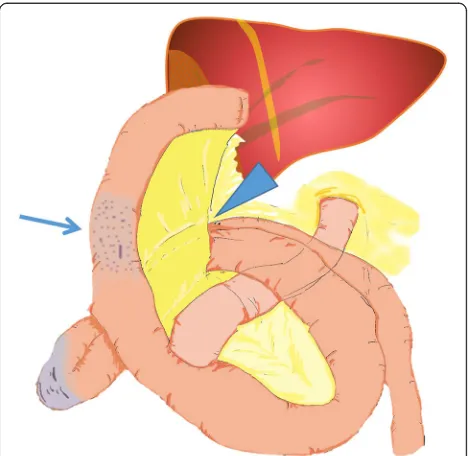
The 44-year-old man in this study was presented with sudden-onset, persistent epigastralgia and had undergone living donor living transplantation (LDLT) for familial amyloid polyneuropathy at 42 years of age, with the left hepatic lobe graft donated by his wife. During LT, biliary reconstruction was performed by hepaticojejunostomy with a Roux limb via the antecolic route as the common bile duct was removed for the sake of the following domino LT. The peritoneal defect related to Roux-en-Y anastomosis was primarily closed with several 4–0 silk interrupted sutures. Although he had experienced repeated episodes of small bowel obstruction, which had all recovered fully following conservative management, at 5, 9, and 14 months post-transplantation, continuous epigastralgia and repeated vomiting for 7 h during the present admission prompted clinical suspicion of bowel strangulation. Abdominal guarding and rigidity in the epigastric region were noted on examination. The body temperature was 37.1 °C. The blood pressure and heart rate were 132/86 mmHg and 105 bpm, respectively. The white blood cell count was elevated at 11,500/μl comprised of 89% neutrophils. However, no significant abnormalities in liver functional tests were observed. We suspected an abdominal emergency and performed an abdominal CT scan that demonstrated dilated, fluid-filled small bowel loops with poor enhancement of the intestinal wall (Fig. 1a). In particular, the Roux limb was markedly dilated (Fig. 1b). Further, a“whirl”appearance and ascites were depicted indicating small bowel volvulus (Fig. 1c). We immediately performed emergent surgery at 6 h after hospital arrival. On laparotomy, no intraperitoneal adhe-sions were observed. We identified an internal hernia through Petersen’s defect (Fig. 2), with a small bowel loop incarcerated beneath the mesenterium of Roux limb through Petersen’s defect causing ischemia of the limb. Following gentle manual repositioning of the incarcerated jejunum, the ischemic color of both the small bowel loop and the Roux limb gradually ameliorated. Although the former recovered completely in color, the appearance of
the latter failed to regain normal color. As a treatment plan, we elected to perform a second-look operation after 48 h for precisely evaluating the condition of the limb. On re-exploration, the Roux limb remained partially dark in color indicating the possibility of remaining ischemic damage. Intraoperative endoscopic examination was performed to determine the most appropriate definitive treatment, revealing partial ischemic injury affecting the mucosa only (Fig. 3) and that the hepaticojejunostomy was intact. On the basis of endoscopic findings, we decided to perform a resection of the portion of limb affected by mucosal necrosis, followed by a jejunojejunost-omy in a side-to-side fashion. Consequently a reanasto-mosis of the hepaticojejunostomy using a newly made limb could be avoided, a procedure that may be compli-cated by technical difficulty and considered a high-risk Fig. 1Enhanced abdominal CT scan on an arrivalaCoronal plane.b,cTransverse plane.a–cCT demonstrated dilated, fluid-filled small bowel loops with poor enhancement of the bowel wall. Arrows indicate a markedly dilated Roux limb. c. The triangle arrow indicates a“whirl”appearance
procedure in emergent settings. The postoperative course was uneventful and the patient was discharged from hos-pital on postoperative day 43. Unfortunately, CT imaging at 1 month after discharge showed dilatation of both the limb and intrahepatic bile ducts (Fig. 4a). We diagnosed stenosis of the limb and performed percutaneous transhepatic
biliary drainage (PTBD) from a dilated terminal branch of B2 using a 7.2 Fr straight tube. Cholangiography via PTBD demonstrated segmental stenosis of the limb likely due to ischemic changes in the serosa; however, the hepaticojeju-nostomy appeared intact. PTBD tube was changed to 12 Fr internal-external drainage tube and placed across the jejunal stenosis (Fig. 4b), followed by balloon dilation with 12 mm diameter (Fig. 4c). Although a balloon dilation procedure was required five times, the stenotic site was successfully dilated and the PTBD tube was eventually re-moved 2 years after the emergency event (Fig. 4d). Recurrence of Roux limb stenosis or intrahepatic bile duct dilatation has not been observed thereafter.
Discussion
Despite recent developments in surgical techniques, biliary reconstruction remains the “Achilles’ heel” of LT. Accordingly, Roux-en-Y or duct-to-duct biliary reconstruction is selected on an individual patient basis. Duct-to-duct biliary reconstruction is preferred even in LDLT due to easy access with endoscopy for treatment of post-transplant biliary strictures and the low incidence of refractory cholangitis [3–6]. Meanwhile, hepaticojejunostomy using a Roux limb remains an alternative option when the recipient bile duct is unusable or the liver graft has multiple hepatic ducts. Roux-en-Y hepaticojejunostomy Fig. 3Intraoperative endoscopy view during re-exploration. Intraoperative
endoscopic findings revealed only partial ischemic injury to the mucosa only
Fig. 4Stenosis of Roux limb at a month after discharge and the treatments.aCT showed dilatation of both the limb and intrahepatic bile ducts.
reportedly has a lower incidence of biliary anastomotic structuring compared to duct-to-duct reconstruction in LDLT [7, 8]. In the present case report, hepaticojejunost-omy was considered the only option as the common bile duct was donated with a recipient liver for a subsequent domino transplantation.
Pertersen’s hernia after Roux-en-Y reconstruction is a known complication of gastrojejunostomy for gastric cancer and bypass surgery for obesity and is observed in 0.2–6.2% of patients [9, 10]. In particular, Pertersen’s hernia reportedly occurs more frequently following laparoscopic Roux-en-Y gastric bypass surgery than open procedures [10]. Pertersen’s hernia is caused by a lack of routine closure of Petersen’s peritoneal defect and fewer abdominal adhesions after laparoscopic procedures [11–13]. Indeed, internal hernia has been rarely reported following major open surgeries such as liver-pancreatic surgery [14]. Fewer surgical adhesions after LT have also been observed, likely due to suppression of surgical adhesions by immunosup-pressants [10, 11]. Blachar and Federle reported that transmesenteric hernias occurred in 20 of 43 LT cases [15]. Additionally, 48 cases of bowel obstruction in 4001 LT patients were caused by surgical adhesions in 19 cases (39.6%) and internal hernia around Roux-en-Y in 18 cases (37.5%) [16], indicating internal hernias including Petersen’s type are a major cause of bowel obstruction after LT, possibly due to fewer intra-abdominal adhesions. Indeed, no intra-abdominal adhesions were observed in the present case.
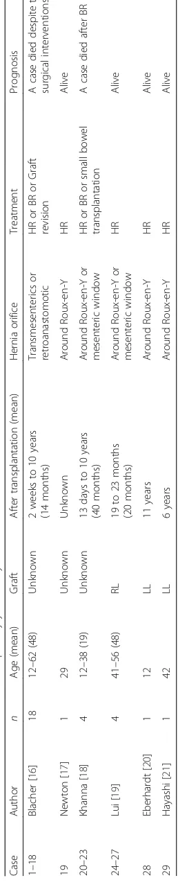
We reviewed the medical literature for cases of in-ternal hernia following hepaticojejunostomy in LT. Table 1 summarizes the clinical features of 29 reported cases [16–21]. All cases underwent biliary reconstruction with Roux-en-Y. It is notable that internal hernias predominantly occurred around the Roux-en-Y limb. Further, 21 of 29 cases were diagnosed by CT imaging, indicating that CT assess-ment is a useful diagnostic tool. Twenty-seven out of 29 cases were recovered by surgery, and a further 21 cases were successfully treated with bowel reposi-tioning followed by closure of peritoneal defects. Meanwhile, among the 6 cases requiring bowel resection, 2 patients underwent extensive resection of the small bowel resulting in a poor prognosis [18].
In the present case, severe complications were avoided by early diagnosis with CT and prompt exploration followed by a second-look operation in which the extent of the Roux limb injury was accurately evaluated using intraoperative endoscopic examination. Consequently, we were able to save the patient without redoing hepaticojejunostomy.
Conclusions
We report a case of Petersen’s hernia observed in a LT patient. Petersen’s hernia is a relatively rare complication
after LT. However, it should be considered as a cause of bowel obstruction in recipients with Roux-en-Y reconstruc-tion. Early diagnosis and prompt and proper surgical intervention including second-look surgery are crucial for the treatment of this type of hernia.
Abbreviations
CT:Computed tomography; LDLT: Living donor liver transplantation; LT: Liver transplantation; PTBD: Percutaneous transhepatic biliary drainage
Acknowledgements
The authors would like to thank Enago (www.enago.jp) for the English language review.
Funding
None of the authors received any funding for this study.
Authors’contributions
SS, RG, NK, YK, MW, TS (Suzuki), DA, KY, TK, and TS (Shimamura) were the attending physicians of the case. SS, RG, MO, AT, and TS (Shimamura) participated in the design of the study. SS, RG, and TS (Shimamura) drafted the manuscript. All authors read and approved the final manuscript.
Consent for publication
Written informed consent was obtained from the patient for publication of this Case Report and all accompanying images. A copy of the written consent is available for review by the Editor-in-Chief of this journal.
Competing interests
The authors declare that they have no competing interests.
Publisher’s Note
Springer Nature remains neutral with regard to jurisdictional claims in published maps and institutional affiliations.
Author details
1Department of Gastroenterological Surgery I, Hokkaido University, Sapporo, Japan.2Division of Organ Transplantation, Hokkaido University Hospital, Sapporo, Japan.3Department of Radiation Medicine, Hokkaido University, Sapporo, Japan.4Department of Transplant Surgery, Hokkaido University, Sapporo, Japan.
Received: 25 May 2017 Accepted: 17 August 2017
References
1. Petersen W. Uber Darmverschlinung nach der Gastroenterostomie. Arch Klin Chir. 1900;62:94–114.
2. Porrett PM, Hsu J, Shaked A. Late surgical complications following liver transplantation. Liver Transpl. 2009;15(Suppl 2):S12–8. doi:10.1002/lt.21893. 3. Qian YB, Liu CL, Lo CM, Fan ST. Risk factors for biliary complications
after liver transplantation. Arch Surg (Chicago, Ill : 1960). 2004;139(10): 1101–5. doi:10.1001/archsurg.139.10.1101.
4. Akamatsu N, Sugawara Y, Hashimoto D. Biliary reconstruction, its complications and management of biliary complications after adult liver transplantation: a systematic review of the incidence, risk factors and outcome. Transpl Int. 2011; 24(4):379–92. doi:10.1111/j.1432-2277.2010.01202.x.
5. Marubashi S, Dono K, Nagano H, Kobayashi S, Takeda Y, Umeshita K, et al. Biliary reconstruction in living donor liver transplantation: technical invention and risk factor analysis for anastomotic stricture. Transplantation. 2009;88(9):1123–30. doi:10.1097/TP.0b013e3181ba184a.
6. Melcher ML, Pomposelli JJ, Verbesey JE, McTaggart RA, Freise CE, Ascher NL, et al. Comparison of biliary complications in adult living-donor liver transplants performed at two busy transplant centers. Clin Transpl. 2010; 24(5):E137–44. doi:10.1111/j.1399-0012.2009.01189.x.
8. Kyoden Y, Tamura S, Sugawara Y, Matsui Y, Togashi J, Kaneko J, et al. Incidence and management of biliary complications after adult-to-adult living donor liver transplantation. Clin Transpl. 2010;24(4):535– 42. doi:10.1111/j.1399-0012.2009.01124.x.
9. Yoshikawa K, Shimada M, Kurita N, Sato H, Iwata T, Higashijima J, et al. Characteristics of internal hernia after gastrectomy with Roux-en-Y reconstruction for gastric cancer. Surg Endosc. 2014;28(6):1774–8. doi:10.1007/s00464-013-3384-7.
10. Steele KE, Prokopowicz GP, Magnuson T, Lidor A, Schweitzer M. Laparoscopic antecolic Roux-en-Y gastric bypass with closure of internal defects leads to fewer internal hernias than the retrocolic approach. Surg Endosc. 2008;22(9):2056–61. doi:10.1007/s00464-008-9749-7.
11. Peker K, Inal A, Sayar I, Sahin M, Gullu H, Inal DG, et al. Prevention of intraabdominal adhesions by local and systemic administration of immunosuppressive drugs. Iran Red Crescent Med J. 2013;15(12):e14148. doi:10.5812/ircmj.14148.
12. Wasserberg N, Nunoo-Mensah JW, Ruiz P, Tzakis AG. The effect of immunosuppression on peritoneal adhesions formation after small bowel transplantation in rats. J Surg Res. 2007;141(2):294–8. doi:10.1016/j.jss.2006.12.541.
13. Higa KD, Ho T, Boone KB. Internal hernias after laparoscopic Roux-en-Y gastric bypass: incidence, treatment and prevention. Obes Surg. 2003;13(3): 350–4. doi:10.1381/096089203765887642.
14. Yu YD, Kim DS, Byun GY, Park BJ, Suh SO. Unexpected internal herniation of the small bowel after right trisectionectomy. ANZ J Surg. 2012;82(9):651–2. doi:10.1111/j.1445-2197.2012.06167.x.
15. Blachar A, Federle MP. Internal hernia: an increasingly common cause of small bowel obstruction. Semin Ultrasound CT MR. 2002;23(2):174–83. 16. Blachar A, Federle MP. Bowel obstruction following liver transplantation:
clinical and CT findings in 48 cases with emphasis on internal hernia. Radiology. 2001;218(2):384–8. doi:10.1148/radiology.218.2.r01ja22384. 17. Newton ER, Turksoy N, Kaplan M, Reinhold R. Pregnancy and liver
transplantation. Obstet Gynecol. 1988;71(3 Pt 2):499–500.
18. Khanna A, Newman B, Reyes J, Fung JJ, Todo S, Starzl TE. Internal hernia and volvulus of the small bowel following liver transplantation. Transpl Int. 1997;10(2):133–6. doi:10.1111/j.1432-2277.1997.tb00555.x.
19. Liu CL, Lo CM, Chan SC, Fan ST, Wong J. Internal hernia of the small bowel after right-lobe live donor liver transplantation. Clin Transpl. 2004;18(2):211–3. doi:10.1046/j.1399-0012.2003.00125.x.
20. Eberhardt CS, Merlini L, McLin VA, Wildhaber BE. Cholestasis as the leading sign of a transmesenteric hernia in a split-liver transplanted child—a case report and review of literature. Pediatr Transplant. 2012; 16(5):E172–6. doi:10.1111/j.1399-3046.2011.01496.x.