OF BREATHING IN THE NEWBORN
A thesis submitted for the degree Doctor of Philosophy
by
Gareth Lewis Ackland
University of London
Departments o f Physiology and Obstetrics & Gynaecology Faculty of Science
University College London
All rights reserved
INFORMATION TO ALL USERS
The quality of this reproduction is dependent upon the quality of the copy submitted.
In the unlikely event that the author did not send a complete manuscript and there are missing pages, these will be noted. Also, if material had to be removed,
a note will indicate the deletion.
uest.
ProQuest 10105222
Published by ProQuest LLC(2016). Copyright of the Dissertation is held by the Author.
All rights reserved.
This work is protected against unauthorized copying under Title 17, United States Code. Microform Edition © ProQuest LLC.
ProQuest LLC
789 East Eisenhower Parkway P.O. Box 1346
Abstract
The newborn ventilatory response to acute isocapnic hypoxaemia is biphasic. An initial increase in breathing (phase 1), mediated by stimulation o f the peripheral chemoreceptors, is followed 1-3 minutes after the onset o f hypoxia by a decline to, or to, below pre-hypoxxc levels (phase 2). The mechanism(s) underlying phase 2 are not known.
This thesis pursues the hypothesis that phase 2 is mediated by CNS mechanisms. First, this hypothesis was tested by investigating the effects o f isocapnic hypoxia on respiratory reflexes in anaesthetized newborn rabbits. These experiments showed that (1) Phase 2 cannot be attributed to a failure in peripheral chemoreceptor function during isocapnic hypoxia. (2) Carotid chemorefiex effects on respiratory output during normoxia are inhibited during isocapnic hypoxia, even though the afferent limb o f the reflex is maintained (3) Somatophrenic reflexes are not affected by isocapnic hypoxia. These findings support the idea that isocapnic hypoxia causes a centrally mediated inhibition o f breathing, which is not attributable to global hypoxic depression.
These findings led to the neurophysiological investigation o f CNS function, in a novel in vivo decerebrate rabbit preparation. Electrical stimulation in the mesencephalon identified a discrete area (the red nucleus), and its efferents, as mediating apnoea; chemical microinjections supported the idea that cell bodies mediate an inhibition o f breathing fi’om such a locus. Furthermore, this inhibitory area was also shown to be involved in mediating the newborn biphasic ventilatory response, since the fall in ventilation was abolished by placing lesions bilaterally in the red nuclei. Pontine inhibitory influences on breathing were also demarcated by electrical stimulation and chemical microinjection, indicating that pontine structures are probably also involved directly in mediating the newborn biphasic ventilatory response.
Personal statement
Except as acknowledged on page 92, the work presented in this thesis was performed solely by the candidate and is original.
Gareth L. Ackland
■ * v
Acknowledgments
Throughout the course o f the last three years I have begun to understand the excitement, challenge and intellectual freedom that pursuing research offers. This is not solely the result of working in such a young (!) and demanding area o f physiology, but is inspired by being in an enthusiastic academic environment which has encouraged questions to be asked, hypotheses to be addressed and to stimulate wide discussion on the topics in question. Furthermore, the value o f conducting basic medical science within the Department o f Obstetrics & Gynaecology has stressed the mutual importance o f clinical medicine and basic science. I thank Professor Charles Rodeck (Obstetrics & Gynaecology) and Professors Roger Woledge and Michael Spyer (Physiology) for the opportunity to conduct my studies in both Departments.
In particular, I am indebted to the following:
Professor Mark Hanson: For his support throughout the three years, strong encouragement for promoting the generation o f independent ideas and for the great times when discussing matters both scientific and not.
Dr. Ray Noble: For the many (late night) discussions, visionary approach, sharing his knowledge o f neurophysiology and the problems associated with investigating the newborn brain stem, and also for discussing general scientific and non-scientific matters.
Dr. Peter Moore: For his teaching, consideration, support and patience.
Dr. Bridget Waites: For her timely arrival and infectious enthusiasm. In addition I am also grateful to the other colleagues who have made the period o f study possible:
Nicole Calder Dr. Dino Giussani Dr. Takanori Watanabe
Clare Crowe Lucy Green Dr. Keith Caddy
LiUian Patterson Fei Li Dr. Juhe Smith
Dr. Laura Bennet Dr. Shiro Kozuma
Table o f contents
INTRODUCTION
CHAPTER 1 - EFFECTS OF HYPOXIA ON CELLULAR AND SYSTEMIC FUNCTION: A REVIEW
General overview
Newborns are particularly susceptible to the occurrence of hypoxia... 32
Why study solely the effects of hypoxia on respiratory control in the newborn? 32 Structure of Chapter 1... 33
SECTION A: EFFECTS OF HYPOXIA ON CELLULAR PHYSIOLOGY Understanding the effects of hypoxia on cellular function is essential for the interpretation of systemic physiological changes... 35
W hat is hypoxia? ...3 5 Several experimental models are used to produce hypoxia... 36
The consequences of hypoxia at the cellular level...37
Ionic changes during hypoxia/anoxia... 37
Membrane potential changes during hypoxia/anoxia...38
Synaptic activity during hypoxia/anoxia...38
Prolonged anoxia leads to anoxic brain damage... 39
Differential sensitivity to hypoxia throughout the nervous system...40
Maturational differences in cellular responses to hypoxia...40
SECTION B: EFFECT OF HYPOXIA ON CARDIORESPIRATORY CONTROL The NEWBORN VENTILATORY RESPONSE TO ACUTE HYPOXAEMIA IS BIPHASIC...43
Stimulationof theperipheralchemoreceptorscauses phase 1 - THE INCREASE IN VENTILATION...44
Cardiovascular response to acute hypoxaemia in newborns... 45
The fetal cardiorespiratory response to acute hypoxaemia... 46
The relevance of adult and fetal respiratory control to the study of the effects of hypoxia on breathing in newborns... 46
Acute hypoxaemia abolishes ‘fetal breathing’ ... 46
Fetal chemoreceptors respond to hypoxaemia but normally do not contribute to the control of FBM...46
Peripheral chemoreceptor sensitivity to Pao^^ but not PacO], increases postnatally...47
Fetal cardiovascular responses to acute hypoxaemia are mediated by the peripheral chemoreceptors... 48
The adult cardiorespiratory response to acute isocapnic hypoxaemia...49
Adults also show BVR to acute isocapnic hypoxaemia...49
THE MECHANISM UNDERLYING PHASE 2 OF BVR
IS UNKNOWN... 50
Hypothesis 1; BVR is duetoperipheralchemoreceptor adaptation TO ACUTE HYPOXAEMIA...50
Direct recordings from carotid chemoreceptor fibres in newborns... 51
Indirect tests of peripheral chemoreceptor function in newborns... 52
Studies of peripheral chemoreceptor fiinction during hypoxaemia in the adult. 54 Hypothesis 2; Central INHIBITION ofrespiratoryoutputbya (HYPOXIA-SENSITIVE) NEURAL PATHWAY LOCATED IN THE BRAIN CAUSES BVR. .56 Fetal inhibitory brain stem influences on FBM during hypoxaemia...56
Inhibitory brain stem influences on breathing in the newborn during BVR...58
Evidence that suggests newborn/ fetal inhibitory brain stem mechanisms are also functional in the adult during hypoxaemia... 59
Hypothesis 3: Directhypoxicdepressionofrespiratoryneurones CAUSES THE FALL IN VENTILATION OBSERVED IN B V R ... .... ... ...62
Hypothesis 4: BVR is causedbythedropinmetabolicrate OBSERVED DURING HYPOXIA... ...64
Metabolism falls during acute hypoxaemia... 64
How much oxygen is essential for aerobic metabolism?...65
Critical effects of environmental temperature on metabolic responses to hypoxaemia...65
Brain stem control of thermoregulation...66
Brown adipose tissue is of particular importance in the newborn... 66
Phase 2 of BVR cannot be explained fully by a fall in metabolism...67
H y p o th e sis 5: BVR is t h e r e s u l t o f CO2 w a s h o u t c au se d by a n in c re a s e IN CEREBRAL BLOOD FLOW DURING HYPOXAEMIA... 67
CBF increases in the fetus and newborn during isocapnic hypoxaemia... 68
CBF also increases in adults during isocapnic hypoxaemia...69
Hypothesis 6: BVR is causedbyrespiratorymusclefailure OR FATIGUE... 70
Hypothesis 8; Phase 2 of BVR is attributabletotheaccumulation
OF INfflBITORY NEUROTRANSMITTERS/NEUROMODULATORS DURING HYPOXIA. ...72
Adenosine and GABA are the most likely neurochemical mediators of BVR ....72
Adenosine... 73
Cellular actions of adenosine... 73
Adenosine is released during hypoxaemia...73
Adenosine inhibits neurotransmitter release...73
Systemic actions of adenosine... 74
Adenosine & adenosine analogue can inhibit breathing... 74
Effects of adenosine receptor antagonists...75
GABA ... 76
Brain GABA concentration increases during hypoxia and inhibits breathing...76
O ther candidates...77
Dopamine... 77
Acetylcholine...77
Opioids... 77
SECTION C: SUMMARY OF INTRODUCTION AND AIMS OF THE PROJECT Summary o f introduction...80
AIMS OF THE PROJECT...81
Aim 1. To develop an anaesthetized newborn animal preparation suitable for a range of neurophysiological experiments...81
RESULTS: SECTION!- EFFECTS OF HYPOXIA ON RESPIRATORY
REFLEXES
Overview,...84
CHAPTER 2: CAROTID CHEMOREFLEXES ARE INHIBITED B Y A CNS MECHANISM DURING ISOCAPNIC HYPOXAEMIA 2.2 Hypotheses... .i ...87
(1) Afferent carotid chemoreceptor discharge is stimulated but does not decline during acute hypoxaemia in anaesthetized newborn rabbits...87
(2) The effect of transient carotid chemoreceptor stimulation on breathing is modulated during hypoxaemia by a central mechanism that inhibits respiratory output in anaesthetized newborn rabbits... 87
2.3 Meth o d s... 88
Animal delivery and care...88
Sedation and anaesthesia...88
Monitoring anaesthesia... 88
Ventilation... 89
Monitoring of pH, blood gases and body temperature...89
Carotid chemoreceptor stimulation...90
2.4 Experimental PROTOCOLS...92
Recording multifibre carotid chemoreceptor afferent activity (n=8 rabbits)...92
Carotid chemoreceptor - phrenic chemorefiex (n= 11 rabbits)... 92
Other preliminary carotid chemorefiex experiments... 93
2.5 Signalprocessinganddatacollection ...94
Blood pressure, heart rate and stimulation marker... 94
Carotid chemoreceptor activity... 96
Phrenic nerve activity...94
Data display and storage... 94
2.6 Analysis... 97
Cardiovascular parameters... 97
Recordings fi'om carotid chemoreceptor multifibres... 97
Carotid chemorefiex test - respiratory parameters... 97
2.7 RESULTS... 99
Carotidchemoreceptorrecordings... ... 99
Blood gas/pH status and cardiovascular parameters... 99
Carotid chemoreceptor fibres do not adapt to acute isocapnic hypoxaemia...99
COj boli increase carotid chemoreceptor discharge during normoxaemia... 100
CO; injections during isocapnic hypoxaemia also increased carotid chemoreceptor discharge...102
CÜ2 boli failed to increase carotid chemoreceptor discharge only during severe hypoxaemia...104
Carotidchemoreflex TEST... 105
Blood gas/pH status and cardiovascular parameters... 105
Isocapnic hypoxaemia elicited BVR... 105
Carotid chemorefiex effects during normoxaemia... 107
Effects of carotid chemoreceptor stimulation on respiratory output were easier to achieve consistently during expiration... 108
Lack of effect of injections on cardiovascular reflexes during normoxaemia.... 108
Carotid chemorefiex effects during isocapnic hypoxaemia -levels of hypoxaemia... 108
Carotid chemorefiex effects seen during normoxaemia disappeared during isocapnic hypoxaemia... 108
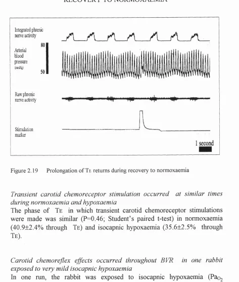
Transient carotid chemoreceptor stimulation occurred at similar times during normoxaemia and hypoxaemia...112
Lack of correlation between the phase of BVR and presence of chemorefiex effect...113
Effect of carotid chemoreceptor stimulation during expiration on other components of the respiratory cycle...118
Inherent variability of controls was not significant ,... 119
Post-stimulus effects on respiratory output...119
Carotid chemorefiex during apnoea... 120
Carotid chemorefiex during severe hypoxaemia...121
Other preliminary carotid chemorefiex experiments... 121
Dithionite injections (n=2 rabbits) support findings of C02-saturated saline chemorefiex experiments...121
Decerebrate rabbit carotid chemorefiex experiments... 123
Lamb carotid chemorefiex experiments... 123
2.8 DISCUSSION... 125
Methods...125
Measurement and control of blood gases, and a direct index of central respiratory output were essential for addressing chemorefiex hypothesis 125 Development of a suitable newborn rabbit preparation... 125
Implementation of a suitable anaesthetic regime... 126
Carotidchemoreceptorrecordings... 127
Methodological drawbacks...127
Lack of baroreceptor effects... 129
Severe hypoxaemia/asphyxia... 130
Carotidchemoreflextest...130
Variable magnitude of BVR... 130
Chemical carotid chemoreceptor stimulation is more appropriate than electrical CSN stimulation in the newborn rabbit preparation...130
Reliability of the transient carotid chemoreceptor stimulus test...131
Perturbation of the respiratory rhythm produces effects on subsequent breaths...132
Dithionite experiments support findings using CO2- saline experiments... 132
Lack of correlation between peak in central respiratory output
and type of carotid chemoreflex effect... 135
CNS studies show that hypoxia inhbits expiratory respiratory neurones... 136
Hypoxia also inhibits expiratory motor activity...137
The application of oscillatory theories to the carotid chemoreflex experiments... 138
Carotid chemoreflex experiments are supported by similar studies in the newborn rabbit... 139
The findings of carotid chemoreflex experiments are also supported by similar studies in adults...139
2.9 Summary... 140
CHAPTER 3 - ISOCAPNIC HYPOXAEMIA DOES NOT INHIBIT SOMATOPHRENIC REFLEXES 3.1 INTRODUCTION...142
3.2 Hypothesis... 143
Phase 2 of BVR is caused by a brain stem mechanism that inhibits respiratory reflexes globally... 143
3.3 Met h o d s...143
Surgical preparation...143
Monitoring anaesthesia...144
Ventilation... 145
Control of pH, blood gases and body temperature...145
Phrenic nerve activity...146
Peripheral nerve stimulation... 146
3.4 Signalprocessinganddatacollection...147
Cardiovascular data...147
Respiratory data...147
Data collection and storage...147
3.5 Experimentalprotocols... 147
3.6 Analysis...149
Cardiovascular parameters... 149
Respiratory parameters...149
3.7 Resu lts...150
Blood gas/pH and cardiovascular data... 150
BVR was present in all preparations... 151
Stimulation of somatic afferents in normoxaemia increases respiratory output. 152 No clear effect on timing of punctate somatic afferent stimuli on respiratory output during normoxaemia... 154
Lack of effect of somatic afferent stimulation on blood pressure/heart rate.... 155
Stimulation of somatic afferents caused respiratory output to increase in isocapnic hypoxaemia... 155
Somatic afferent stimulation also increases respiratory output in recovery... 156
Severe hypoxaemia/asphyxia did not abolish effect of stimulating somatic afferents on respiratory output... 156
3.8 DISCUSSION... 157
M ethods... 157
Decerebration... 157
Results...157
The biphasic ventilatory response is present in both anaesthetized and decerebrate, decerebellate preparations...157
No clear effects of somatic afferent stimulation on respiratory timing... 158
Comparable results from a recent similar study...158
The effect of changing stimulation intensity... 159
Group I and II fibres are stimulated at the threshold used for the somatophrenic reflex...160
Evoking somatophrenic reflexes at threshold does not elicit strong cardiovascular reflexes due to stimulation of group I and II fibres... 161
Stimulation of hindlimb afferents is unlikely to affect carotid chemoreceptor discharge...162
Hypoxia and hypoxia-related substances do not affect somatic afferents... 162
RESULTS: SECTION 2
-
THE ROLE OF THE BRAIN STEM AND
MID-BRAIN IN MEDIATING THE EFFECTS OF HYPOXIA ON
BREATHING IN NEWBORNS
Overview o f Results: Section 2... 167
CHAPTER 4: THE DEVELOPMENT OF A NEWBORN RABBH PREPARATION FOR NEUROPHYSIOLOGICAL STUDIES INVESTIGATING THE ROLE OF THE BRAIN STEM AND MESENCEPHALON IN MEDIATING BVR 4.1 Introduction... 168
4.2 AIM: TO DEVELOP NEWBORN DECEREBRATE PREPARATION ...169
4.3 Met h o d s...169
General methods... 169
Decerebration... 171
Electrical stimulation...173
Chemical stimulation...173
Reversible focal cooling...175
Histological methods...176
4.4 Signalprocessinganddatacollection ... 178
4.5 Analysis __________________________________ ...178
Central respiratory output...178
Electrical stimulation...178
Microinjectate stimulation... 179
Cardiovascular parameters:...179
4.6 Experimental PROTOCOL...179
Respiratory response to isocapnic hypoxaemia...179
4.7 Results ...___ ...____ ... 180
Decerebrate newborn rabbits exhibit BVR (n=32 rabbits)... 180
Sham rage occurred in one rabbit during hypoxaemia... 182
Halothane depresses cardiorespiratory output markedly in newborn decerebrate rabbits (n=2) and lambs (n=2)... 183
Histological techniques...183
4.8 Discussion...184
M ethods...184
Decerebration conferred stability... 184
Histological techniques proved suitable... 184
Results... 185
Decerebrate newborn rabbits exhibit BVR... 185
Sham rage occurred in only one rabbit...185
Arterial blood pressure showed “biphasic response”to isocapnic hypoxaemia... 185
4.9 Summary...186
CHAPTER 5 - BVR IS MEDIATED BY STRUCTURES IN THE MESENCEPHALON
5.1 DfmODUCTION... ... . ... .. ... 188
5.2 Hypotheses ... 189
(1) Phase 2 of BVR is abolished by the destruction of a group of mesencephalic cell bodies, demarcated by electrical and chemical stimulation, in newborn ‘decerebrate rabbits ... 189
' (2) Any putative mesencephalic inhibitory mechanism is only functional during hypoxia... 189
5.3 Methods ...190
5.4 Pro to cols... 190
Identification of areas that mediate inhibition of respiratory output...190
The respiratory response to isocapnic hypoxaemia after areas that had been demonstrated to inhibit respiratory output are destroyed by electrolytic lesions...190
Does inhibition of a inhibitory mesencephalic area increase respiratory output?...191
5.5 Analysis... 191
5.6 Results ...__ ... 192
Identification of inhibitory mesencephalic areas by electrical stimulation (n=38 rabbits)... 192
hficroinjection expèriments (n=6 rabbits)...197
Lesioning (n=14 rabbits)... 203
Lesioning mesencephalic areas that produced an inhibition of respiratory output during normoxaemia abolished phase 2 of BVR (n=5 rabbits)... 203
Abolition of BVR attributable to maintenance of respiratory fi*equency...204
Bilateral lesions in areas where apnoea was elicited abolished BVR (n=3 rabbits)...206
Kainic acid injection (n=l rabbit)...211
Midline electrolytic lesion (n=l rabbit)...212
Phase 2 of BVR persisted when lesions were made in mesencephalic areas that failed to produce an inhibition of respiratory output during normoxaemia (n=9 rabbits)...213
Decline in respiratory fi’equency accounted for post-lesion BVR in animals where bilateral/midline apnoea could not be elicited... 216
Microinjection of GABA increased respiratory output during isocapnic hypoxaemia (n=3 rabbits) but not normoxaemia (n=4 rabbits)...217
5.7 Discussion... 219
Electrical stimulation... 219
Chemical stimulation... 219
EAA microinjection... 221
Extensive bilateral electrical stimulation in the mesencephalon could cause deleterious cardiovascular changes...222
Bilateral electrolytic lesions made only in the red nucleus did not affect subsequent respiratory output during normoxaemia...222
Bilateral electrolytic lesions made only in the red nucleus abolished phase 2 of BVR (lesioned(non BVR animals)... 223
The failure of other lesions to abolish phase 2 of BVR (lesioned(BVR)) animals was due to placement of the stimulating electrode outside the red nucleus/rubrospinal tract... Z... 223
Higher incidence of sham rage during hypoxaemia in lesion (BVR) and lesion (non-BVR) rabbits compared to controls... 224
Lesioning had no clear effect on phase 1 increase... 224
Biphasic arterial blood pressure response not affected by lesions... 224
GABA microinjections indicate that an inhibitory mesencephalic inhibitory area is only active during isocapnic hypoxaemia...225
Abolition of BVR by lesions in the red nucleus reconciles apparently contradictory supra- and rostral pontine lesioning/transection studies... 226
The structure of the red nucleus... 227
Nucleus ruber magnocellularis...227
Nucleus ruber parvicellularis... 228
The red nucleus controls motor output and spinal cord processing... 228
Red nucleus inhibitory efferents have been identified in adult cats... 229
Respiratory related afferents to the mesencephalon and red nucleus... 230
5.8 Summary. ...___ ...___ ...232
CHAPTER 6 - THE ROLE OF THE PONS m MEDIATING INHIBITION OF RESPIRATORY OUTPUT IN NEWBORN DECEREBRATE RABBITS
6.1 Introduction...234
6.2 Hypotheses ... ...234
(1) Electrical stimulation of areas in the pons causes inhibition of respiratory output...234
(2) Microinjection of EAA into dorsal pons at the level of the middle cerebellar peduncle causes an inhibition of respiratory output...234
(3) Cooling of upper rostral pontine regions reverses phase 2 of BVR...234
6.3 Me t h o d s... 235
6.4 Analysis... 235
6.5 Resu lts... 236
Experiment 1: Electrical stimulation of pontine structures (n=20 rabbits) 236 Loci where respiratory output was inhibited... 237
Locus coeruleus (n=5 rabbits ) ...237
Reticular formation (n=10 rabbits)... 238
Raphe nuclei (n=3 rabbits)... 239
Experiment 2: Microinjections into the dorsal pons, including locus coeruleus (n=8 rabbits)...241
Experiment 3: Effect of cooling at level of middle cerebellar peduncle on respiratory output during isocapnic hypoxaemia (n=l rabbit)... 244
Cooling of dorsolateral pons reversed phase 2 by increasing peak phrenic activity... 244
6.6 Discussion... 247
Pontine reticular formation... 247
Raphe nuclei...248
Locus coeruleus...249
Microinjections...250
Confirmation that cooling the upper rostral pons reverses phase 2 of BVR in the rabbit... 251
CHAPTER 7 - FINAL DISCUSSION: HYPOXIA AND BVR FROM AN EVOLUTIONARY PERSPECTIVE
7.1 Mainfindingsofthethesis ...254
Results: Section A ... 255
Results: Section B ... 256
7.2 Hypoxiaand BVR fromanevolutionaryperspective... ...257
Numerous strategies exist across species for countering the effects of hypoxic environments...257
The evolution of an oxygenated atmosphere... 257
Diversity of mechanisms for counteracting hypoxia... 258
Aquatic breathers...258
Air-breathers/air-aquatic breathers...258
Intermittent air-breathers... 259
Metabolic considerations... 260
Evidence for a hypoxia-sensitive mechanism at the molecular/genetic level....261
Evidence from cellular_m vivo studies for hypoxia-sensitive mechanism 262 Potential role of the mesencephalon, red nucleus and rostral brain stem 263 Clinical and pathophysiological implications... 264
In conclusion...264
7.3 Final Summary ...___ ...___ ...__...__...265
REFERENCES,
... 266APPENDICES
Appendix 1 - Calibrations...308Appendix 2 - Solutions... 312
Appendix 3 - Histological atlas of newborn rabbit brainstem/mesencephalon 314 Appendix 4 - Individualized carotid chemoreceptor recording results (Chapter 2)... 329
Appendix 5 - Map of mesencephalic area, from electrical stimulation experiments, that elicit inhibition of respiratory output... 332
Appendix 6 - Method of ventilation... 334
Table o f figures
CHAPTER 1 - INTRODUCTION
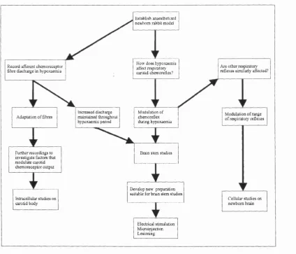
Figure 1.1 Schematic diagram illustrating the potential development of the project, depending on the outcome of the initial experiments
investigating carotid chemoreflexes in newborn anaesthetised rabbits... 82
CHAPTER 2 - CAROTID CHEMOREFLEXES ARE INHIBITED BY A CNS MECHANISM DURING ISOCAPNIC HYPOXAEMIA
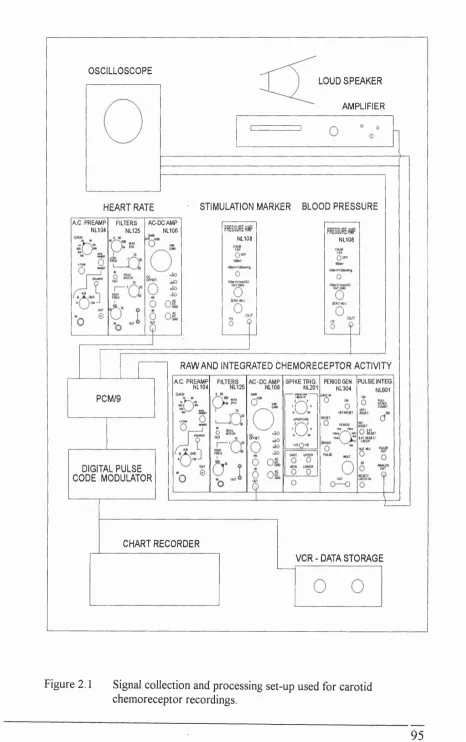
Figure 2.1 Signal collection and processing set-up used for carotid
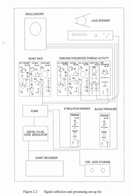
chemoreceptor recordings... 95 Figure 2.2 Signal collection and processing set-up for
carotid chemoreflex experiments...96 Figure 2.3 Method of analysis for carotid chemoreflex test... 98 Figure 2.4 Carotid chemoreceptor discharge increased and remained
elevated throughout hypoxaemia...99 Figure 2.5 CO2 -saturated saline bolus increased carotid chemoreceptor
discharge during normoxaemia. After 10 minutes of isocapnic hypoxaemia, there was a similar increase in carotid chemoreceptor discharge on
CO2 -saturated saline bolus injection. Also note increase in baseline carotid
chemoreceptor discharge... 100 Figure 2.6 Group results showing of effect of C02-saturated saline
on multifibre carotid chemoreceptor discharge during normoxaemia
and isocapnic hypoxaemia...101 Figure 2.7 Effect of Ringer’s saline (pH 7.38) on multifibre carotid
chemoreceptor discharge during normoxaemia... 101 Figure 2.8 C02-saturated saline injections increased carotid chemoreceptor
discharge 1 min before the onset of isocapnic hypoxaemia... 102 Figure 2.9 C02-saturated saline bolus increased carotid chemoreceptor
discharge 1 min after the onset of isocapnic hypoxaemia. Note increased
basal carotid chemoreceptor discharge due to fall in Paoj... 103 Figure 2.10 C02-saturated saline injection continued to increase carotid
Figure 2.11 COz-saturated saline increased carotid chemoreceptor discharge 1 min after the end of isocapnic hypoxaemia. Note increased basal carotid chemoreceptor discharge has declined due to the return to normpxaemic
Paoj values...104 Figure 2.12 Isocapnic hypoxaemia elicited BVR in anaesthetized, paralysed and vagotomized newborn rabbits... 105 Figure 2.13a PPA during control (normoxaemia), and phase 1 and
phase 2 of BVR... 106 Figure 2.13b Respiratory frequency during control (normoxaemia), and
phase 1 and phase 2 of BVR...106 Figure 2.14 Transient carotid chemoreceptor stimulation (using COz-saturated saline) during expiration prolonged expiration during normoxaemia...107 Figure 2.15 Transient carotid chemoreceptor stimulation (using C02-saturated saline) during early inspiration increased PPA during normoxaemia... 107 Figure 2.16a The effect of prolongation of Te produced by carotid
chemoreceptor stimulation during expiration disappeared during isocapnic hypoxaemia, as shown by the % change in Te for the stimulation breath
as compared with control breaths... 109 Figure 2.16b The effect of prolongation of Te produced by carotid
chemoreceptor stimulation during expiration disappeared during isocapnic hypoxaemia, as shown by the difference in Te between stimulation and
control breaths...109 Figure 2.17 Transient carotid chemoreceptor stimulation (using C02-saturated saline) made during expiration prolonged the duration of expiration during
normoxaemia... 110 Figure 2.18 Transient carotid chemoreceptor stimulation fails to pertub
respiratory output during isocapnic hypoxaemia in the same rabbit... I l l Figure 2.19 Prolongation of Te returns during recovery to normoxaemia... 112
Figure 2.20a Individual rabbit data for transient carotid chemoreceptor
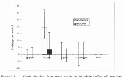
stimulation in amnaesthetized, paralyzed and vagotomized rabbits... 114 Figure2.20b Individual runs for transient carotid chemoreceptor stimulation in a decerebrate lamb and decerebrate rabbit... 117 Figure 2.21 Effect of transient carotid chemoreceptor stimulation during
normoxia and hypoxia on (1) Te, (2) comparison between Ti of pre-stimulation
breath (PSC) and Ti (poststimulation breath) (3) PPA of precontrol breath and poststimulation breath...118
Figure 2.22 Comparison of the pre-stimulation breath and pre-control during normoxaemia and isocapnic hypoxaemia showed that there was no tendency for breaths to be prolonged spontaneously... 119 Figure 2.23 Following perturbation of Te by transient carotid chemoreceptor stimulation, there was a compensatory increase in respiratory frequency for the next breath during normoxaemia and early hypoxaemia only...119 Figure 2.24a, b Carotid chemoreceptor stimulation could elicit phrenic nerve activity during apnoea, but effect disappeared with prolonged apnoea... 120 Figure 2 .2 5 Transient carotid chemoreceptor stimulation shortened Ttot
during gasping/very slow breathing... 120 Figure 2.26 Sodium dithionite injections stimulated respiratory output in
normoxaemia. ABP also fell during stimulation of respiratory output... 121 Figure 2.27 Stimulatory effect of sodium dithionite on respiratory output
and ABP persisted at 180s of isocapnic hypoxaemia...121 Figure 2.28. Stimulatory effect of sodium dithionite on respiratory output
and ABP persisted after 240s of isocapnic hypoxaemia... 122 Figure 2.29 On recovery to normoxaemia, sodium dithionite produced
hyperpnoea again. ABP is not shown... 122 Figure 2.30 Three possible interpretations for the carotid
chemoreflex experiments... 133
CHAPTER 3 - ISOCAPNIC HYPOXAEMIA DOES NOT INHIBIT SOMA TOPHRENIC REFLEXES,
Figure 3.1 Diagram showing electronic set-up for signal processing and data collection for somatophrenic reflex experiments... 148 Figure 3.2 BVR was elicited in anaesthetised rabbit experiments (n=7 rabbits), (peak and nadir of respiratory output during isocapnic hypoxaemia shown)... 151 Figure 3.3 BVR in decerebrate lamb experiments (n=4 lambs), showing
peak and nadir .of respiratory output (during isocapnic hypoxaemia
compared to normoxaemic controls... 151 Figure 3.4 Stimulating sciatic nerve both during normoxaemia and after
5 min isocapnic hypoxaemia increased respiratory output (decerebrate lamb response)... 152 Figure 3.5 Somatic afferent stimulated respiratory output during normoxaemia and after 5 min isocapnic hypoxaemia in anaesthetised rabbits. Similar results were seen for decerebrate rabbit experiments...153 Figure 3.6 Somatic afferent stimulation increased respiratory output during
normoxaemia and after 5 min isocapnic hypoxaemia in decerebrate lambs... 153 Figure 3.7 Sustained or transient stimulation of somatic afferents caused
CHAPTER 4 - THE DEVELOPMENT OF A NEWBORN RABBIT PREPARATION FOR NEUROPHYSIOLOGICAL STUDIES INVESTIGATING THE ROLE OF THE BRAIN STEM AND MESENCEPHALON IN MEDIATING BVR
Figure 4.1 Cooling probe used to cool the dorsorostral pons... 175 Figure 4.2 Newborn decerebrate rabbits show a biphasic response in
respiratory output to 8 min of isocapnic hypoxaemia (n=32 rabbits)... 181 Figure 4.3 PPA during control, phase 1 and phase 2 of BVR (n=32 rabbits).... 181 Figure 4.4 Respiratory frequency during control, phase 1 and phase 2
of BVR (n=3 2 rabbits)...182 Figure 4.5 Sham rage in one decerebrate rabbit was characterized by ABP
increase (70% over control ABP), plus an initial augmentation of PPA,
followed by apnoea...182
CHAPTER 5 - BVR IS MEDIATED BY STRUCTURES IN THE MESENCEPHALON
Figure 5.1 Electrical stimulation in the mesencephalon at the level of the superior coUiculus could produce rapid increases in respiratory output,
due to either increased frequency and/or PPA...192 Figure 5.2 Electrical stimulation, within a discrete loci 7-8mm from the
surface of the mesencephalon at the level of the superior colliculi, inhibited respiratory output. Apnoea was observed usually...193 Figure 5.3 \fixed effects on respiratory output could also be elicited by
electrical stimulation of mesencephalic areas at the level of the
superior/inferior colliculi...193 Figure 5.4 Histology confirmed that electrical stimulation of the
rubrospinal tract, as well as the red nucleus, inhibited respiratory output... 194 Figure 5.5 Discrete inhibitory area illustrated by showing the effects of electrical stimulation on respiratory output in one rabbit. Stimulatory effect of electrical stimulation on respiratory output at 6mm depth from surface is followed 1mm below, at depth 7mm, by profound inhibition of respiratory output. Partial inhibition (mixed effect) occurs at a depth of 7.3mm, but having driven the electrode through the area of inhibition, electrical stimulation again causes
rapid increases in respiratory output...196 Figure 5.6 Stimulation of respiratory output due to microinjection of
glutamate in the periaqueductal grey... 198
Figure 5.7a Transient inhibition of two breaths (fall in PPA) due to microinjection of glutamate at depth where electrical stimulation inhibited
respiratory output at the level of the superior coUiculus... 200 Figure 5.7b Transient prolongation of Te foUowing microinjection of
glutamate in area of mesencephalon at level of superior coUiculus where
electrical stimulation had inhibited respiratory output...200 Figure 5.7c In another rabbit, transient inhibition of breath (PPA decreased) followed microinjection of glutamate in the red nucleus... 201 Figure 5.7d Histological trace showing site of microinjection in red nucleus (lateral border) for figure 5.7c. Two microinjections were made at this site,
both causing transient inhibition of respiratory output (decreased PPA)... 201 Figure 5.8 BUateral electrolytic/chemical lesions in the red nucleus or its
efferents abolished phase 2 of BVR (n=5 rabbits)...203 Figure 5.9 For non-lesion experiments PPA increased during phase 1 of
BVR but did not alter during phase 2 (n=5 rabbits)... 205 Figure 5.10 Respiratory fi*equency did not contribute to phase 1 of BVR but declined sharply during phase 2 in non-lesioned experiments. In marked contrast, respiratory frequency was maintained throughout isocapnic hypoxaemia in lesioned rabbits where (bilateral) apnoea had been elicited previously by
electrical stimulation (n=5 rabbits)... 205 Figure 5.11a Rabbit 090394. Isocapnic hypoxaemia elicited BVR in the intact rabbit. Apnoea was elicited subsequently on both sides of the midline of the mesencephalon at the level of the superior coUiculus, by electrical stimulation. Histological trace shows that lesions were made in these apnoeogenic areas in both the red nucleus and in the rubrospinal tract, at the level of the inferior coUiculus. Here lesioning increased the relative magnitude of phase 1,
Figure 5.12 Unilateral electrical stimulation demarcated an area in the red nucleus which produced apnoea. Kainic acid was injected subsequently into this area, but spread medially to the other side of the mesencephalon at the level of the superior coUiculus. Thus bilateral destruction at the depth of the red nucleus in the mesencephalon at the level of the red nucleus abolished
phase 2 of BVR...211 Figure 5.13 A midline electrolytic lesion abolished phase 2 of BVR. Lesioning increased the magnitude of phase 1, in comparison to the peak respiratory output during hypoxaemia in the intact rabbit. Histology showed that the midline lesion had been made in the region of the decussation of
rubrospinal fibres...212 Figure 5.14a BVR persisted in all rabbits where bilateral respiratory
inhibition was not found. (n=9 rabbits)...214 Figure 5.14b Example of rabbit where bilateral lesions were made, but
where BVR persisted on exposure to isocapnic hypoxaemia after lesioning/electrical stimulation. In this case, apnoea was only elicited unilaterally fi'om red nucleus. Apnoea was only found on the left side: the stimulating electrode was placed more laterally. Hence, the inhibitory
region in the red nucleus was not lesioned on the left side...214 Figure 5.15 Respiratory frequency did not contribute to the rise in respiratory output during hypoxaemia, but fell sharply thereby causing phase 2 for lesion (BVR) rabbits (n=9 rabbits)... 216 Figure 5.16 An increase in PPA caused the peak in respiratory output during hypoxaemia in lesion (BVR) rabbits (n=9 rabbits). This increase was greater in pre-lesion than post-lesion rabbits. PPA did not fall during phase 2...216 Figure 5.17 Microinjection of GABA, into an area at the level of the superior coUiculus where electrical stimulation made during normoxaemia had eUcited inhibition of respiratory output, reversed apnoea, by initiating gasping... 217 Figure 5.18 GABA, microinjected into an area at the level of the superior
coUiculus where electrical stimulation made during normoxaemia had eUcited inhibition of respiratory output, could increase respiratory output
transiently during hypoxaemia...218
CHAPTER 6 - THE ROLE OF THE PONS IN MEDIATING INHIBITION OF RESPIRATORY OUTPUT IN NEWBORN DECEREBRATE RABBITS
Figure 6.1 Diagram showing rostro-caudal positions where tracks were
made with the electrical stimulating electrode...236 Figure 6.2 Stimulation of locus coeruleus could cause inhibition
of respiratory output... 237 Figure 6.3 Electrical stimulation of the reticular formation produced apnoea ....238 Figure 6.4 Electrical stimulation produced inhibition in the region
of the raphe/midline nuclei... 239 Figure 6.5 Compilation of sites where electrical stimulation produced
inhibition of breathing or apnoea...240 Figure 6.6 Inhibition of respiratory output due to microinjection of
glutamate in region of locus coeruleus... 242 Figure 6.7 Stimulation of respiratory output in dorsorostral pons, again at
level of locus coeruleus, stimulated respiratory output during normoxaemia and isocapnic hypoxaemia. The example shown illustrates the effect of
glutamate injected during hypoxaemic apnoea... 242 Figure 6.8 Microinjection of glutamate into parabrachial region
produced apneusis...243 Figure 6.9 Phase 2 of BVR was reversed by cooling the floor of the IV
ventricle at the level of the middle cerebellar peduncle... 243 Figure 6.10 The restoration of phase 1 levels of respiratory output on
cooling (probe temperature (°C) indicated by dotted line) was attributable
solely to an increase in PPA, with no effect on respiratory fi-equency... 245
CHAPTER 7 - FINAL DISCUSSION: HYPOXIA AND BVR FROM AN EVOLUTIONARY PERSPECTIVE
Table o f tables
Table 1.1 The biphasic ventilatory response has been reported in all mammals studied, in both the anaesthetized and conscious states...43 Table 2.1 Blood gas, pH, HR and ABP values for carotid chemoreceptor
recordings (n=8 rabbits)... 99 Table 2.2 Blood gas, pH, HR and ABP values for anaesthetized rabbit carotid
chemoreflex experiments (n=10 rabbits)... 105 Table 3.1 Blood gas, pH, HR and ABP values for anaesthetized rabbits
exposed to isocapnic hypoxaemia (n=7 rabbits)... 150 Table 3.2 Blood gas, pH, HR and ABP values for decerebrate rabbits
exposed to isocapnic hypoxaemia (n=4 rabbits) ...150 Table 3.3 Blood gas, pH, HR and ABP values for decerebrate lambs
exposed to isocapnic hypoxaemia (n=4 lambs)... 150 Table 3.4 In preliminary experiments, no consistent effects were seen
with punctate stimuli...154 Table 4.1 Blood gas, pH, HR and ABP values for decerebrate rabbits
exposed to isocapnic hypoxaemia (n=32 rabbits)... 180 Table 5.1 Blood gas, pH, HR and ABP values for lesion (non-BVR)
rabbits exposed to isocapnic hypoxaemia experiments
(n=5 rabbits)...204 Table 5.2 Blood gas, pH, HR and ABP values for lesion (BVR) rabbits
exposed to isocapnic hypoxaemia experiments
(n=9 rabbits)...213
Table o f plates
Plate 2.1 Photograph showing arrangement of catheters for injection of COj- saturated saline to stimulate carotid body chemoreceptors... 91 Plate 2.2 Photograph showing recording electrode used for both carotid
chemoreceptor fibre and phrenic nerve recordings...91 Plate 3.1 Photograph showing custom-made stimulating electrode for
somatophrenic experiments... 146 Plate 4.1 Photograph showing recording electrode used for most experiments reported in Results: Section 2 ...171 Plate 5.1 Photograph of Cresyl Violet stained slice showing lesions made in rubrospinal tract and adjacent reticular formation, at level of inferior coUiculus. Electrical stimulation of the the two most lateral tracks elicited apnoea/inhibition
of respiratory output...195 Plate 5.2 Photograph of Cresyl Violet stained slice showing site of
microinjection of glutamate into PAG, that stimulated respiratory output 199 Plate 5.3 Photograph of Cresyl Violet stained slice showing site where
microinjection of ^utamate elicited transient inhibition of respiratory output. This was located on the lateral border of the red nucleus... 202 Plate 5.4 Photograph showing bilateral lesions placed in red nucleus, which abolished phase 2 of BVR (rabbit 090394). The lesions were made at sites where respiratory output had been inhibited/abolished by electrical stimulation...207 Plate 5.5 Photograph showing slice illustrating start of stimulating electrode tracks made in one rabbit where BVR was not abolished...215 Plate 6.1 Photograph showing site of cooling probe (locus coreuleus) where ventilatory fall during isocapnic hypoxia was reversed...246
Abbreviations
The following list includes the symbols and abbreviations used in respiratoiy physiology. These comply with the standards approved by the Commission on Respiratory Physiology (1980). For main histological abbreviations, refer to Appendix 3.
[ li Intracellular concentration of ion [ ]o Extracellular concentration of ion
P fla Hydrogen ion concentration in arterial blood
% Percentage
°c
Degrees Celsiusa Alpha
5-HT Serotonin
ABP Arterial blood pressure
ACh Acetylcholine
ATP Adenosine triphosphate BAT Brown adipose tissue
bpm Beats per minute
BVR Biphasic ventilatory response C {number} Cervical nerve root
ca. Circa
Calcium ion
cr
Chloride ionCo. Company
CO2 Carbon dioxide
CBF Cerebral blood flow
cm CNS CSF CSN DA EAA ECG EMG EPSP f FRC FBM
F 1 0 2
GA HR lAA i.m. IPSP IQR i.v. K+
L {number} Ltd
MAP n
Centimetre
Central nervous system Cerebrospinal fluid Carotid sinus nerve Dopamine
Excitatory amino acid Electrocardiogram Electromyogram
Excitatory postsynaptic potential Frequency
Functional residual capacity Fetal breathing movements Inspired oxygen fraction Gestational age
Heart rate
Inhibitory amino acid Intramuscular
Inhibitory postsynaptic potential Interquartile range
Intravenous Potassium ion Lumbar nerve root Limited
n Number
N2 Nitrogen
Na^ Sodium ion
NaCN Sodium cyanide
NE Noradrenaline
NTS Nucleus tractus solitarius
O2 Oxygen
P Probability
P0 2 Partial pressure of oxygen
Pc02 Partial pressure of carbon dioxide PAG Periaqueductal grey
PCr Phosphocreatine
Paco2 Partial pressure of carbon dioxide in arterial blood Pao2 Partial pressure of oxygen in arterial blood
pH Logjo hydrogen ion concentration in a solution pHa Logio hydrogen ion concentration in arterial blood PPA Peak phrenic activity
RVLM Rostro-ventrolateral medulla S {number} Sacral nerve root
s.c. Subcutaneously
SD Standard deviation
SEM Standard error of the mean
Te Expiratory time
Ti Inspiratory time
UK United Kingdom
USA United States of America Vco2 Carbon dioxide production
Ve Ventilation
VLM Ventrolateral medulla V0 2 Oxygen consumption
Vt Tidal volume
SI units
Length m cm mm pm Metre Centimetre Millimetre Micrometres M ass kg g mglig
Kilograms Grams Milligrams Micrograms Tim e hr min s ms Hz Hour Minute Second MillisecondCycles per second
Volume 1 ml
pi
ni
Litre Millilitre Microlitres Nanolitres Electrical mA pA mV Q Milliamps Microamps Millivolts Resistance ChemicalM Moles
mM Millimoles
pM Micromoles
Pressure is expressed as mmHg (millimetres o f mercury) throughout the thesis, rather than in SI pressure units (Pascals)
CHAPTER 1
Ge n e r a l o v e r v ie wo fin t r o d u c t io n
Newborns are particularly susceptible to the occurrence
of
hypoxia
Newborn babies, especially those bom prematurely, are particularly prone to apnoea (Miller & Smull, 1955; Henderson-Smart, 1981). Apnoea results in asphyxia - a fall in Paoj and increased Paco2* Apnoeas may be central or obstmctive. Central apnoea, the cessation o f airflow and inspiratory effort, is particularly common in premature babies (Milner & Greenough, 1988). Immaturity o f sleep state patterns and inhibitory laryngeal respiratoiy reflexes have been imphcated in central apnoea (Marchai, Bairam & Vert, 1987). Obstructive apnoea, the cessation o f airflow despite continued respiratory efforts, is the result o f poor co-ordination o f the upper airway musculature. This occurs during early infancy in both term and pre-term infants, thereby increasing the risk o f hypoxic spells. This occurs during sleep (Henderson-Smart, 1980). About 50% o f apnoeas in premature babies are mixed i.e. both central and obstructive (Brazy, Kinney & Oakes, 1987).
Although it is not clear whether SIDS victims have a higher incidence o f disturbed respiratory patterns, such as increased incidence and severity o f apnoeic attacks, clearly respiratory arrest must occur at some stage. Whether hypoxaemia ehcits inappropriate physiological responses as a result o f immature development o f respiratory control in SIDS victims compared to age-matched babies is not known.
Why study solely the effects o f hypoxia on respiratory control in the newborn?
To understand the basic physiological mechanisms that underhe the newborn response to asphyxia requires separating out the effects o f Paoj and Pacoz- The consequences o f hypoxia in the newborn become more clearly hazardous when the cardiorespiratory responses are considered. Although hypoxia ehcits a number o f cellular and systemic physiological effects, this thesis concentrates on the apparently paradoxical ventilatory response o f newborn mammals that is
observed in hypoxia.
The terms "newborn" and "neonate" are often used loosely in the literature: in clinical medicine neonate refers to infants less than one month old. For clarity the term newborn is used throughout this thesis to indicate that the postnatal age under investigation is around one month old, and not yet an adult.
Structure o f Chapter 1
Section A o f this introduction considers the effects o f hypoxia on cellular function. Because the integration o f the factors that control respiratory output occurs in the central nervous system, section A focuses on the responses o f brain tissue to hypoxia, including how the cellular properties o f the newborn central nervous system indicate why newborns may tolerate systemic hypoxia better than adults.
Section B examines a variety o f hypotheses that have been proposed to explain the effect o f hypoxia on the control o f breathing in the newborn, adult and fetus. This section also considers how hypoxia affects neurotransmitter and neuromodulator release and hence cellular processes, and the consequences o f these changes for systemic physiological fimction.
SECTION A
EFFECTS OF HYPOXIA ON CELLULAR PHYSIOLOGY
1.1
Understanding the effects of hypoxia on cellular function is
essential for the interpretation of systemic physiological changes
While the experiments conducted in this thesis can be classified broadly as “systems” or “whole animal” physiology, it is important to note that the cellular mechanisms that are involved in the response to hypoxia are also considered. Hypoxia is an excellent example o f how the study o f cellular and systemic physiology are interdependent. Experiments investigating cellular mechanisms can help elucidate systemic physiological processes. For example, this is particularly clear when considering the role o f neurotransmitters/neuromodulators on cellular fimction and systemic hypoxic responses (see section B). Conversely, the systemic effects o f hypoxia provide important markers for future work conducted at the cellular level.
1.2
What is hypoxia?
Hypoxia is an insufhcient supply o f oxygen to match the O2 demand o f a cell, tissue or organ. The critical Pqj is that P0 2 at which the demand for oxygen uptake falls below its normoxic level. A reduction in Poj o f arterial blood (Paoj), which will be referred to mostly throughout this thesis, is termed hypoxaemia. Unmeasured or unknown Paoj levels in experiments where inspired oxygen is reduced are referred to as hypoxia.
Oxidative metabohsm may be impaired, but not abohshed, under hypoxic conditions. In contrast, anoxia, where the oxygen supply is completely cut off, results in the cessation o f oxidative phosphorylation once mitochondrial P0 2 falls below a critical level. When oxygen levels fall below critical levels, but not necessarily under anoxic conditions, the process o f glycolysis occurs solely. This is the anaerobic degradation o f glucose to lactate and pyruvate. Anaerobic metabohsm produces one-nineteenth o f ATP per mole o f glucose as compared to aerobic metabolism.
and systemic physiological processes paying particular attention to the levels o f hypoxia used. Anoxia has been commonly used in cellular studies, presumably because it should ehcit maximal changes in cellular function. Caution must therefore be exercised in relating work at the cellular level to aspects o f systems physiology.
Several experimental models are used to produce hypoxia.
Hypoxic hypoxia occurs when the Pqj o f the inspired gas mixture supplied is reduced, usually by replacing O2 with N2.
Anaemic hypoxia occurs when the oxygen-carrying capacity o f the blood is reduced. This form of hypoxia can be achieved by:
a) Removal o f red blood cells, (e.g. Koos, Murray & Doany, 1992) b) Dietary iron deficiency (e.g. Koos & Doany, 1990)
c) Poisoning the haemoglobin o f red cells with carbon monoxide, which displaces oxygen and shifts the oxygen dissociation curve to the left (see Haab, 1990 )
d) Oxidizing haemoglobin to methaemoglobin using NaN02 (Hudak, Koehler, Rosenberg, Traystman & Douglas-Jones, 1986)
Histotoxic hypoxia occurs when oxygen utilization is inhibited, for example by cyanide, which inhibits cytochrome oxidase, (e.g. Mitra, Dev, Romaniuk, Trivedi, Prabhakar, & Chemiack, 1992)
Stagnant hypoxia occurs when the supply o f blood is inadequate (Barcroft, 1920a,b). Severe congestive heart failure results in circulatory hypoxia, while shock or infusion o f a drug that dramatically lowers blood pressure can cause hypotensive hypoxia.
1.3 The consequences of hypoxia at the cellular level.
The severe impairment o f oxidative phosphorylation produces hydrogen and lactate ions. These waste products o f anaerobic metabohsm are retained within the brain unlike CO2 and H2O, the end products o f aerobic metabohsm. This is due to the relative impermeability o f the blood-brain barrier to charged ions. Consequently, an intraceUular acidosis ensues. Outside the brain, both hydrogen and lactate ions escape into the circulation, causing "lactacidosis". Because tissue P0 2 is extremely difficult to measure, metabohtes such as lactate can be useful in determining the state o f oxygenation o f an organ.
Ionic changes during hypoxia/anoxia.
Membrane potential changes during hypoxia/anoxia.
Initially the membrane potential o f nerve cells undergoes a transient depolarization in hypoxic/anoxic conditions. This is followed by hyperpolarization, accompanied by a reduction in input (membrane) resistance, before a fast depolarization occurs due to the rapid extracellular changes described above (Hansen, Hounsgaard & Jahnsen, 1982; Leblond & Kmjevic, 1989). Ghal cell membrane potentials also fall during hypoxia (KufQer, Nicholls & Orkand, 1966). The early hyperpolarization associated with a reduction in input resistance is due to the activation o f K+ channels in the postsynaptic membrane. The dependence o f intracellular Ca^+ homeostasis on ATP may be involved, since anoxia dismpts the sequestration o f calcium by internal stores. This [Ca^+Ji rise increases membrane K+ conductance leading to decreased excitabihty (Kmjevic, 1975; Brinley, Tiffert, Scarpa & Mullins, 1977). However, since ATP channel blockers have httle effect on the anoxic hyperpolarization, it seems unlikely that K+ ATP channels are involved. A rise in Ca^^, or cychc AMP that is triggered by anoxia (Schmidt, Scmidt & Robinson, 1973) or a dechne in locahsed ATP could also result in the phosphorylation or dephosphorylation o f membrane proteins, thereby increasing membrane permeabihty. The rapid depolarization is mediated in part by release o f glutamate from synaptic terminals, which is modulated by presynaptic ATP-sensitive K+ channels (Ben-Ari, Kmjevic & Crepel, 1990).
Synaptic activity during hypoxia/anoxia.
Neuronal cell discharge briefly increases at the start o f anoxia
(Grossman & Williams, 1971), even though postsynaptic potentials can still be ehcited (Speckmann & Caspers, 1974; Hansen, Hounsgaard & Jahnsen, 1982). Anoxia rapidly abohshes IPSPs, which precede the depression o f EPSPs. For example, when hippocampal neurones are hyperpolarized under anoxic or hypoxic conditions evoked excitatory post-synaptic potentials are affected less than evoked inhibitory potentials (Fujiwara, Higashi, Shimoji & Yoshimura, 1987; Rosen & Morris, 1993).
Even though the synthesis rate o f neurotransmitters dependent on glucose is reduced even during mild hypoxia (Gibson & Blass, 1983), it is unlikely that neurotransmitter synthesis plays the key role in the blockade o f synaptic transmission. Rather the blockade o f synaptic responses is due to the impairment o f presynaptic Ca^+ currents required for synaptic transmission (Kmjevic, Chembini & Ben Ari,
1989; Kmjevic & Leblond, 1989).
Prolonged anoxia leads to anoxic brain damage.
Recovery from anoxia is characterised by a post anoxic hyperpolarization, which starts after the retum to normoxia, due to the reoxygenation o f a Na+/K+ ATPase (Leblond & Kmjevic, 1989). This coincides with the gradual recovery o f potentials, resistance, excitability and IPSP/EPSP to control values. Often a temporary rebound effect can also be observed.
Differential sensitivity to hypoxia throughout the nervous system.
Peripheral neurones do not seem to be as vulnerable to hypoxia or anoxia (Gerard, 1930; Adrian, 1933; Lehmann, 1937) as are areas o f the central nervous system. In the brain, there is a differential sensitivity to anoxia, hypoxia or ischaemia. For example, from histological studies in the cerebellum, the Purkinje cells are highly vulnerable to anoxia, the neighbouring granular cells show less sensitivity while the interspersed Golgi cells are resistant to the insult (Scholz, 1963). Electrophysiological studies also show that the cells o f the hippocampal shce preparation are differentially sensitive to anoxia (Kmjevic & Leblond, 1989). Differential sensitivity to hypoxia has been attributed to different metabolic and biochemical properties o f particular brain regions. For example, brain structures that show the highest rates o f blood flow and glucose utihsation usually seem to be the most vulnerable to anoxic insults (Myers, 1977). Differential lactic acid accumulation during hypoxia or anoxia may also contribute to making some regions more susceptible than others (Myers, 1979).
Maturational differences in cellular responses to hypoxia.
There are marked differences between adult and neonatal cellular responses to hypoxia. In vitro intracellular recordings from adult and neonatal rat hypoglossal motor neurones reveal that adult neurones exhibit a threefold greater depolarization and an increased input resistance compared to the neonate on exposure to tissue P0 2 o f 10-15 mmHg (Haddad & Donnelly, 1990). A much slower rate o f [K^]o accumulation has been reported in the newborn cerebral cortex during anoxia (Mares, Kriz, Brozek and Bures, 1976; Hansen, 1977). Moreover, in the young rat hippocampus, smaller changes in synaptic transmission, membrane potential and input resistance are observed in comparison to the adult (Cherubini, Ben-Ari & Kmjevic, 1989).
A number o f factors have been proposed to account for the abihty o f young/immature neurones to tolerate a fall in P0 2: to a greater extent than the adult.
(a) Different membrane properties (Hochachaka, 1986),
(b) Slower rise in [K^q accumulation, and higher [K^]g levels
attained, in younger animals in comparison to adults (Haddad & Donnelly, 1990),
(c) Smaller demands for ion pumping in the immature brain. The neurones o f young mammals are smaller, less branched (Aghajanian & Bloom, 1967) and also have larger interstitial spaces (Bondareflf & Psyh, 1968).
(d) The rate o f energy metabolism (per unit weight) in newborns is reported to be 5-20% o f that in the adult and glycolytic capacity is sufficient to generate twice the amount o f ATP at the normal rate. Global ischaemia depletes ATP and PCr more rapidly in older animals. (Dufiy, Kohle & Vannucci, 1975; Hansen & Nordstrom, 1979).
These findings at the cellular level support the fi*equently cited and long held observation that newborn mammals are more tolerant to hypoxia/anoxia than adults (Boyle, 1670; Le Gallois, 1812; Bert, 1870; Kabat, 1940; Fazekas, Alexander & Himwich, 1941). The use o f newborn CNS tissue for some in vitro preparations illustrates further this greater tolerance to hypoxic/anoxic conditions (see Mitchell,
SECTION B
EFFECT OF HYPOXIA ON CARDIORESPIRATORY CONTROL
Th e n e w b o r n v e n t il a t o r y r e s p o n s e t o a c u t e is o c a p n ic
HYPOXAEMIA IS BIPHASIC
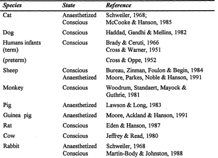
The newborn ventilatory response to acute hypoxia is termed biphasic; an initial increase in ventilation (phase 1) is followed approximately 1-3 minutes after the onset o f hypoxia by a decrease to, or to below, pre-hypoxia levels o f ventilation (phase 2). Respiratory pauses or apnoea can also be observed during phase 2. The biphasic ventilatory response is observed in all mammals investigated and is seen in both anaesthetized and unanaesthetized preparations (table 1.1).
Species State Reference
Cat Anaesthetized
Conscious
Schweiler, 1968;
McCooke & Hanson, 1985 Dog Conscious Haddad, Gandhi & Mellins, 1982 Humans infants
(term)
Conscious Brady & Ceruti, 1966 Cross & Warner, 1951
(preterm) Cross & Oppe, 1952
Sheep Conscious
Anaesthetized
Bureau, Zinman, Foulon & Begin, 1984 Moore, Parkes, Noble & Hanson, 1991 Monkey Conscious Woodrum, Standaert, Mayock &
Guthrie, 1981
Pig Anaesthetized Lawson & Long, 1983
Guinea pig Anaesthetized Moore, Ackland & Hanson, 1991 Rat Conscious Eden & Hanson, 1987
Cow Conscious Jeffrey & Read, 1980
Rabbit Anaesthetized
Conscious
Schweiler, 1968
Martin-Body & Johnston, 1988
Table 1.1 The biphasic ventilatory response has been reported in all newborn mammals studied, in both the anaesthetized and conscious states.
Wh a t a r e t h e m e c h a n is m s t h a t m e d ia t e B V R ?
St im u l a t io n o f t h e p e r ip h e r a l c h e m o r e c e p t o r s c a u s e s p h a s e 1 -
THE in c r e a s e IN VENTILATION.
The principal peripheral arterial chemoreceptors, the carotid and aortic bodies, transduce changes in pH^, Pacoj and Paoj (Heymans & Bouckaert, 1930; Heymans, Bouckaert & Dautrebande, 1930). The carotid body is composed o f two parenchymatous cell types. Type 1 cells, or glomus cells, are chemoreceptive and are surrounded by sustentacular (type 2) cells forming clusters that are richly supphed by fenestrated capillaries (Verna, 1979). Preganghonic sympathetic fibres from the superior cervical ganghon synapse onto the type 1 cells (Floyd & Neil, 1952; Eyzaguirre & Lewin, 1961). Chemoreceptor discharge may also be modulated by other efferent fibres, apart from preganghonic sympathetic efferents (Fidone & Sato, 1970; N eil & ORegan, 1971a,b). Blood flow through the carotid body is the highest per unit mass in the body, with a perfiision rate ten times higher than would be predicted from its metabohc requirements.
Decreases in Paoj, or increases in Paco2 or are transduced into increased action potential discharge. Conversely, alkah or high Paoj levels diminish carotid chemoreceptor discharge. The mechanism of chemotransduction is not fully understood. The two most likely, but conflicting, theories both agree that an elevation in intracellular calcium is necessary for the release o f neurotransmitter vesicles and subsequent generation o f action potentials in the carotid sinus nerve. However, the mechanism that triggers this increase in cytosohc calcium remains elusive. The key event in promoting synaptic vesicle release could be due to the closing o f O2 sensitive K'*' channels, thereby allowing calcium influx which in tum would further release internal calcium stores (Gonzalez, Almaraz, Obeso & Rigual, 1992). Alternatively, the hypoxic sensing mechanism could hinge on the dismption o f ATP production in mitochondria which would initiate release o f calcium from mitochondria, and internal calcium stores (Biscoe & Duchen, 1990; Duchen & Biscoe, 1992). However, more recently, data has emerged that supports strongly the idea for voltage-gated calcium entry (Buckler & Vaughan-Jones, 1994).
Spyer, 1984). Aortic and carotid chemoreceptors perform a similar role (Comroe, 1939), although it seems that carotid chemoreceptors make a larger contribution to the hypoxic ventilatory response (Martin-Body, Robson & Sinclair, 1985). Aortic afferents travel in the aortic branch o f the vagus nerve (Nonidez, 1935). Other chemoreceptors sensitive to changes in blood gas status have been located but mostly they play a minor or undefined role in the regulation o f respiration; it is possible that they perform local regulatory functions. Peri-adventitial chemoreceptor tissue has been found outside the carotid body region (Clarke & Daly, 1985). Chemoreceptive activity has also been recorded firom the abdominal vagal paraganglia in adult rats (Howe, Pack & Wise, 1981) and fi*om the aortico pulmonary region in young kittens (Coleridge, Coleridge & Howe, 1967). Pulmonary neuroepithehal cells (Lauweryns, de Bock, Guelinckx & Decramer, 1983; Lauweryns, Van Lommel & Dom, 1985), cultured firom fetal rabbit lungs also exhibit, perhaps hke type 1 carotid body cells (Gonzalez, Almaraz, Obeso & Rigual, 1992), 02-sensitive potassium channels (Youngson, Nurse, Yeger & Cutz, 1993).
A sudden reduction in Fiqj initially causes increased ventilation due to
peripheral chemoreceptor stimulation (Schweiler, 1968; Blanco, Hanson, Johnson & Rigatto, 1984; Lawson & Long, 1983). Peripheral chemodenervation abohshes the increase in ventilation (Schweiler, 1968). Indeed, the peripheral chemoreceptors play a vital role in ensuring normal respiratory control during early development under normoxic conditons because peripheral chemodenervation markedly decreases the survival rate, and causes prolonged apnoea, in newborn piglets (Donnelly & Haddad, 1990), lambs (Bureau, Lamarche, Foulon & Dalle, 1985) kittens and rabbits (Schweiler, 1968).
Cardiovascular response to acute hypoxaemia in newborns.