INTRODUCTION
The first contact of microbial plant parasites with their host occurs at the plant cell wall surface. During penetration and colonisation, the pathogen secretes plant cell wall degrading enzymes such as endopolygalacturonases. These enzymes are primarily involved in the nectrotrophic stage of pathogenesis24. Many cell wall degrading enzymes have been reported from fungal pathogens1,2,9,12,15,26. There are some reports that specifically associate the production of endopolygalacturonases in pathogens with pathogenesis10,24,15,21.
PRODUCTION OF POLYGALACTURONASES IN ISOLATES OF
Cryphonectria cubensis OF DIFFERING PATHOGENICITY
Percy M. Chimwamurombe1*, Brenda D.Wingfield2, Anna-Maria Botha2 & Michael J. Wingfield2
¹*Department of Biology, University of Namibia, Post Bag - 13301, Windhoek (Namibia) 2Forest and Agricultural Biotechnology Institute (FABI),
University of Pretoria, Pretoria - 0002, (South Africa)
(Received: December 03, 2005; Accepted: December 30, 2005)
ABSTRACT
Cryphonectria cubensis causes a serious stem canker disease on Eucalyptus in tropical and subtropical parts of the world. Previously, it was shown that isolates of C. cubensis display varying levels of pathogenicity. The aim of this study was to consider whether a relationship exists between the production levels of polygalacturonases and pathogenicity in isolates of C. cubensis. Seven isolates of C. cubensis known to have differing levels of pathogenicity were assessed for polygalacturonase production using agarose gel diffusion and reducing sugar assays. Our results showed that the levels of polygalacturonase production are not significantly different in natural isolates of C. cubensis that have different pathogenicity levels. However, in one hypovirulent isolate, CMW6009, which has been artificially transfected with hypovirus CHV1-713 from Cryphonectria parasitica, a delay of six days in the production of polygalacturonases was observed. We conclude that polygalacturonases probably have a minor role in determining the pathogenicity of the C. cubensis. Furthermore, the hypovirus CHV1-713 that causes hypovirulence in C. cubensis, has a major role in controlling pathogenicity and its mechanism of action may involve disruption of the production of polygalacturonases and other cell wall degrading enzymes.
Key words: Eucalyptus, Cryphonectria cubensis, polygalacturonases, host defence, hypovirulence, Cryphonectria parasitica.
elicitor-active oligogalacturonides. It also increases the residence time of such molecules to act as defence signal molecules3, 4.
Host defence responses include increased production of PGIPs, production of chitinases and glucanases, phytoalexin production, stimulation of the phenylpropanoid pathway, superoxide peroxidations and hypersensitive responses3. These events halt the progress of disease in an infected plant. The efficacy of plant defence responses to a pathogen depends strongly on the extent and speed of the onset of the defence signals9.
Cryphonectria cubensis is a well-known and important canker pathogen of Eucalyptus spp.14,27. This pathogen is most important where susceptible Eucalyptus spp. are grown in tropical and subtropical countries14,27. Isolates of C. cubensis have been shown to display different levels of pathogenicity in both greenhouse and field inoculation trials23.
In another study, the hypovirus CHV1-713, isolated from the chestnut blight pathogen Cryphonectria parasitica17 was shown to reduce pathogenicity in a highly virulent South African C. cubensis isolate, namely CMW211322. Furthermore, the transfected isolate produced a bright yellow orange mycelium compared to the white mycelium of the non-transfected isolate.
The economic importance of Eucalyptus has justified studies on pathogens such as C. cubensis. Of particular interest is a need for knowledge pertaining to infection. In this regard, we have considered the role of cell-wall degrading enzymes, such as polygalacturonases, in pathogenesis. In this study, we repor t on the production of polygalacturonases in vitro by isolates of C. cubensis known to have different levels of pathogenicity.
MATERIALS AND METHODS Fungal Isolates
Seven South African isolates of Cryphonectria cubensis, (CMW2113, CMW6103, CMW6106, CMW6097, CMW6087, CMW6111 and
CMW6009) were grown at 25 °C on malt extract agar (MEA; 2 % w/v malt extract, 2 % w/v agar) plates for 6 days. The cultures are maintained in the culture collection of the Forestry and Agricultural Biotechnology Institute (FABI), University of Pretoria. Isolates CMW2113, CMW6103 and CMW6106 have been shown to have high levels of pathogenicity and isolates CMW6087, CMW6097, and CMW6111 are known to have low pathogenicity 23. Isolate CMW6009 represents the hypovius transfected form of the highly pathogenic isolate CMW2113, which is routinely used in field screening trials to select disease tolerant planting stock. This isolate has been transfected with the hypovirus CHV1-713 from Cryphonectria parasitica22. The transfected isolate has subsequently also been shown to be hypovirulent.
Polygalacturonase production in C. cubensis To induce production of polygalac-turonases in vitro, five mycelial plugs (4 mm2) were taken from the actively growing margins of cultures on 2 % MEA. These plugs were grown in minimum salts liquid medium (100 ml). The medium contained: 0.5 g yeast extract (Merck); 1.0 g NaOH, 3.0 g DL-Malic acid; 2.0 g NH4NO3; 1.0 g KH2PO4; 0.1 g MgSO4 and supplemented with 0.5 % w/v polygalacturonic acid (PGA) (Sigma Chemical Company) as a carbon source in a litre of sterile distilled water11. Cultures were incubated with shaking at 100 rpm at 25 °C in the dark for ten days. Samples from the culture vessels were collected on each of the ten days. Mycelium was separated by suction filtration through Whatman No. 113 filter paper using a Buchner funnel. The filtrates were then filter-sterilised through 0.22-micron disposable syringe filters (Millipore, USA) and stored at 4 °C. Each time a total protein concentration was determined to ensure the equal amounts were used. All samples were assayed for polygalacturonase activity in triplicate.
Cup-plate agarose diffusion assay
PGA (0.01 %) was used as substrate. The medium was transferred to Petri dishes (20 ml per plate). A 4 mm cork borer was used to punch three wells 2.5 cm apart in the solid gel. Each well was filled with 30 µl of either endoPG standard or blank control or the different filtrates to be tested. The plates were incubated overnight at 30 °C. After incubation, the gel was developed by flooding the plates with 10 ml of 0.05 % ruthenium red (Sigma Chemical Co., USA) for 2 h at 25 °C. Excess dye was removed by washing the plates several times with distilled water. A distinct clear zone on the stained agarose gel indicated PG activity. Two diameter readings (at right angles to each other) of the zones were taken from duplicate plates and the average value was calculated. Each isolate was independently tested three times. Production of polygalacturonases was calibrated against a dilution series of Aspergillus niger endoPG (418 unitsml-1, Sigma Chemical Co., USA, one unit equals the amount of enzyme required to catalyse the production of a reducing sugar per minute). Assays without enzyme served as controls. The mean diameter readings were compared for all the isolates and for each day. Differences in the ability of isolates to produce polygalacturonases were analysed using Tukey ‘s multiple comparison method from SAS software18.
Reducing sugar assay
Polygalacturonase activity in the different filtrates was determined by measuring the reducing-end groups using the p-hydroxybenzoic acid hydrazide (PAHBAH) method29. This measurement was done to confirm the outcome of agarose gel diffusion assays. The PAHBAH assays were calibrated against a dilution series of D-galacturonic acid.
The activity of polygalacturonases were determined by incubating 50 µl of the different samples in a 1 ml solution containing 0.25 % w/v PGA and 40 mM sodium acetate (pH 5.0) for 1 h at 30 °C. This reaction was terminated by addition of 1.5 ml freshly prepared 5 % PAHBAH. The sample tubes were boiled for 10 minutes and cooled to room temperature before taking absorbance readings at 410 nm using a spectrophotometer (Pharmacia LKB. Ultrospec III, Sweden). The assays were performed in triplicate. Statistical analyses of data were similar to those for the agarose diffusion assays. One unit of enzyme activity was defined as the amount of enzyme producing 1 µmol of reducing group per minute at 30 °C in 40 mM of sodium acetate (pH 5.0).
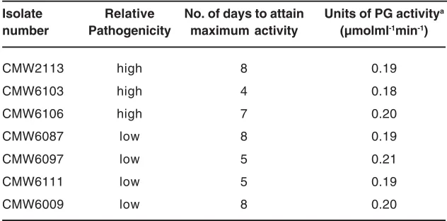
Table -1. Polygalacturonase production in isolates of C. cubensis differing in pathogenicityA
Isolate Relative No. of days to attain Units of PG activitya
number Pathogenicity maximum activity (µmolml-1min-1)
CMW2113 high 8 0.19
CMW6103 high 4 0.18
CMW6106 high 7 0.20
CMW6087 low 8 0.19
CMW6097 low 5 0.21
CMW6111 low 5 0.19
CMW6009 low 8 0.20
a values are an average of three repeats, and not significantly different (F=6.21, DF=179, P<0.0001).
Fig. - 1. Graphical representation of the trend of polygalacturonase production by seven isolates of C. cubensis in minimum salts medium for 10 days.
Diameter values are averages of three repeats.
Fig. - 2. The trend of production of polygalacturonases for seven isolates of C. cubensis cultured for 10 days in minimum salts medium. Absorbance values (A410) are averages of three repeats.
There is a characteristic slow rise in absorbance values for isolate CMW6009. The other six isolates have statistically identical rates of production of polygalacturonases.
RESULTS AND DISCUSSION Cup-plate agarose diffusion assay
All the isolates of C. cubensis produced polygalacturonases. The maximum polygalacturonase activity was reached at different times, although the amounts produced were statistically similar (F=6.21, DF=179, P<0.0001) (Table 1) for all except the transfected isolate CMW6009. The transfected isolate showed a delay of 6 days before polygalacturonases were produced. This was considerably longer than for the remaining isolates (Fig. -1). The weakly pathogenic isolate CMW6087 behaved differently. It displayed a gradual increase in poly-galacturonase production until 8 days, which was different to the behaviour of other isolates.
Reducing sugar Assay
No significant differences were observed in the production of polygalacturonases for naturally occurring C. cubensis using the reducing sugar assay (F=6.21, DF=179, P<0.0001). However, the transfected isolate CMW6009 showed a delayed production of polygalacturonases. A similar trend had been observed with agarose diffusion assays described above. The mean units of PG activity in all the isolates were 0.2 µmolml-1min-1 (Table -1). PG production during the course of this study is illustrated in Fig -2.
Results of this study show that there are no significant differences in the ability of C. cubensis isolates that differ in pathogenicity to produce polygalacturonases in vitro. However, the hypovirulent isolate CMW6009 displayed an obviously delayed production of poly-galacturonases. These results are consistent with those of previous studies where production of polygalacturonases in pathogens has not been tightly linked to varying levels of pathogenicity19,13,7. Our findings also suggest that polygalacturonases are not an important determinant in the pathogenicity of C. cubensis.
In a previous study5, we observed that the DNA sequences of polygalacturonase-inhibiting proteins (PGIPs) in selected Eucalyptus species have very similar amino acid sequence. This suggests a low diversity of the PGIPs in the host
plant. A high variability and diversity of PGIPs suggests that the host plant is constantly under evolutionary pressure to adapt to a greater diversity of endoPGs16,20. Since the PGIPs of Eucalyptus have low diversity, it can be expected that the endoPGs of the pathogen, C. cubensis, would also have a low diversity. Furthermore, this may suggests that there are few isoforms of polygalacturonases in C. cubensis.
The role of endopolygalacturonases in pathogenicity is known to differ for different fungal species6,13,21,19. In some species, there is a strong correlation between the production of endopolygalacturonases and pathogenicity, while in others there is no such relationship6,7,25. In C. parasitica, a close relative of C. cubensis, targeted disruption of the endopolygalacturonase (enpg-1) gene resulted in no reduction of pathogenicity on American chestnuts13. This implies that the role of endoPGs in the pathogenicity of C. parasitica is minor. However, the results of Gao and co-workers (1996)13 require careful interpretation. Only one isoform of the endoPG was disrupted in their study and this did not result a reduction in pathogenicity. The possibility that other isoforms may still be functional must be considered, because fungi can produce different isoforms of endoPGs at the onset of infection12,28.
In the present study, all the possible isoforms of polygalac-turonases were collectively assayed. We thus feel confident in our suggestion that polygalacturonases are not a determinant in the pathogenicity of C. cubensis.
Results of this study showed that the C. parasitica hypovirus (CHV1-713) results in a decrease in the production of polygalacturonases in C. cubensis. This provides support for the view that the virus could be useful in biological control of Cryphonectria canker. In planta, the isolate has reduced pathogenicity on Eucalyptus grandis22. In C. parasitica, the same hypovirus has been reported to decrease the accumulation of enzymes such as laccases, cutinases, cellobiohydrolases and polygalacturonases and it is associated with hypovirulence17. Therefore, the mechanism of action of hypovirulence in C. cubensis by the
C. parasitica hypovirus may involve interaction with polygalacturonase genes or gene products.
ACKNOWLEDGEMENTS
We acknowledge the National Research Foundation, the Forestry Molecular Biology Cooperative Programme and the THRIP programme of the Department of Trade and Industry, South Africa for financial support. We are also grateful to Schalk Van Heerden for his assistance in this study.
1. Boudart G, Lafitte C, Barthe JP, Frasez D, Esquarre-Tugaye M-T. Differential elicitation of defense responses by pectic fragments in bean seedlings. Planta 206: 86-94 (1998) 2. Centis S, Guillas I, Sejalon N,
Esquarre-Tugaye M-T. Endopolygalacturonase genes from Colletotrichum lindemuthianum: Cloning of CLPG2 and comparison of its expression to that of CLPG1 during saprophytic and parasitic growth of the fungus. Molecular Plant-Microbe Interactions 10: 769-775 (1997)
3. Cervone F, Castoria R, Leckie F, De lorenzo G. Perception of fungal elicitors and signal transduction. In: Aducci P, ed. Signal transduction in Plants, Basel, Switzerland, 153-177 (1997)
4. Cervone F, Hahn MG, De Lorenzo G, Darvill AG, Albersheim P. Host-Pathogen Interactions: A plant protein converts a fungal pathogenesis factor into an elicitor of plant defence responses. Plant Physiology 90: 542-548 (1989)
5. Chimwamurombe PM, Botha A-M, Wingfield MJ, Wingfield BD. Molecular relatedness of polygalacturonase inhibiting protein genes in Eucalyptus species. Theoretical and Applied Genetics (in press).
6. Cleveland TE, Cotty PJ. Invasiveness of Aspergillus flavus isolates in wounded cotton bolls is associated with the production of a specific fungal polygalacturonase. Phytopathology 81: 155-158 (1991)
REFERENCES
7. Di Pietro A, Roncero MI. Cloning, expression, and role of pathogenicity of pg1 encoding the major extracellular endopoly-galacturonase of the vascular wilt pathogen Fusarium oxysporum. Molecular Plant-Microbe Interactions 11: 91-98 (1998) 8. Dingle J, Reid WW, Solomons GL. The enzymatic degradation of pectin and other polysaccharides II- Application of the “Cup-plate” assay to the estimation of enzymes. Journal of Science and Food Agriculture 4: 149-155 (1953)
9. Dixon RCA, Lamb CJ. Molecular
communication in interactions between plants and microbial pathogens. Annual Reviews in Plant Molecular Biology 41: 339-367 (1990)
10. Durrands PK, Cooper RM. The role of pectinases in vascular wilt disease as determined by mutants of Verticillium albo-atrum. Physiological and Molecular Plant Pathology 32: 362-371 (1988)
11. Errampalli D, Kohn LM. Comparison of pectic zymograms produced by different clones of Sclerotinia sclerotiorum in culture. Phytopathology 85: 292-298 (1995) 12. Fraissinet-Tachet L, Reymond-Cotton P,
Fèvre M. Characterisation of a multigene family encoding an endopolygalacturonase in Sclerotinia sclerotiorum. Current Genetics 29: 96-99 (1995)
the major in vitro endopoly-galacturonase of the chestnut blight fungus, Cryphonectria parasitica. Applied Environmental Microbiology 62: 1984-1990 (1996) 14. Hodges CS. The taxonomy of Diaporthe
cubensis. Mycologia 72: 542-548 (1980) 15. Le Cam B, Massiot P, Rouxel F. Cell wall
polysaccharide-degrading enzymes produced by isolates of Mycocentrospora acerina differing in aggressiveness on carrot. Physiological and Molecular Plant Pathology 44: 187-198 (1994)
16. Leckie F, Mattei B, Capodicasa C, Hemmings A, Nuss L, Aracri B, De Lorenzo G, Cervone F. The specificity of polygalacturonase-inhibiting protein (PGIP): a single amino acid substitution in the solvent-exposed b-strand/ b-turn region of the leucine-rich repeats (LRRs) confers a new recognition capability. EMBO Journal 18: 2352-2363 (1999) 17. Nuss DL. Using hypoviruses to probe and
perturb signal transduction processes underlying fungal pathogenesis. The Plant Cell 8: 1845-1853 (1996)
18. SAS Statistical software. SAS/STAT Users guide. Version 6, Forth Edition: volume 1 and 2. SAS Instutite Inc, Cary. NC. (1989) 19. Scott-Craig JS, Panaccione DG, Cervone F,
Walton JD. Endopolygalacturonase is not required for pathogenicity of Cochliobolus carbonum on maize. The Plant Cell 2: 1191-1200 (1990)
20. Stotz HU, Bishop JG, Bergmann CW, Koch M, Albersheim P, Darvill AG, Labavitch JM. Identification of target amino acids that affect interactions of polygalacturonases and their plant inhibitors. Physiological and Molecular Plant Pathology 56: 117-30 (2000) 21. Ten Have A, Mulder W, Visser J, van Kan J.
The endopolygalacturonase gene Bcpg1 is
required for full virulence of Botrytis cinerea. Molecular Plant-Microbe Interactions 11: 1009-1016 (1998)
22. Van Heerden S, Geletka LM, Preisig O, Nuss DL, Wingfield BD, Wingfield MJ. Characterisation of South African Cryphonectria cubensis isolates infected with a Cryphonectria paratisitica hypovirus. Phytopathology, (accepted) (2001)
23. Van Heerden S, Wingfield MJ. Genetic diversity of Cryphonectria cubensis isolates of South Africa. Mycological Research (in press) (2000)
24. Walton JD. Deconstructing the cell wall. Plant Physiology 104: 1113-1118 (1994) 25. Walton JD, Cer vone F.
Endopoly-galacturonase from the maize pathogen Cochliobolus carbonum. Physiological and Molecular Plant Pathology 36: 351-359 (1990)
26. Wattad C, Freeman S, Dinoor A, Prusky D. A nonpathogenic mutant of Colletotrichum magna is deficient in extracellular secretion of pectate lyase. Molecular Plant-Microbe Interactions 8: 621-626 (1995)
27. Wingfield MJ, Swart WJ, Abear B. First record of Cryphonectria canker of Eucalyptus in South Africa. Phytophylatica 21: 311-313 (1989)
28. Yao C, Koller W. Diversity of cutinases from plant pathogen fungi: Different cutinases are expressed during saprophytic and pathogenic stages of Alternaria brassicola. Molecular Plant-Microbe Interactions 8: 122-130 (1995)