Manning, David J., Barker-Mill, Susan and Donovan, Tim (2006)
Time-dependent observer errors in pulmonary nodule detection. British Journal of
Radiology, 79 (940). pp. 342-346.
Downloaded from: http://insight.cumbria.ac.uk/id/eprint/167/
Usage of any items from the University of Cumbria’s institutional repository ‘Insight’ must conform to the following fair usage guidelines.
Any item and its associated metadata held in the University of Cumbria’s institutional repository Insight (unless stated otherwise on the metadata record) may becopied, displayed or performed, and stored in line with the JISC fair dealing guidelines (available here) for educational and not-for-profit activities
provided that
• the authors, title and full bibliographic details of the item are cited clearly when any part of the work is referred to verbally or in the written form
• a hyperlink/URL to the original Insight record of that item is included in any citations of the work
• the content is not changed in any way
• all files required for usage of the item are kept together with the main item file.
You may not
• sell any part of an item
• refer to any part of an item without citation
• amend any item or contextualise it in a way that will impugn the creator’s reputation
• remove or alter the copyright statement on an item.
The full policy can be found here.
SHORT COMMUNICATION
Time-dependent observer errors in pulmonary nodule detection
1
D MANNING,
PhD
,
FInstP,
1S C BARKER-MILL,
PhD,
1T DONOVAN,
MScand
2T CRAWFORD,
PhD1
School of Medical Imaging Sciences, St Martin’s College, Lancaster LA1 3JD and
2Department of
Psychology, Lancaster University, Lancaster, UK
ABSTRACT.The work was carried out to investigate differences in visual search characteristics between groups of observers with different levels of experience in the task of pulmonary nodule detection in chest radiology and we report here on these differences in respect of time related decisions. Volunteer observers were divided into three groups depending on their level of expertise. There were eight radiologists, eight radiographers and eight novices. Their task was to detect pulmonary nodules in a test bank of 120 digitized posteroanterior (PA) chest radiographs. Five of the eight radiographers were tested twice: once before and once after a 6-month training programme in interpretation of the adult chest radiograph. During each test session the observers’ eye movements were tracked. Data on the observers’ decisions through Alternate Free Response Operating Characteristic (AFROC) methodology were
correlated to their eye-movement and fixation patterns. True negative decisions from all observers were associated with shorter fixation times than false negative decisions. No correct negative decisions were made after fixations exceeding 3 s.
Received 2 March 2005 Revised 24 August 2005 Accepted 30 September 2005
DOI: 10.1259/bjr/13453920
’2006 The British Institute of Radiology
In previously reported studies we have investigated observer experience [1, 2] and the effects of lesion conspicuity [3] on performance in nodule detection from plain chest radiology. Comparisons of diagnostic perfor-mance through alternate free response receiver operating characteristic (AFROC) [4] showed that after training and extensive caseload experience radiographers’ detection rate improved to approach that of the experts. AFROC is a variation on receiver operating characteristic methodo-logy that takes into account the location in the image of the observer decision. Through this technique the observer must indicate not only his decision on the disease status of the image (positive or negative for the presence of a pulmonary nodule) but also the location of the lesion.
The observer groups were as follows:
N
eight first-year student radiographers considered asnovices to chest interpretation tasks;
N
eight experienced clinical radiographers before train-ing in chest interpretation;N
five of the eight radiographers after their training in chest interpretation; andN
eight radiologists.Eye-tracking these observers gave insight into diffe-rences between the groups in terms of their visual search strategies and we concluded that, amongst other things, the experts were more economical in their patterns of search, carried out fewer fixations and spent less time on the task. After a training period that included a minimum of 500 cases but no specific instruction in
search patterns, the radiographers developed
spontaneously similar search strategies to those of the
radiologists. But both radiologists and trained radio-graphers had a false negative error-rate in excess of 40% [2]. We accept that the requirements of AFROC are stringent (a false negative is defined by a missed lesion rather than an incorrect case decision); but this is still a significant miss-rate and in this paper we report our observations on the relationship between the experience of the observer, the duration of visual attention and the probability of a false negative error in the task.
Eye movement, visual attention and visual scan paths
The field of view in humans extends over 180
˚
but onlythe centre of the field provides sharp details.
opposed to the parts that receive no attention at all. It has been noted that the same details in an image can be accessed using quite different scanpaths but similar fixation clusters [6]. As a result, analysis of fixation cluster location and dwell time gives a better opportunity
for performance comparisons than the scanpath
sequence if the research question is focused on whether image features are being hit. However, scanpath data can give fascinating insights into how individuals prioritise locations of semantic interest although the analysis of these paths is difficult and sometimes inconclusive.
Errors in radiology are known to be multifactorial but a sizable proportion are known to be perceptual in origin [7, 8]. This has led to interest in the possible sources of perceptual error and the visual strategies of radiologists in their task [9]. Early work on the visual dwell behaviour of radiologists during film reading was carried out by Kundel et al [10–12] who went on to later describe survival curve methods for analysing time-related decision-making in those observers [13, 14]. Our aim here is to report on the timing of decisions, their accuracy and their relationship to experience by the survival analysis of decisions from eye-tracking data.
Aim
The aim of this study was to report an observed relationship between the experience of the observer, the duration of visual attention and the probability of incorrect decisions in pulmonary nodule detection.
Materials and methods
Observers
Eight volunteers were recruited from a cohort of first year radiography students who acted as the novice group of film readers. Eight experienced radiographers volunteered from a post-graduate course on chest image interpretation at the commencement of their course. Five of these radiographers then offered themselves for repeat testing at the end of their 6 months training and 500 cases of experience. Eight radiologists with extensive and current experience in chest radiology volunteered to act as the most experienced group of observers. All observers gave their consent and all performance data were made anonymous. Recruitment of all the observers and the conduct of the experiments followed the ethical guidelines for experiments involving volunteer human subjects at St Martin’s College and Lancaster University. At the time when the data for these experiments were collected (in 2001) there was no requirement of COREC approval for NHS employees who were acting as volunteer participants in this type of research.
Detectable nodules
Each observer viewed a bank of 120 digitized chest images of adults. The images contained 81 pulmonary nodules agreed as significant in pathological appearance from confirmed radiological reports. Nodules were
roughly circular and ranged in diameter size from 5 mm to 20 mm with varying degrees of measured conspicuity [3]. Nine films contained more than one nodule and 30 nodules were located in these multi-nodule films. Normal films were included in the observer task and the complete test bank was divided into three sets of 40 images with prevalence-rates of 12%, 50% and 82%.
Observer performance measurement
Alternate free response operating characteristic meth-odology (AFROC) was used [4]. This required observers to indicate a location to a decision for a lesion and to assign a score between 1 and 4 on their level of confidence in that decision. A zero score was allocated to all decisions of ‘‘no nodule present’’. In AFROC methodology false positive decisions are treated in the following way: the highest scoring false positive decision is the only one recorded per image which avoids the possibility of infinite values in summing false positive responses.
Observer test sessions were never longer than 1 h to avoid the effects of fatigue on performance. There was a minimum 6-month interval between the before- and after-training observer tests on the radiographers to give an effective case-memory washout period.
Parameters
All eye-tracking data comparing the performance of the observer groups were processed through the ASL (Applied Science Laboratories, Bedford, MA), software
EYENALH. The measured parameter from the eye
tracking for use in the present study was the accumu-lated dwell time at each decision point.
A fixation
We defined a fixation (visual dwell) as a point of gaze
remaining continuously within a 1
˚
area for at least100 ms. A 1
˚
angle subtended from the eye to the imageat a viewing distance of 40 cm approximates to a circle the size of a 2.5 cm diameter disc at arms length. Re-fixations were summed to give cumulative fixation times to clusters when there was overlap of the 1
˚
areas up to a5
˚
area. These definitions are ones that are commonlyused in work of this kind [9, 14]. The observers were allowed to search freely and no limit was imposed on the duration of inspection for any given image.
A true negative decision was defined as a timed fixation of a lesion-free zone of the chest image that elicited a zero response from the observers on the AFROC scale.
The dwell-time data for all fixations related to positive and negative decisions were analysed through the statistical package SPSSHto provide information on the percentage survival of decisions over time.
These data allowed us to characterize the observers’ decisions in greater detail than true and false negative and true and false positive outcome, giving an opportunity to
identify time-related features of decision outcomes. The time related information on decisions gave an opportunity to analyse time differences between correct and incorrect decisions whether positive or negative.
Results
Time related decisions: survival analysis
We investigated how the four possible decision outcomes of true and false positive (TP FP) and true and false negative (TN FN) related to the duration of gaze through a survival analysis of the fixation data. The results are presented in Tables 1–4 and Figures 1–4. Survival analysis is used in this context as a means of showing the proportions of decisions that are completed for each category (TP FP TN FN) at increasing accumulated time intervals of visual attention. It is analogous to the cell survival curves used in radio-biology to indicate the proportion of cells surviving increasing radiation doses [15]. So in Table 1 and Figure 1 for example, 50% of all true positive decisions made by the radiologists survived 2200 ms of accumu-lated visual attention on the tumour location but none of
their true positive decisions took more than 5000 ms of visual scrutiny.
In the tables we have highlighted the time values for the completion of all the observer decisions in their outcome categories to draw attention to the differences in duration between the true and false decisions in each case. For all observers the false negative decisions took them significantly longer than true negatives. For positive decisions, correct decisions were made faster than incorrect ones except in the case of the novices who dwelt longer on genuine lesions before deciding they were pathologies.
The survival curves show that for all observers 50% of all their true negative decisions were made within 1000 ms of gaze duration and the false negative decisions have dwell-times somewhere between the positives (true and false) and the true negatives. This is consistent with the findings of others who have carried out this type of analysis [14] but our results indicate extension along the time axis with decreasing levels of experience.
Discussion
The aim of this work was to analyse eye-tracking data to classify the false negative errors made when observer groups with different levels of experience are asked to detect pulmonary nodules. The different levels of experience in the groups give some insight into how, if at all, errors vary with expertise. The time related data provided measurable differences in the fixation times associated with observers making correct or incorrect decisions.
Survival analysis
Observers’ decisions are made over time periods that can be related to the duration of visual fixations from eye-tracking data. Mean or median dwell times over trials and between readers can be calculated but dwell times are not normally distributed, making statistical comparisons difficult to interpret. The technique of survival analysis has been found useful in these circumstances [13, 14] and this operation on the data demonstrated some consistent findings for categories of decisions.
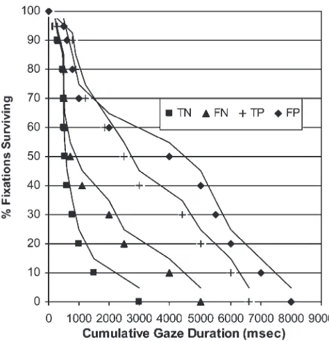
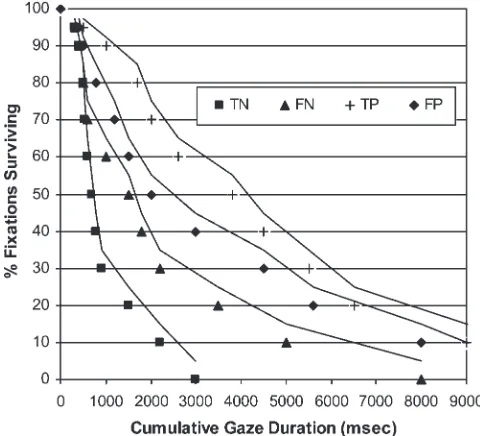
Figures 1 and 2 show the family of decisions survival curves for radiologists and radiographers with chest interpretation training derived from the data in Tables 1 and 2. Virtually all the true negative decisions made by these, the most experienced observers, were made within 2 s of cumulative fixation time on an image feature. False negative decisions were characterized by longer dwell times but no negative decisions (either true or false) were made by radiologists after 4.75 s fixation. Figures 3 and 4 and Tables 3 and 4 show similar findings for the low experience groups but there is a tendency towards longer fixation times for incorrect negative decisions with decreasing levels of experience. In the case of the novices (Table 4) lesions were missed after a cumulative gaze duration of up to 8 s. The time difference in the proportions of true and false negative decisions made by these observers can be summarized by saying that in
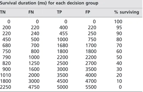
Table 1. Radiologists
Survival duration (ms) for each decision group
TN FN TP FP % surviving
0 0 0 0 100
200 220 400 220 95
220 240 455 250 90
450 500 1000 750 80
680 700 1680 1700 70
750 800 1800 1800 60
790 1000 2200 2200 50
820 1250 2500 2700 40
900 1600 3000 3500 30
1010 2000 3500 4000 20
1800 3000 4500 4700 10
2250 4750 5000 5500 0
TN, true negative; TP, true positive; FN, false negative; FP, false positive.
Table 2. Radiographers post training in chest interpretation Survival duration (ms) for each decision group
TN FN TP FP % surviving
0 0 0 0 100
200 220 300 500 95
300 330 800 600 90
450 500 900 800 80
500 500 1200 1000 70
500 550 1850 2000 60
550 700 2500 4000 50
600 1100 3000 5000 40
800 2000 4400 5500 30
1000 2500 5000 6000 20
1500 4000 6000 7000 10
3000 5000 6600 8000 0
this experiment, all negative decisions made after gaze duration of 3 s were incorrect.
It seems that correct negative decisions (correct deci-sions that normal features are not nodules) tend to be made rapidly after fixation occurs and that this is a feature of expert performance. Conversely, incorrect negative decisions are characterized by extended gaze duration especially in novice readers. When areas of the images hold their attention for several seconds of accumulated fixation time, observers show a semantic interest in the appearances. This suggests that they are suspicious of the feature and they are operating on the information at a perceptual/cognitive level. These errors are not failures of detection but of recognition and decision and can be explained partly by the visual ambiguities of the image and partly by the level of experience of the reader. The finding may be important because of: (a) its potential for reducing false negative error if visual dwell information is fed back to the reader; and (b) its potential use in training schemes for radiologists and monitoring the effects of caseload experience.
If these results are reproducible in other settings and our interpretation of their meaning is correct there are several ways that they may help in the improvement of observer performance. A simple expedient is to inform film readers
that their negative decisions are more likely to be incorrect when they are made after a period of indecision over a particular image feature. In short, we suggest informing observers that if a feature looks suspicious enough to warrant more than 2 s of their attention it is probably not innocent. More sophisticated aids linked to these findings
Table 3. Radiographers pre training in chest interpretation Survival duration (ms) for each decision group
TN FN TP FP % surviving
0 0 0 0 100
300 300 350 350 95
330 400 800 820 90
330 500 1100 1200 80
400 600 2000 2000 70
560 1200 3000 3100 60
700 1600 3300 3800 50
850 2000 3800 4800 40
1000 2400 4200 5500 30
1200 3000 5100 6500 20
1600 4500 6500 8000 10
2400 6500 8200 9000 0
TN, true negative; TP, true positive; FN, false negative; FP, false positive.
Table 4. Novices
Survival duration (ms) for each decision group
TN FN TP FP % surviving
0 0 0 0 100
300 300 500 400 95
400 400 1000 500 90
500 500 1700 800 80
550 600 2000 1200 70
600 1000 2600 1500 60
680 1500 3800 2000 50
800 1800 4500 3000 40
900 2200 5500 4500 30
1500 3500 6500 5600 20
2200 5000 9000 8000 10
3000 8000 12000 10000 0
TN, true negative; TP, true positive; FN, false negative; FP, false positive.
Figure 1. Time-related decisions for radiologists. Compared with less experienced observers the radiologists made their decisions faster. The separation between the positive and negative decision pair curves is closer than for all other groups.
Figure 2. Time-related decisions for radiographers post-training. After experience and training over 6 months and 500 cases the radiographers have speeded their decision making and markedly reduced the separation of their true and false negative curves. Compare with Figure 3.
might involve computer aided feedback to observers in real-time to give indication of the gaze duration for individual locations in the image.
Conclusion
The data and their analysis from an eye-tracking experiment have given insights into the errors made by observers with different levels of experience. The most notable outcomes of this are that the duration of fixation on a feature in the chest image may be an effective
discriminator in predicting whether a negative decision will be correct or incorrect.
We consider this observation to be potentially impor-tant for feedback strategies for education purposes and to aid performance.
Acknowledgment
We wish to thank the radiologists, radiographers and students, all of whom are associated with the School of Medical Imaging Sciences at St Martin’s College, Lancaster, UK, who kindly agreed to take part in this study. Eye-tracking equipment for this research was supplied through a grant from the Peter Barker-Mill Memorial Trust.
References
1. Manning DJ, Ethell S, Crawford T. An eye-tracking AFROC study of the influence of experience and training on chest x-ray interpretation. Medical imaging 2003; Image Perception Observer Performance and Technology Assessment. Editors Dev Chakraborty and Elizabeth Krupinski, Proc SPIE 2003;5034:257–266.
2. Manning DJ, Ethell SC, Crawford T, Donovan T. How do radiologists do it? The influence of experience and training on searching for chest nodules. Radiography Available online 23 March 2005. Item 49, www.sciencedirect.com/ science/journal/10788174.
3. Manning DJ, Ethell SC, Donovan T. Detection or decision errors? Missed lung cancer from posteroanterior chest radiograph. Br J Radiol 2004;77:231–5.
4. Chakraborty DP. Statistical power in observer performance studies: comparison of the receiver operating characteristic and free response method in tasks involving localization. Acad Radiol 2002;9:147–56.
5. Yarbus AL. Eye movements and vision. New York, NY: Plenum Press, 1967.
6. Kundel HL. Reader error, object recognition and visual search. In: Chakraborty D, Eckstein M, editors. Medical imaging 2004: image perception, observer performance and technology assessment. Proceedings of SPIE 2004;5372: 1–9.
7. Fitzgerald R. Error in radiology. Clin Radiol 2001;56:938–46. 8. Chief Medical Officer. Learning from failure: evidence and experience. An Organisation with a Memory. London: Stationery Office, 2000:1–7.
9. Gale AG. Human response to visual stimuli. In: Hendee WR, Wells PNT, editors. The perception of visual informa-tion, 2nd edition. New York, NY: Springer, 2000:134. 10. Kundel H, Nodine C, Carmody D. Visual scanning, pattern
recognition and decision making in pulmonary nodule detection. Invest Radiol 1978;13:175–81.
11. Kundel H, Nodine C, Krupinski E. Searching for lung nodules: visual dwell indicates locations of false positi-veand false negative decisions. Invest Radiol 1989;24:472–8. 12. Kundel H, Nodine C, Toto L. Searching for lung nodules: the guidance of visual scanning. Invest Radiol 1991;26: 777–81.
13. Anderson S, Auquier A, Hauk WW, Oakes D, Vandaele W, Weisberg HI. Statistical methods for comparative studies. New York, NY: John Wiley & Sons, 1980.
14. Kundel H. Visual search in medical images. In: Beutel J, Kundel H, Van Metter R, editors. Handbook of medical imaging Vol 1 Physics and psychophysics. Bellingham WA: SPIE, 2000:837–58.
15. Motulsky H. Intuitive biostatistics. Oxford: Oxford University Press Inc., 1995:Chapter 6.
Figure 3. Time-related decisions for radiographers pre-training.