_____________________________________________________________________________________________________ *Corresponding author: E-mail: [email protected];
Substrate Specificity at the Molybdenum Site in
Xanthine Oxidase Enzyme
Temesgen Nurlign Chekol
1*1
Department of Chemistry, College of Natural and Computational Science, Wolkite University, P.O.BOX 07, Ethiopia.
Author’s contribution
The sole author designed, analysed, interpreted and prepared the manuscript.
Article Information
DOI: 10.9734/IJBCRR/2019/v26i430102 Editor(s): (1) Dr. Enrico Sanjust, Associate Professor, Department of Internal Medical Sciences, University of Cagliari, Italy. Reviewers: (1) Roshan Keshari, Delhi Pharmaceutical Sciences and Research University, India. (2) B. R. Paital, Odisha University of Agriculture and Technology, India. Complete Peer review History:http://www.sdiarticle3.com/review-history/45542
Received 10 October 2018 Accepted 24 December 2018
Published 20 July 2019
ABSTRACT
Xanthine oxidase is one of the most useful molybdenum containing enzymes, which catalyzes a wide range of purine derivative heterocyclic substrates. In order for the interaction between the reactants to take place, the substrates are expected to enter the binding pocket and attain a proper orientation with the help of binding pocket amino acid residues. In addition to the binding pocket amino acids, there are several factors that affect the progression of substrates. Therefore, the study is mainly focused to identify the factors affecting the binding stage of catalysis. The activity of xanthine oxidase family enzymes greatly depends on the proper orientation of the substrates and their interaction sites. Therefore, the rate of formation of substrate- enzyme complex is proposed to be affected by the proper orientation and the interaction site of the substrate. Moreover, the keto and enol forms of substrates as well as the existence of the substituent groups affect the reactivity of xanthine oxidase. Thus, the rate of the reaction is proposed to be affected by these factors. The variable activities of the substrates towards xanthine oxidase enzyme are largely due to the factors that affect the reductive half-reaction such as proper orientation of substrates, binding sites, activation of the active site, toutomeric nature of substrates and the inductive and steric effects. This work is used to provide valuable information that may have a mechanistic importance in establishing the substrate preferences of bmXOR to RcXDH and AOR type of enzymes in order to relate electronic structure contributions to enzymatic catalysis.
Keywords: Xanthine oxidase; proper orientation; interaction site; substrate.
1. INTRODUCTION
1.1 Survey of Molybdo-enzymes
In a natural environment, most enzymes are distributed within living cells either in the cytoplasm or membrane. Although most enzymes are proteins, some of them require the presence of additional non-protein components, such as cofactors. The presence of cofactors such as metals and/or biomolecules may have purely structural or functional roles or the cofactor may possess both roles. One of the interesting metals that serve as a cofactor in the chemistry of life, although less commonly occurring, is the molybdenum (Mo) ion. The presence of Mo in the active sites of some proteins represent a group of proteins known as ‘molybdo-proteins’ or ‘molybdo-enzymes’ [1,2,3]. Molybdo-enzymes are important classes of enzymes, found in several organisms [1,2,3] such as microorganisms [1,3], plants [4,5,6], animals [1,6] as well as human beings [1,2,3]. Like iron, zinc, copper, manganese, and cobalt, molybdenum can be utilized as a stably bound, variably coordinated cofactor in proteins, and is found in more than 50 molybdenum-containing enzymes [4,6,7].
Molybdenum as a metal is inert in the redox reactions or biological processes, and it requires pyranopterin cofactor to give the active Moco cofactor [1]. The pterin cofactor has been proposed to participate in assisting the transfer of electrons from or to the active site containing molybdenum metal. Generally, the role of Moco is to position Mo correctly within the active site, control the redox behavior of the enzymes, allow the enzyme to gain its biological activity, and participate with its pterin ring system in the electron transfer to or from the molybdenum atom [7]. In mononuclear enzymes, molybdenum is part of the active sites of a much more diverse group of enzymes that in general function catalytically to transfer an oxygen atom either to or from a physiological acceptor/donor molecule [1]. It is on the basis of this commonly encountered aspect of catalysis that these enzymes are frequently referred to as oxotransferases [1,8,9,10]. Similarly, the vast majority of these enzymes possess a “Mo=O unit” in their active sites and are often referred to as oxomolybdenum enzymes [1,7]. Some (polysulfide reductase, for example, and possibly formate dehydrogenase) do not catalyze oxygen
atom transfer, and others do not possess a “Mo=O unit” [1,6]. Enzymes that possess molybdenum in their active sites catalyze biological processes that are essential to the organisms, indeed neither plants nor animals can survive without molybdenum [1,8].
Xanthine oxidoreductase enzymes have been isolated from a wide range of organisms, such as from bacteria [8,11] to man [1,11], and catalyze the hydroxylation of a wide variety of Purine [2,3,12], pyrimidine [6,7,13], pterin [3,4], and aldehyde [1,4,7] substrates. Xanthine oxidoreductase belongs to Xanthine Oxidase family enzymes, a family that encompasses a wide variety of enzymes that have similar arrangements and composition of redox centers [14,15,16].
1.2 Physiology and Biochemistry of Xanthine Oxidase Family Enzymes
The XOR enzymes are known to catalyze the final two steps of Purine metabolism by converting hypoxanthine to Xanthine and Xanthine to uric acid [10,11,12]. That means, in purine metabolism, the final two steps are catalyzed by XOR to convert hypoxanthine into Xanthine and then Xanthine into uric acid. Monoxygenase enzymes are known to utilize water rather than oxygen molecule as the source of oxygen and generating rather than consuming reducing equivalents [11,12,13]. Although the mammalian Xanthine oxidoreductase enzymes are synthesized in the form of Xanthine dehydrogenase (XDH), the XDH form of XOR enzyme is readily converted into the Xanthine Oxidase (XO) form by sulfhydryl oxidation or limited proteolysis [6,7,12]. In mammalian organs, the highest level of XOR activity is expressed in liver [3]. The presence of Xanthine oxidoreductase enzymes in the liver can be used as a marker for a hepatic damage, through the XOR enzyme circulating in the blood [3,17,18]. However, the most observable disease in humans is the deposition of uric acid, known to be responsible for gouty conditions. This condition is more pronounced in the joints, through the deposition of sodium urate crystals [3,6,19].
enzymes (such as XDH, EC: 1.1.1.204 and XO, EC: 1.1.3.22) are organized in a linear
ideal for electron transfer [2,11,21]. However, the typical feature of the XO family enzymes is the reductive half-reaction active site. The XO family enzymes are mono-nuclear since they contain a single Mo ion at their reductive half
active sites ([MoSO] (Sdithiolene
[11,22,23].
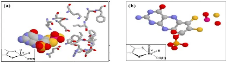
The reductive half-reaction active site, as shown in Fig. (2b), is defined by three environments [11]. The active site environment, for XO family enzymes, is composed of the two coordination spheres (the primary and secondary) and the solvent access channel (that is directed towards the two coordination spheres) [1,3]. The resting state geometry for the primary coordination sphere, as described by R. Hille [1], is a distorted penta-coordinate [1,4,11]. As shown in Fig
Fig. 1. The overall structure (center
enzyme (PDB accession code of 1V97) [24]. cofactors: The FAD, pairs of [2Fe
the application of protein explorer [13]
Fig. 2. The reductive half-reaction active sites for the
1V97) [24] showing the (a) primary coordination sphere environment and (b) reductive half-reaction active site. The illustration is prepared the same as Fig
enzymes (such as XDH, EC: 1.1.1.204 and XO, EC: 1.1.3.22) are organized in a linear fashion, ideal for electron transfer [2,11,21]. However, the typical feature of the XO family enzymes is the reaction active site. The XO family nuclear since they contain a single Mo ion at their reductive half-reaction
dithiolene)2 OH(H))
reaction active site, as shown b), is defined by three environments [11]. The active site environment, for XO family enzymes, is composed of the two coordination heres (the primary and secondary) and the solvent access channel (that is directed towards the two coordination spheres) [1,3]. The resting state geometry for the primary coordination sphere, as described by R. Hille [1], is a distorted ,4,11]. As shown in Fig. (2b),
the MoVI ion is tethered by axial and equatorial ligands located about 0.5 Å above the equatorial plane. In XOR enzymes, the equatorial plane of the primary coordination sphere is defined by ligands such as a single pyranopte
sulfido (SMo), and hydroxide (HOeq)) [4,6].
The ligands coordinated to Mo ion in the primary coordination sphere, as reviewed by R.
are proposed to participate during the oxidative hydroxylation reaction [1]. The proposed roles the equatorial ligands range from promoting nucleophilicity to serving as electron transfer conduits. In addition to the equatorial ligands, the terminal apical oxo may also play a role in increasing a charge donation from the S
to the redox states of the Mo ion. The redox states of Mo ion in the catalytic cycle are Mo MoV and MoIV, respectively, in the Mo d d2 electron count systems.
center) and cofactor compositions (left and right) of the
enzyme (PDB accession code of 1V97) [24]. Left and right display the linear arrangement of the cofactors: The FAD, pairs of [2Fe–2S], and Moco cofactors. The illustration is prepared with
the application of protein explorer [13]
reaction active sites for the bmXOR (PDB accession code of 1V97) [24] showing the (a) primary coordination sphere environment and (b) reductive
reaction active site. The illustration is prepared the same as Fig.
the MoVI ion is tethered by axial and equatorial ligands located about 0.5 Å above the equatorial plane. In XOR enzymes, the equatorial plane of the primary coordination sphere is defined by ligands such as a single pyranopterin dithiolene,
)) [4,6].
The ligands coordinated to Mo ion in the primary coordination sphere, as reviewed by R. Hille [1], are proposed to participate during the oxidative hydroxylation reaction [1]. The proposed roles of the equatorial ligands range from promoting nucleophilicity to serving as electron transfer conduits. In addition to the equatorial ligands, the terminal apical oxo may also play a role in increasing a charge donation from the SMo ligand
tates of the Mo ion. The redox states of Mo ion in the catalytic cycle are MoVI, , respectively, in the Mo d0, d1 and
) of the bmXOR display the linear arrangement of the 2S], and Moco cofactors. The illustration is prepared with
XOR (PDB accession code of 1V97) [24] showing the (a) primary coordination sphere environment and (b) reductive
As shown in Fig. (3a), the coordination geometry for the reductive half-reaction active sites of xanthine oxidase family enzymes is a distorted square pyramidal coordination sphere consisting of MoVI ion at the center. The four equatorial ligands are the terminal Mo=S group with bond distance of approximately 2.455 Å, the two dithiolene sulfurs from a single pyranopterin cofactor of the molybdenum center with bond distances 2.488Å (which is trans to the sulfido terminal) and 2.277Å (which is trans to the equatorial hydroxyl terminal), and the equatorial hydroxyl terminal which has a metal-oxygen bond distance of approximately 1.855 Å and the fifth ligand is the apical Mo= O group with bond distance of approximately 1.774 Å (Fig. 3) [1,4].
The X-ray crystallographic studies on bmXOR enzymes [2] revealed that the apical position is occupied by a Mo=O group, which represents the strong-field ligand that defines the molecular z-axis of the center. The apical oxo (OMo=O)
ligand, in XO, has a bond distances of 1.774 Å (Fig. 3) [1]. The short bond distances exhibited by Mo=O reflects a multiple bond characteristic. The multiple bonds may contain a bond along the z-axis and two bonds. The
bonds may be formed through the overlap of oxygen (px or py) and Mo (dxz or dyz) orbitals
(Fig. 4).
The ligand field of these Mo ions is governed by the different energy levels possessed by the Mo orbitals (dxz, dyz and dxy). As shown in Fig. 4, the
Mo (dxy) is the orbital that is involved in the redox
process because Mo (dxz and dyz) orbitals are
influenced by a strong field relative to the dxy
orbital.
1.3 Survey of Reducing Substrates for Xanthine Oxidase
Purine has three interaction sites at C2, C6 and
C8 position, which could be oxidized by xanthine
oxidase, when these positions are available. Purine could be oxidized at its C6-pyrimidine
position to give hypoxanthine which is a physiological substrate. Hypoxanthine can also be oxidized at its C2 position to yield xanthine
which can also be oxidized at its C8 position to
produce uric acid. Similarly, purine could be methylated at its C6-pyrimidine position to give
6-methylpurine which may be oxidized at its C2
position to yield 2-hydroxy-6-methylpurine. Like
(a)
Mo
O
S
S
S
VI
OH
1.855 2.488
1.774
2.277 2.455
1.747
1.766
(b)
Mo
O
S
S
S
VI
OH
95.6 79.585.7 85.4
110.0
106.4
Mo
O
S
S
S
VI
OH
100.8
106.8
100.7 107.2
Fig. 3. The bonding description for the reductive half reaction active site of bmXOR (PDB accession code of 1V97) [24], showing bond distance (Å) and bond angles ().The
(a)
M d orbitals M d + oxo M d + oxo + 4 Leq
xy, x2y2
xz, yz
z2 z2
xz, yz
xy x2y2
E
ne
rg
y
(b) Homo Lumo
bmXOR-Moco-Mo(VI)
bmXOR-Moco-Mo(IV)
Fig. 4. The feature of Mo in the active site, showing (a) the d splitting with oxo ligand and four other ligands and (b) the frontier orbitals depicting the dxy orbitals. The view is oriented down the Mo=O bond vector (z-axis) and prepared using GaussView software
package
xanthine, 2-hydroxy-6-methylpurine could be oxidized at its C8 position to yield 2,
8-dihydroxy-6-methylpurine. The orientation of the heterocyclic substrates inside the binding pocket could be compared using purine as the smallest precursor for all purine derivatives. Therefore, four types of substrates were shown as follows: Substrates containing –CH3 (6MP), –OH
(hypoxanthine), -OH/-OH (xanthine), and CH3
/-OH (HMP). The activities and rates of reactions determined using these substrates revealed that hypoxanthine was by far the most reactive and the substrates with –CH3 group such as 6-Methyl
purine were shown to be less reactive for XOR enzymes [4,7,25]. Generally, purine and its analogs exhibit some potent medicinal properties in the vitro and in the vivo thus suggesting a new rationale for its use in the management of various diseases and in maintenance therapy [17].
1.4 The Substrate-xanthine Oxidase Binding Stage
On the basis of the orientation, the Mo bound hydroxide (HOMo) terminal is poised for
nucleophilic attack on the electron deficient carbon of the physiological substrates (xanthine) (Scheme 1). Since this type of nucleophilic reaction is believed to be mediated by XO family enzymes, the same principle is proposed to take place if xanthine or hypoxanthine is replaced by other purine derivatives that have electron deficient carbon centers as their sites of oxidation. Thus, orientation of substrates, the redox sites, binding pocket environment, nature
of the substrates, inductive and steric effects, and the toutomeric nature of substrates and the overall architecture of the enzymes are some of the factors affecting the specificity of the substrates towards the XO family enzymes.
The catalytic transformation of the substrates (CRH) to their respective products (O-CCR), as
“Very rapid” species Mo S O : S S O VI H N N N N H H O O H Mo SH O S S O IV N N N N O H Mo S O S S O V N N N N H O H
HO-,H+
e-, H+
2H+, 2e -O -O Glu1261 H H H O N N N N O H O O H H H O OH O Glu1261 OH O Glu1261 Re ROH E ROH E RH E RH E RED lease RED Catalysis OX Binding
OX
Scheme 1. The postulated catalytic mechanism for xanthine oxidase. The Scheme shows the active site Glutamic acid (Glu1261) assisted hydride transfer hydroxylation reaction at
the C8 position of xanthine. The circle indicates the substrate-enzyme binding stage [15,19] NH H N N H 8C N O O -O Glu1261 H Mo S S O: O S VI H O
Mo
S
S
O
O
S
VI
H
NH
N
CH
N
H
2
C
N
O
O
-O
Glu
1261
H
(a)
(b)
Scheme 2. The initial stage of catalysis mediated by XOR enzymes in the presence of reducing substrates such as (a) xanthine, (b) hypoxanthine (adapted from references 3)
of this work, is the substrate-enzyme binding stage, shown in Scheme (1).
Substrate Specificity and the factors affecting reactivity: Unlike other enzymes, the XO enzymes are known to show a wide range of substrate specificity to catalyze several structurally unrelated substrates, such as purine derivatives, pteridine derivatives, and simple aldehydes. These substrates have similar bonding description and sites of hydroxylation/oxidation to that of xanthine and hypoxanthine; they are believed to behave the same as xanthine (Scheme 2).
The events taking place during the initial stage of catalysis (as shown in Scheme 2) mediated by XOR enzyme in the presence of substrates are the abstraction of acidic hydrogen by Glu1261,
nucleophilic attack Mo-O-, transfer of HRH from
CRH to Mo=S and transfer of 2e’s to MoVI center.
The broad substrate specificity of XOR enzymes was exploited to derive a valuable tool for characterizing the enzymes’ catalyzed-reactions. On the basis of the orientation, as shown in Scheme (1 and 2), the terminal water of XOR enzymes was poised for nucleophilic attack on the C2 or C8 positions of hypoxanthine or
provide insight into the mechanistic similarity transpired during the initial stage of catalysis (Scheme 1). The specificity of substrate molecules is usually determined under several factors that influence the enzyme catalyzed-reactions.
The kinetic parameters (such as vmax, km, kcat,
and kcat/Km) for the overall reaction are used to
compare the properties of the enzyme as well as their specificity towards the respective substrates. The order of the activities
recorded for bmXOR (from highest to lowest) was as follows: Hypoxanthine > xanthine > HMP > 6MP > purine. The reactivity of xanthine oxidoreductase enzyme with a range of substrates could be described using steady state enzyme kinetic constants such as Michaelis constant (km), turn over number (kcat),
maximum velocity (vmax) and efficiency, kcat/ km
[7,14,16]. The enzymes have the ability to catalyze a broad range of reducing substrates and alternate between the reduced and oxidized forms of enzymes [18].
2. MATERIALS AND METHODS
2.1 Materials
The structures of interest (such as purine derivatives, the active site, and binding pocket amino acids) were sketched using Chem Draw Ultra 2003, version 8.0 (Cambridge software corporation, Cambridge, MA. U.S.A.). These structures were also sketched using GaussView 3.0 (Gaussian, Inc., Pittersburgh, PA. U.S.A.) Software package. This software was also used to develop the input geometries, calculate the bond distances, as well as visualize the optimized geometries and frontier orbitals. The input geometries prepared using GaussView 3.0 software program were optimized using Gaussian 03W (2003), version 6.0 (Gaussian, Inc, Pittersburgh PA, USA) software package, on Dell Optiplex780 model computer, 2011 (Dell, Inc; Wilhie Sdh Bhd; Penang, Malaysia). In addition, AOMix 2011/2012, 6.6 (Centre for
Catalysis Research and Innovation, University of Ottawa, Ottawa, Canada) software package was
used to determine the composition of atomic orbitals.
2.2 Methods
2.2.1 Determination of interaction sites
The charge density on the interaction sites were probed by optimizing the free (unbound) substrates, shown in Fig. (2.2.1). The structures
of free substrates were constructed using GaussView 3.0 software package. All geometry optimizations on the “keto” and “enol” forms of the substrates (Fig. 2.2.1) were performed using Gaussian 03W (version 6.0) software program, by applying a density functional theory (DFT) method of Beck’s three-parameter exchange functional combined with (B3YP) [6].
The geometries of the free substrates (Fig. 2.2.1) were optimized using the 6-31G (d’, p’) basis set with a polarization function, used for the atoms (C, H, O, N, S) and “# B3LYP gen #P opt pop=full geom=connectivity gfprint gfinput pseudo=read iop (6/7=3)” key words and job type [The definition of the key words are provided at the end of this chapter]. The Mulliken atomic charges and the total energies for the free substrates were computed from the output files of the optimized substrates.
Characterization of the optimized structures: The Mulliken atomic charges on the locus of interactions (as shown in Fig. 2.2.1) and hydrogen atoms bound to the interaction sites were computed in order to characterize the electrophilicity of the interaction sites of the
respective substrates. In addition to the Mulliken atomic charges, the total energies were computed from the optimized geometries,
in order to characterize the stability of the optimized structures. The molecular orbital
analyses for the constituent chemical fragments were performed using AOMix software program, in order to generate the percentage compositions of different molecular fragments.
2.2.2 Definition of keywords used in Gaussian job[26,27]
The key word “B3LYP” was used to describe Beck’s three-parameter exchange functional combined with the Lee, Yang, and parr’s – correlation functional [26,27]. The key word ”gen” was used to provide a separate basis set
input section and specify an alternate density fitting basis set [26,27]. The key word
“#P” was used to describe additional output generated, which included messages at
the beginning and end of each link giving assorted machine department information, The key word ”opt” was used to describe the geometry optimization to be performed. The key
energies. Finally, the key word “geom. = connectivity” was used to indicate the source of
input files.
General structures Substrates
N1
N3 NH
C8 N R1
R2
R1 R2
Purine (PU) -H -H
Hypoxanthine (HY) -OH -H
Xanthine (XA) -OH -OH
6-Methylpurine (6MP) -CH3 -H 2-Hydroxy,6-methylpurine (HMP) -OH -CH3
N
N NH
C N
H
PURINE H
H 1
2
3 4
5 6
7 8 9
HN
C2
N NH
C8
N
HYPOXANTHINE
HN
N H
N H
C8
N O
O
XANTHINE
N
C2
N NH
C8
N
6-METHYL PURINE CH3
H
N
N H
N H
C8 N CH3
O
2-HYDROXY-6-METHYL PURINE
[O] [CH3]
[O]
[O]
H
O
H H
H H
9 3. RESULTS
3.1 Determination of Interaction Sites
Fig. 3.1.1. The total energy obtained from optimization purine derivatives (PU = Purine, HY E = Hypoxanthine enol, HY= Hypoxanthine keto, 6MP = 6-Methylpurine, XA E = Xanthine enol, and XA K = Xanthine keto). The plot was generated from the raw data shown in Appendix (A, Table A.II.2)
Fig. 3.1.2. The total energy obtained from optimization of the oxidized forms of purine derivatives (HMP E, HMP K, HY E, HY K, XA E and XA K)
PU 6MP HY K HY E XA K XA E
Total Energy (kcal/mol) -2.5849E+05 -2.8317E+05 -3.0571E+05 -3.0571E+05 -3.5294E+05 -3.5291E+05
-6.0000E+05
-4.0000E+05
-2.0000E+05
0.0000E+00
T
o
ta
l
E
n
er
g
y
(K
ca
l/
m
o
l)
HMP E HMP K HY E HY K XA E XA K Total Energy (kcal/mol) -3.304E+05 -3.304E+05 -3.057E+05 -3.057E+05 -3.529E+05 -3.529E+05
-4.000E+05
-3.500E+05
-3.000E+05
-2.500E+05
T
0
ta
l
E
n
er
g
y
(
K
ca
l/
m
o
l
10
Fig. 3.1.3. The Mulliken atomic charges probed at the most favorable interaction sites of purine derivatives. The plot was developed from the raw data shown in Appendix (A, Table A.II.1).
C
2N
C
6N
NH
C
8N
Fig. 3.1.4. Comparison of Mulliken atomic charges on the interaction sites of purine (Pu). The plot was
HMP E HMP K HY E HY K XA E XA K Mulliken Atomic Charges 0.143985 0.13369 0.094573 0.161378 0.15219 0.152253
0
0.1
0.2
M
u
ll
ik
en
A
to
m
ic
C
h
a
r
g
es
(
A
.U
)
Pu C2 Pu C6 Pu C8
Mulliken atomic Charges 0.079933 0.009193 0.153024
0
0.1
0.2
M
u
ll
ik
en
A
to
m
ic
C
h
a
rg
es
(
A
.U
11
developed from the raw data shown in Appendix (A, Table A.II.1)
Fig. 3.1.5. Comparison of Mulliken atomic charges, on the interaction sites at C2, for the keto and enol toutomers of purine and its derivatives (MP, HMP, and HY). The plot was developed from the raw data shown in Appendix (A, Table A.II.1)
Fig. 3.1.6. Comparison of Mulliken atomic charges, on the interaction sites at C8, for the keto and enol toutomers of purine and its derivatives (MP, HMP, HY, and XA). The plot was developed from the raw data shown in Appendix (A, Table A.II.1)
Pu 6 MP HY K HY E HMP E
Mulliken atomic Charges 0.079933 0.078185 0.161378 0.094573 0.143985
0
0.1
0.2
M
u
ll
ik
e
n
A
to
m
ic
C
h
a
rg
es
(A
.U
)
Pu 6 MP HMP E HMP K XA E XA K
Mulliken atomic Charges 0.153024 0.150114 0.143985 0.13369 0.15219 0.152253
0
0.05
0.1
0.15
0.2
M
u
ll
ik
en
A
to
m
ic
C
h
a
rg
es
(
A
.U
12
Fig. 3.1.7. Comparison of Mulliken atomic charges, on the interaction sites at C2 and C8, for purine derivatives (purine, hypoxanthine, 6-methylpurine, xanthine and 2-hydroxy-6-6-methylpurine, respectively). The plot was developed from the raw data shown in Appendix (A, Table A.II.1)
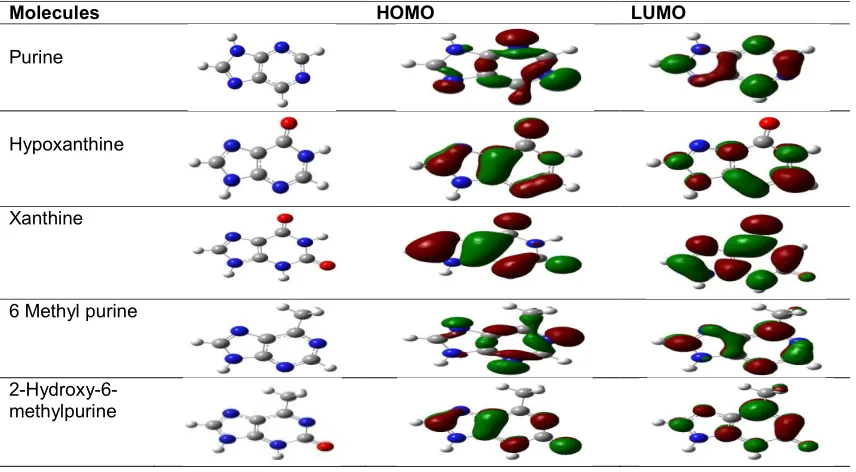
Fig. 3.1.8. The percent compositions of carbon, in the frontier orbitals (HOMO and LUMO), determined for all interaction sites (at C2 and C8). The data were obtained from AOMix software package
PU-C2 PU-C6 PU-C8 HY-C2 HY-C8 6MP-C2 6MP-C8 XA-C8 HMP-C8
Mulliken atomic Charges 0.079933 0.009193 0.153024 0.161378 0.138284 0.078185 0.150114 0.152253 0.13369
0 0.05 0.1 0.15 0.2
M
u
ll
ik
en
A
to
m
ic
C
h
a
r
g
es
(A
.U
)
PU-C2 PU-C6 PU-C8 HY-C2 HY-C8 6-MP-C2 6-MP-C8 XA-C8 HMP-C8
%C Homo 0.93 4.96 3.68 17.19 9.02 5.11 0.91 20.65 17.42
%C Lumo 18.63 11.61 31.12 8.5 41.44 10.74 18.77 4.81 2.57
0 10 20 30 40 50
C
a
rb
o
n
C
o
m
p
o
si
ti
o
n
(
%
13
Fig. 3.1.9. The percent compositions of carbon, on HOMO, determined on the interaction sites (at C2 and C8). The data were obtained from AOMix software package
PU-C2 PU-C6 PU-C8 HY-C2 HY-C8 6-MP-C2 6-MP-C8 XA-C8 HMP-C8
%C Homo 0.93 4.96 3.68 17.19 9.02 5.11 0.91 20.65 17.42
0 5 10 15 20 25
C
a
rb
o
n
C
o
m
p
o
si
ti
o
n
(
%
Table 3.1.1. The frontier orbitals for purine derivatives. Molecular orbital and electronic structure visualization was performed from the check point files using GaussView 3.0 software
program
Molecules HOMO LUMO
Purine
Hypoxanthine
Xanthine
6 Methyl purine
2-Hydroxy-6-methylpurine
4. DISCUSSION
Xanthine oxidase is one of the most useful molybdenum containing enzymes which catalyze a wide range of substrate during the catalyze reactions. The objective of this research was to describe the factors affecting the binding stage of catalysis. The structures of free substrates and the geometry of the active site in the presence of substrate in the binding pocket amino acid residues were constructed using Gauss View 3.0 software package. All geometry optimizations were performed using Gaussian 03W 6.0 software package by applying a density functional theory (DFT) method to generate several parameters such as total energy, Mulliken atomic charges, orbitals and percent composition. The Mulliken atomic charges and the total energies were compiled from the output files to characterize the interaction sites and the stability of the optimized structures. The factors that are proposed to influence the progression of substrates to products and affect the reactivity of substrate-enzyme binding stage are: the orientation of substrates inside the binding pockets, substrate-binding sites, the activation of the active site, geometric distortion of the active site, nature of substrates and nature of amino acid residues as well as the toutomeric nature of substrates. In this section, the orientation of
substrates, the interaction sites of substrates will be discussed.
4.1 Substrate Specificity
4.1.1 Substrate orientation inside the binding pockets
In order to verify the factors that affect the reactivity of the enzymes, the behavior of the enzymes in the presence of the simplest heterocyclic substrate (purine) is described below. Proper substrate orientation inside the binding pocket is essential in enzyme catalysis so that the active site amino acid residues can be aligned to most effectively lower the overall activation barrier of the reaction and to stabilize the transition state. The mechanistic study for the oxidation of purine substrate allows to assume three possible orientations (interaction sites) in the binding pocket. The C2, C6 or C8 positions of
the substrate are oriented towards the molybdenum center for the nucleophilic attack in the presence of the most important conserved amino acid residues such as Glu1261, Glu802 and
Arg880, suggesting that purine could be
hydroxylated at the C8 (Fig. 4.1.1, a), C6 (Fig.
4.1.1, b) or C2 (Fig. 4.1.1, c) position during
Mo S S O S O Glu1261 O O O OH Glu802 C2 N C6 N H N C8 N
H2N
H2N
Arg880 - H :
a)
Mo S S O S O O OH Glu802 C2 N C6 NHN C8
N
H2N
H2N
Arg880 H : Glu1261 O O -b) Mo S S O S O Glu1261 O O O OH Glu802
H2N
H2N
Arg880
- H :
C2 N C6
N NH
C8 N
c)
Fig. 4.1.1. The orientation of purine in the binding pocket of xanthine oxidoreductase enzyme. Purine has three interaction sites, at a (C8), b (C6) and c (C2) sites
As shown in Fig. (4.1.1), it is proposed that purine might interact with the active site is one of the three interaction sites. Amongst these sites, the C6-pyrimidine (qc = 0.009193) (Table A.II.1)
is considered to be the favored site one as shown by its oxidation product (hypoxanthine). This C6-pyrimidine position might be attained
when purine is oriented as shown in Fig. (4.1.1, b) because of the involvement of the binding pocket amino acid residues. The charge distribution over the carbon atoms of purine varies on each interaction site at C2 position it is
0.079933, at C6 position it is 0.009193 and at C8
position it is 0.153024 (Fig. 3.1.4). Even though the C8-pyrimidine position is the most
electrophile for the nucleophilic attack, the finding suggested that the C6-pyrimidine position is the
most favorable interaction site. This may be due to the unique orientation of purine in the binding pocket amino acid residues during interaction of the active site and the contribution of the HOMO orbital of C6 position is the most dominant over
the other interaction sites when it interacts with the active site. As shown in Fig. 4.1.1, the role of the binding pocket amino acid residues is proposed to create hydrogen bonding microenvironments in providing the proper orientation of substrates during the near attack conformation. However, purine does not create hydrogen bonding microenvironments, since C6
carbonyl projects into the hydrophobic end of the binding pocket amino acid residue that may interact with Arg880. Since the orientation of
purine in the binding pocket is expected to lack a hydrogen bonding microenvironment contributed from the binding pocket amino acid residues, there is no evidence for purine to be affected by a hydrogen bonding network or activated by the binding pocket amino acid residues. Therefore, the decrease in activity and change in substrate specificity of XOR enzymes could largely be due to conformational change.
As shown in Fig. (4.1.2), hypoxanthine is the oxidized product of purine when it is oxidized at its C6 position, on substrate that could be further
oxidized to xanthine. Hypoxanthine is proposed to have two alternative orientations (interaction sites) in the enzyme active site. The two orientations differing in that either the C2 or C8
position of substrate is oriented towards the molybdenum center of the enzyme, it can be suggested that hypoxanthine could be hydroxylated at C8 or C2 position. The
crystallographic studies [14] revealed that hypoxanthine is more readily hydroxylated at its C2 position than that of its C8 position. This is
because hypoxanthine at C8 lies further below
the same plane of the square pyramidal molybdenum center (1Å) than xanthine (0.4Å) and this orientation might not promote enzyme catalysis. The effect may be due to the role of the binding pocket amino acid residues such as Glu802, Arg880 and Glu1261 known to be important
position of hypoxanthine relative to its C8 position
may be less effective stabilization of the negative charge that accumulates on the heterocycle in the course of nucleophilic attack at C8 position
or, due to the formation of C6=O- and better
stabilization of the negative charge accumulating during the transition state through the interaction with the positively charged Arg880. The third
reason why hypoxanthine is selectively hydroxylated at C2 rather than at C8 is that the
inherent electronic structure of the substrate in which a computation shows that a partial charge of 0.161378 rests on C2 and that of 0.138224 on
C8. These factors indicated that the C2 position is
more susceptible to nucleophilic attack than C8
position. The hydrogen bonds formed between the C6-pyrimidine and Glu802 may serve as an
anchor to allow the C2-pyrimidine (qC =
0.161378) of hypoxanthine to be oriented towards HOMo for a nucleophilic attack. In addition to the oxidation of the C6-pyrimidine of
purine, the interaction site could also be methylated to give 6-methylpurine (6MP). Similar
to the C6-carbonyl of hypoxanthine, the -CH3 at
the C6-pyrimidine of 6MP could be oriented
towards Glu802 to assume similar orientation as
hypoxanthine. Like hypoxanthine, the 6MP interacts at the C2 (qC = 0.078185) position
with the HOMo terminal of the enzymes active site.
As shown in Fig. 4.1.3, both hypoxanthine and 6-methylpurine substrates are proposed to bind the active site enzyme through the same interaction site at C2 position. Here, the most important
anticipated factor which affects the reactivity of hypoxanthine and 6-methylpurine is that their difference in the ability to form hydrogen bonding microenvironments as well as their proper orientation when they interact with the conserved amino acid residues. In the case of hypoxanthine, the carbonyl oxygen is oriented towards the Arg880 amino acid residue. This
carbonyl oxygen is able to form hydrogen bonding microenvironments with the H donor NH2 group of the Arg880 amino acid residue. This
N
N NH
C N
H
PURINE H
H 1
2
3 4
5 6
7 8 9
HN
C2
N NH
C8
N
HYPOXANTHINE
HN
N H
N H
C8
N O
O
XANTHINE
N
C2
N NH
C8
N
6-METHYL PURINE
CH3
H
N
N H
N H
C8
N
CH3
O
2-HYDROXY-6-METHYL PURINE
[O] [CH3]
[O]
[O]
H O
H H
H H
Fig. 4.1.2. Purine and its derivatives. When purine oxidized and methylated at its C6 position, it gives hypoxanthine and 6-methyl purine, respectively. Xanthine and HMP are the oxidized
Mo S S O S O Glu1261 O O O OH Glu802
H2N
H2N
Arg880 - H : C2 N N NH C8 N CH3 Mo S S O S O Glu1261 O O O OH Glu802
H2N
H2N
Arg880 - H : C2 HN N NH C8 N O
a)
b)
Fig. 4.1.3. The orientation of 6-methylpurine and hypoxanthine inside the active site of xanthine oxidase enzyme
Mo S S O S O Glu1261 O O O OH Glu802
H2N
H2N
Arg880 - H : Mo S S O S O Glu1261 O O O OH Glu802
H2N
H2N
Arg880 - H : NH H N N H C8 N CH3 O NH H N N H C8 N O O
Fig. 4.1.4. The orientation of xanthine and 2-hydroxy-6-methylpurine inside the active site of xanthine oxidase enzyme with the conserved binding pocket amino acid residues. Both xanthine and HMP bound to the Molybdenum center through the same
interaction site at C8
leads to the formation of the most stable tetrahedral Michaelis-Menten intermediate during the initial stage of catalysis by neutralizing the negative charge accumulated on the ring of this substrate as well as the formation of hydrogen bond through the carbonyl at C2-pyrimidine with
Glu802 that may gain the most favorable
interaction. On the contrary, even though the CH3 group of 6-methylpurine is oriented towards
the Glu802 amino acid residue; it cannot form any
hydrogen bond to stabilize the negative charge accumulated on the ring of this substrate. As a result, 6-methylpurine could form less stable tetrahedral Michaelis-Menten intermediate during the initial stage of catalysis than that of hypoxanthine. This is because the ineffective utilization of the substrate for the binding pockets amino acid residues as well as the steric effect created by the CH3 group.
As shown in Fig. (4.1.4), HMP is oriented in such a way that the C2=O oxygen interacts with Arg880
and the C6 CH3 group points away from it. This
orientation is more reasonable for the low reactivity of 2-hydroxy-6-methyl purine in bovine milk enzyme. Here, we have suggested that HMP binds in an inverted orientation to that seen with xanthine and for this reason is unable to utilize Arg880 effectively for the transition state
stabilization. This account for the low reactivity of substrates such as HMP and 6-methylpurine with wild type enzyme and at the same time their relative insensitivity to mutation of the active site amino acid residue, Arg880 to methionine [4]. As
shown in Fig. (4.1.4), the carbonyl oxygen of xanthine could form hydrogen bonding microenvironments with the H donor NH2 group
of Arg880 binding pocket amino acid residue.
However, the substituent group CH3 in HMP
could not form hydrogen bonding microenvironments with Arg880. Both xanthine
and 2-hydroxy-6-methylpurine are able to bind with the equatorial hydroxyl terminal of the active site of xanthine oxidase enzyme through their C8
xanthine and HMP substrates in xanthine oxidase enzyme is the proper utilization of the conserved binding pocket amino acid residues during the initial stage of catalysis. Moreover, the methyl group for 2-hydroxy-6-methylpurine at C6
position could prevent the electrostatic interaction with Arg880, which cannot form
hydrogen bonding microenvironments through methyl group and thus it gives less stable product than that of xanthine. This is mainly because HMP is unable to stabilize the negative charges accumulated on the heterocycle through an electrostatic interaction of C6 carbonyl oxygen
with Arg880 in which the negatively charged
carbonyl oxygen is a hydrogen acceptor and the NH2 group of Arg880 is a hydrogen donor [15,16].
Generally, we can suggest that methylation of the reducing substrates at their respective positions may interfere with the interaction site which causes the low reactivity of these reducing substrates as shown in (Fig. 3.1.1). This is due to the fact that methylated substrate is too large to fit into the binding pockets for the progression of the reaction. The low reactivity of purine and HMP may be due to the interaction site at C6
carbonyl binds with the enzyme active site in the inverted manner. Thus, these substrates cannot interact with the amino acid residue, Arg880 which
plays a great role in stabilizing the transition state and in making the hydrogen bonding microenvironments. The formation of hydrogen bonding is important in providing the proper orientation of the intended substrate that reacts with the active site in the course of the reaction. In other words, because of the inverted position, the active site amino acid residue, Arg880
interacts at C2 carbonyl position in which the
accumulation of charges at the transition state cannot be stabilized by this amino acid residue. This situation is accountable for the catalytic effect to stabilize the building up of charges on OC2 at the transition state since these molecules
are oriented with the C2 rather than the C6
position oriented towards the amino acid residue, Arg880. Therefore, it is suggested that good
substrates are oriented in the active site with respect to Arg880 when their C6 functional groups
are oriented towards this residue, in order to utilize its stabilizing contribution to the transition state whereas poor substrates may orient opposite to C6 position, projecting their C6
functional groups into the hydrophobic binding pocket on that side of the active site. As a summary, the orientation of substrates in the active site of xanthine oxidoreductase plays a critical role in determining the catalytic
effectiveness of Glu802 and Arg880 in
hydroxylation of xanthine at its C8 position.
Properly oriented with N7 towards the Glu802 and
C6=O towards the Arg880, the reaction proceeds
much more rapid than when the substrate binds in the inverted orientation as indicated in the case of HMP. The tautomerization of purine derivatives has been proposed as an alternative means of stabilizing the accumulation of negative charges over a carbon atom and increasing the electrophilicity of substrates at their respective interaction site of the deficient carbon atoms. That means, the arrangement of these keto and enol forms could reduce the activation barrier by destabilizing the reactant state. The enol toutomeric forms of hypoxanthine and xanthine are less stable than their keto forms shown in (Fig. 3.1.3). This may be due to the repulsive forces between the incoming hydrogen atom/ proton and the oxygen atom during interaction to form a single H-O bond by breaking the double bond of oxygen. Therefore, it is suggested that the keto toutomeric forms of these physiological substrates are relatively more stable than their enol forms. On the other hand, the enol form of HMP is more stable than its keto form in which the enol form may have the probability of forming hydrogen bonding microenvironments with the binding pocket amino acid residues as shown in (Fig. 3.1.3). As shown in (Fig. 3.1.3), we can suggest that the keto toutomeric forms of the physiological substrates are energetically more favorable than their enol forms. That means, the keto forms of hypoxanthine and xanthine are more stable than their enol forms because their keto forms of these physiological substrates were involving more vigorous reactions than their enol forms do. However, the enol toutomeric form of HMP is energetically more favorable than its keto form. In other words, the more energetic the reaction, the more stable the product formed since the amount of energy absorbed to break the bonds of the molecule is the same as the amount of energy released during bond formation/ product formation.
4.1.2 Interaction sites
unbound molecules are determined based on the value of Mulliken atomic charges and percent composition on their carbon centers. The interaction sites of substrates which possess more positive partial charges are usually considered good electrophiles. The more the electrophile interaction sites are the more susceptible for nucleophilic reaction during the initial stage of catalysis. More electrophile species are expected to undergo better nucleophilic reactions than those bearing less positive charges on their carbon atoms of the interaction sites. However, this may not be always true to determine the most favorable interaction sites of the unbound substrates based on their partial charges as shown in (Fig. 3.1.1). In this case, the orientation of substrates in the binding pocket amino acid residues plays a great role to determine the most favorable interaction sites. This is the reason why the most favorable interaction site of purine is at its C6 position (its
partial charge, qc = 0.009193), rather than the most electrophile interaction site at its C8 position
(its partial charge, qc = 0.153024) as indicated in (Fig. 3.1.4). Even the interaction site of purine at its C2 position is more electrophile than its C6
position. The charge and bonding at the site of interaction could be important during the formation and dissociation of the activated state. However, there is no tangible proof for selectivity of interaction based on charges. It is better to consider the % compositions of carbon on its HOMO orbitals for purine and its derivatives as shown in (Fig. 3.1.9) at their respective interaction sites. For instance, the percent composition of carbon on HOMO orbital of purine at its three interaction sites is given as Pu C2 =
0.93, Pu C6 = 4.96 Pu C8 = 3.68. This indicates
that the interaction site at C6 position contributes
more during interaction. Therefore, C6 position of
purine is the most favorable interaction site among its three interaction sites. This confirms that the electrophilicity nature of the interaction sites could not be determined based on the Mulliken atomic charges alone.
Similarly, the % compositions of carbon on HOMO orbitals for purine derivatives at different interaction sites are given as HY C2 = 17.19 and
HY C8 = 9.02, 6-MP C2= 5.11 and 6-MP C8 =
0.91, XA C8 = 20.65 and HMP C8= 17.42. From
this, it is suggested that hypoxanthine is selectively interacted at its C2 position during
hydroxylation rather than at C8 position likely to
support the electrophilicity nature determined based on partial charges mentioned earlier in Section (4.1.1) (qc = 0.161378 at C2 and qc
= 0.138284 at C8). Like that of hypoxanthine,
6-methylpurine is selectively hydroxylated at its C2
position rather than C8 position which contradicts
the electrophilicity nature determined based on partial charges (qc = 0.078185 at C2 and qc =
0.150114 at C8) as shown in (Fig. 3.1.7).
Similarly, xanthine and 2-hydroxy-6 methylpurine could be hydroxylated at the same interaction site at C8 position, but the contribution of carbon
on HOMO orbitals for xanthine (20.65) is greater than that of 2-hydroxy-6 methylpurine (17.42) as shown in Fig. (3.1.9) which is in a good agreement with the electrophilicity nature determined based on partial charges (qc= 0.152253 for xanthine and qc= 0.133690 HMP) as shown in Fig. (3.1.7).
As shown in Fig. (3.1.3), the keto-enol toutomerism could influence the electrophilicity of the binding site of carbon atom, due to the variation of the partial charges. Hypoxanthine enol is the least electrophile (qc = 0.094573) and hypoxanthine keto is the most electrophile (qc = 0.161378) among the keto-enol toutomerism of purine derivatives, such as HMP keto and enol forms, and xanthine keto and enol forms. Thus, hypoxanthine keto is expected to exhibit the highest reactivity towards XO enzyme and hypoxanthine enol shows the least affinity towards XO enzyme. Moreover, both the keto and enol forms of HMP exhibit a higher affinity towards XO enzyme than hypoxanthine enol does Fig. 3.1.3). From this, it is suggested that
the keto-enol toutomerism as well as the formation of hydrogen bonding
microenvironments are expected to affect the initial stage of catalysis. Therefore, the keto-enol toutomerism and the presence of the substituent groups could be considered as the other factors that affect the binding sites of purine and its derivatives. As shown the in Fig. 3.1.3, the value of Mulliken atomic charges on the carbon atoms of their respective interaction sites for purine and its derivatives, such as purine (C6 ), 6MPU (C2),
hypoxanthine keto (C2), and hypoxanthine enol
(C2) , respectively are: qc = 0.009193, qc=
0.078185, qc = 0.161378, and qc = 0.094573. From this, we can say that purine is considered as the least electrophile (qc = 0.009193) of all purine derivatives and hypoxanthine keto is considered as the most electrophile (qc = 0.161378) of all purine derivatives. This indicates that hypoxanthine keto can undergo the fastest rate of reaction with the active site of XO enzyme at its C2 position whereas purine could undergo
substituent groups for the variation of reactivities towards XO enzyme: the –CH3 group may serve
as an electron donating group by inductive effects. On the other hand, -OH may serve as an electron withdrawing group inductively and electron donating group through resonance. Some substrates were shown to have methylated and others carbonylated C6-pyrimidine sites
which causes some substrates to lose the most favorable interaction. This effect was shown with substrates such as hypoxanthine and 6MP. Although they both have substitution on the C6
-pyrimidine, the only difference is the substitution of –CH3 and –OH, respectively, on 6MP and
hypoxanthine. They both interact with HOMo at their C2-pyrimidine site and the same orientation
with their C6-CH3 and C6-OH oriented towards
Glu802. The activities and rates of reactions
determined using these substrates revealed that hypoxanthine was by far the most specific for XOR enzymes. On the other hand, the substrates with –CH3 group such as 6MP were
shown to be less specific for XO enzyme. In addition, hypoxanthine keto is more electrophile (qc = 0.161378) than its enol form (qc = 0.094573) and may undergo faster rate of reaction than hypoxanthine enol during their interaction with XO enzyme at the same interaction site (C2). Therefore, other than
Mulliken atomic charges, the presence of substituent groups on purine and its derivatives as well as the keto-enol toutomerism are the most expected factors that affect the initial stage of catalysis.
5. CONCLUSIONS
The physiological importance of XO in areas of human health and the oxidative degradation of organic metabolites in the cell emphasizes the need to understand the mechanism of XO activity. The activity of xanthine oxidase family enzymes greatly depends on the proper orientation of the substrates. The involvement of the binding pocket amino acid residues for the near attack conformation affects the interaction sites of the substrates. As a result, the rate at which the substrates interacted with the active site of xanthine oxidase enzyme is proposed to be slow. This leads to the decrease in the rate of the reaction. In addition to substrate proper orientation, the interaction sites of substrates have more pronounced effect on the reactivity of xanthine oxidase enzymes during the progression of the given reaction. When an enzyme binds with a particular interaction site of the precursor substrate, it leads to the formation
of substrate- enzyme complex. Therefore, the rate of formation of this substrate- enzyme complex is proposed to be affected by the interaction site. Moreover, the keto and enol forms of the particular substrates as well as the existence of the substituent groups on the intended substrates affect the charge density of the interaction sites of the carbon atoms. This affects the affinity of the substrates to bind with the equatorial oxygen of the active site of xanthine oxidase enzyme. As a result, the rate of the reaction is proposed to be affected by these factors.
In order for the interaction between the reactants (substrate and enzyme) to take place, the substrates are proposed to enter the binding pocket amino acid residues and attain a proper orientation to bind through their favorable interaction sites with the enzyme active site. Therefore, the catalytic transformations of the substrates to their products are expected to begin with the substrate binding stage and formation of the tetrahedral Michaelis-Menten type intermediate.
The kinetic parameters [7,14,16] for the overall reactions are used to compare the properties of the enzymes as well as their specificity towards the substrates. Therefore, the order of activities recorded for bm XOR (from the highest to the lowest) is given as: hypoxanthine > xanthine > HMP > 6MP > Purine. Thus, the reactivity of xanthine Oxidase enzyme with a range of substrates could be described using the steady-state enzyme kinetic constants such as Km-Michaelis constant, Kcat-reduced constant, Vmax and Kcat/ Km.
Generally, the reactivities of substrates towards xanthine oxidase family enzymes is influenced by several factors such as the redox sites, the binding pocket environment, orientation of substrates inside the binding pockets, geometric distortion of the active site, nature of the amino acids, nature of substrates, the overall architecture of the enzyme, inductive and steric effects and the toutomeric nature of substrates.
ACKNOWLEDGEMENTS
Department’s Post Graduate Computation Center.
COMPETING INTERESTS
Author has declared that no competing interests exist.
REFERENCES
1. Hille R. The mononuclear molybdenum enzymes. Chem. Rev. 1996;96:2757- 2816.
2. Voityuk AA, Albert K, Roman JM, Huber R, Rosch N. Substrate oxidation in the active site of xanthine oxidase and related enzymes. A model density functional study. Inorg. Chem. 1997;37:176-180. 3. Bayse AC. Density-functional theory
models of xanthine oxidoreductase activity: Comparison of substrate tautomerization and protonation. J. Chem. Soc. 2009;29: 2306-2314.
4. Pauff JM, Cao H, Hille R. substrate orientation and catalysis at the molybdenum site in xanthine oxidase. J. Biol. Chem. 2009;284:8760–8767.
5. Garattini E, Menedel R, Romao MJ, Wright R, Terao M. Mammalian molybdo– flavoenzymes, an expanding family of proteins: Structure, genetics, regulation, function and pathophysiology. Biochem. J. 2003;372:15-32.
6. Hernandez B, Luque JF, Orozco M. Tautomerism of xanthine oxidase substrates hypoxanthine and allopurinol. J. Org. Chem. 1996;61:5964-5971.
7. Greenlee L, Handle P. Xanthine oxidase: Influence of pH on substrate specificity. J. Biol. Chem.1964;239:1090-1095.
8. Amano T, Ochi N, Sato H, Sakaki S. Oxidation reaction by xanthine oxidase. theoretical study of reaction mechanism. J. Am. Chem. Soc. 2007;129:8131-8138. 9. Zhang HX, Wu D. A theoretical study on
the mechanism of the reductive half-reaction of xanthine oxidase.Inorg. Chem. 2005;44:1466-1471.
10. Bayse AC. Theoretical characterization of the “Very Rapid” Mo (V) species generated in the oxidation of xanthine oxidase. Inorg. Chem. 2005;45:2199-2202.
11. Enroth C, Eger TB, Okamoto K, Nishino T, Nishino T, Emil F, Pai EF. Crystal structures of bovine milk xanthine dehydrogenase and xanthine oxidase: Structure-based mechanism of conversion. J. Biol. Chem. 2000;97:10723-10728.
12. Yamaguchi Y, Matsumura T, Ichida K, Okamoto K, Takeshi Nishino T. Human xanthine oxidase changes its substrate specificity to aldehyde oxidase type upon mutation of amino acid residues in the active site: Roles of active site residues in binding and activation of purine substrate. J. Biol. Chem. 2007;141:513–524.
13. Martz E. Protein explorer: Easy yet powerful macromolecular visualization, Trends in Biochem. Sci. 2002;27:107-109. Available:http://proteinexplorer.org.
14. Pauff JM, Cao H, Hille R. Substrate orientation and catalytic specificity in the action of xanthine oxidase: The sequential hydroxylation of hypoxanthine to uric acid. J. Biol. Chem.2010;284:8760-8767. 15. Pauff JM, Zhang J, Bell CE, Hille R.
Substrate orientation in xanthine oxidase. J. Biol. Chem. 2008;283:4818–4824. 16. Pauff JM, Hemann CF, Hemann NJ,
Leimkuhler S, Hille R. The role of argenine 310 in catalysis and substrate specificity in xanthine dehydrogenase from Rhodobacter capsulatus. J. Biol. Chem. 2007;282:12785–12790.
17. Romao MJ. Molybdenum and tungsten enzymes: A crystallographic and mechanistic Overview. J. Royal. Soc. 2009;17:4053-4068.
18. Hille R. Structure and function of xanthine oxidoreductase. Eur. J. Inorg. Chem. 2006;36:1913-1926.
19. Dinesh S, Shikha GW, Bhavana GW, Nidi S, Dileep S. Biological activities of purine analoges. Rev. J. Pharm. and Sci. Innov. 2012;1(2):29-34.
20. Pauff JM, Cao H, Hille R. Substrate orientation and the origin catalytic power in xanthine oxidoreductase. In. J. Chem. 2010;50:355-362.
21. Hille R, Nishino T. Xanthine oxidase and xanthine dehydrogenase. J. Bio Chem. 1995;9:995-1003.
22. Voityuk AA, Albert K, Stlmeier SK, Nasluzov VA, Neyman KM, Hof P, Huber R, Romao JM, Rosch N. Prediction of alternative structures of the molybdenum site in the xanthine oxidase-related aldehyde oxido reductase. J. Am. Chem. Soc. 1997;119:3159-3160.
24. Matsumoto K, Okamoto K, Hille R, Eger BT, Pai EF, Nishino T. The crystal structure of XOR during catalysis: Implications for reaction mechanism and enzyme inhibition. Proc. Nat. Acad. Sci. 2004;101:7931-7936.
25. Pauff JM, Zhang J, Bell CE, Hille R. Substrate orientation in xanthine oxidase crystal structure with HMP. J. Biol. Chem. 2007;6:1-16.
26. Becke ADJ. Density functional thermo chemistry 3: The role of exact exchange. J. Chem. Phys. 1993;98:5648-5652.
APPENDICES
APPENDIX I: Survey of the reducing substrates
N1
N3
N1
H N3
N1
H N2
N1
N4
Pyrimidine
Pyrazole
Imidazole Pyrazine
N1
N3 NH
C8
N
N
N1 N
C7
C6
N N1
N3 NH
N8
HN1
N3 NH
N8
HN
N
H N
C7
C6
N N1
N3 NH
C8
N
Allopurinol Lumazine
Pteridine Pyrazolopyrimidine
Purine R1
R2
O O
O Purine Derivatives
(i)
(ii)
(iii)
Purine derivatives: purine (R1 and R2 = H), hypoxanthine (R1 = OH and R2 = H), 6-methylpurine (R1 = CH3 and R2 = H), 2-hydroxy-6-6-methylpurine (R1 = CH3 and R2 = OH),
and xanthine (R1 = OH and R2 = OH).
R1 R2
O
R1 R2
O
R1 R2
O
R1 R2
O
Carbonyl
Aldehydes Formate Formamide
(i) (ii) (iii)
R1 or R2 = H for all substrates, and R1 or R2 could be H (formaldehyde), CH3 (acetaldehyde), CH2CH3 (Propionaldehyde) CH3(CH2)2 (Butylaldehyde), phenyl
(benzaldehyde), OH (formate), and NH2 (Formamide)
Fig. A. I.1.The classification of reducing substrates. Upper panel (i) represents the purine derivatives, upper panel (ii) represents the pyrazolopyrimidine derivatives (such as allopurinol), upper panel (iii) represents the pteridine derivatives (such as Lumazine).
![Fig. 3. The bonding description for the reductive half reaction active site of bmXOR (PDB accession code of 1V97) [24], showing bond distance (Å) and bond angles ().The illustration is adapted from reference [24]](https://thumb-us.123doks.com/thumbv2/123dok_us/8380430.1678472/4.612.118.508.397.660/description-reductive-reaction-accession-distance-illustration-adapted-reference.webp)