Article Type: Specialties Paediatric Surgery DOI: 10.1102/1470-5206.2013.0007 ß2013 e-MED Ltd
Missed small bowel obstruction that
complicated an acute appendicitis:
a misdiagnosis
Mahmoud Abdullah AlReefi
aand Nasser Shukri
b aSaudi Board of Otorhinolaryngology, King AbdulAziz University, Rabigh Branch, Jeddah 21488, Saudi Arabia;bDepartment of Pediatric Surgery, Maternity and Children’s Hospital, Jeddah 21434,
Saudi Arabia
Corresponding address: Dr Mahmoud Abdullah AlReefi, MBBS, Saudi Board of Otorhinolaryngology, King AbdulAziz University, Rabigh Branch, PO Box 35046 Jeddah 21488, Saudi Arabia.
Email: m_alreefi@yahoo.com
Date accepted for publication 26 March 2013
Abstract
We present a case of an 11-year-old boy who had an abdominal pain for 4 days, which was found to be caused by an acute appendicitis that ruptured. The second day after an open appendectomy, he was found to have a small bowel obstruction that was not found during the procedure. The reasons for this misdiagnosis are discussed in this article to help other health care providers avoid this dangerous complication.
Keywords
Small bowel obstruction; intra-abdominal inflammatory process, perforated appendicitis.
Case report
An 11-year-old boy presented to our hospital’s emergency department complaining of an abdominal pain for 4 days. The patient was in his usual state of good health until 4 days before presenting to the emergency room where he started to have gradual onset of diffuse abdominal pain, progressive course, scoring 7/10 in severity, stabbing nature, not relieved or aggravated by any factors, not referred to any location. The pain radiated to the right lower quadrant 10 h after the onset of the pain.
The pain was associated with nausea, vomiting (twice a day, non-bilious, non-projectile) and anorexia. No urinary symptoms were present and his bowel habits were normal. Undocumented fever was reported with no chills. He has no previous medical issues and no previous hospital admission. There was no history of previous surgeries, no known allergies and he not taking any medications. There was no family history of chronic illnesses, and the parents did not drink alcohol and were non-smokers.
The boy went to multiple polyclinics where only pain killers were prescribed for him; no further investigations were done until he arrived at our center. On examination, the patient was lying still in the bed, looking sick, in severe pain, and was mildly dehydrated. His vital signs were stable except his temperature, which was high (38.58C).
ileum. The appendix was separated from the ilium by very gentle manipulation using a gauze and warm saline. After separating, we examined the bowel starting from the ileocecal valve for approximately 60 cm proximally, a standard procedure in such cases; neither perforation nor signs of adhesion formation were found. Appendectomy was then carried out.
We sent the appendix for histopathologic evaluation. The report stated: 4-cm long appendix with swelling of the distal portion and fecalith in the lumen, clear points of perforation. Microscopically, the section showed diffuse suppurative acute appendicitis with areas of necrotic tissue.
The patient was then sent to the ward and was kept on intravenous fluids and antibiotic coverage. His vital signs and general condition were monitored.
On the second postoperative day, the patient was doing fine, still on intravenous fluids and antibiotics. Twenty-four hours after the surgery, we decided to start sips of water, which he could not tolerate without vomiting. So feeding was delayed. Later that day, the patient had multiple attacks of greenish vomiting, severe nausea and vague generalized abdominal pain which was colicky in nature, moderate severity (5/10 on pain scale), not relieved or aggravated by any factors. He was passing gas and small amount of normal-colored stool.
On examination the boy was resting in bed, in moderate pain, looked sick, but no signs of dehydration were noted. All vital signs were stable and normal.
Abdominal examination revealed a mildly distended abdomen, with pain all over but mostly in the epigastric and umbilical areas (there was some pain at the surgical site), no rigidity, no rebound tenderness and sluggish bowel sounds. Rectal examination showed normal stool.
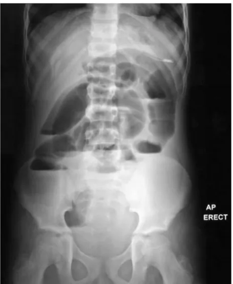
A nasogastric tube was inserted, which drained around 300 ml of greenish fluid and the patient reported a moderate relief of pain. An erect and supine abdominal radiograph at that time showed the nasogastric tube in place, multiple air fluid levels and generalized distension of the small bowel (Figs. 1 and 2). The radiograph was interpreted as paralytic ileus by the radiologist, considering the fact that the patient started to have the symptoms on his second postoperative day. Blood investigations were only remarkable for a high lipase level (256 U/L). Abdominal ultrasonography was unremarkable.
Taking into consideration the history, the examination and the investigation results (especially the abdominal radiograph), a diagnosis of paralytic ileus was made and we decided to continue supportive care after excluding all the causes that might lead to the development of ileus.
Unfortunately, the patient did not improve clinically; the nasogastric tube drained 900 ml of greenish content in the next 24 h. He passed small amounts of gas but no stool. The same scenario continued for the next 2 days with 400–500 ml of greenish secretions; there were no changes in the abdominal radiograph (Figs. 3 and 4) but the intensity and frequency of the colicky abdominal pain were increasing.
We kept the patient under nil by mouth status, with strict input–output monitoring and followed his daily laboratory tests, all of which were within normal limits.
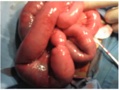
On the sixth day after appendectomy and failure of conservative management, we started to doubt that this was caused by paralytic ileus, and decided to take the patient to the operating room for an explorative laparotomy to find and treat the cause of the obstruction.
The small bowel was found to be dilated markedly about 60 cm proximal to the ileocecal valve, where a stenotic point (which was most likely missed when we ran the bowel in the first surgery) was found causing partial obstruction of the small bowel, most likely formed by fibrinous adhesions (Fig. 5). The bowel was of normal caliber distal to it. Complete exploration of the bowel revealed no other anomalies. No ischemic bowel or gangrenous changes were found. There was no intussusception and no pathology was identified at the site of the appendectomy. The bowel was milked through the narrow area; all the content passed smoothly and a normal caliber was
Fig. 1. Supine abdominal radiograph on the second postoperative day.
restored. The abdominal wall was closed and the patient was sent back to the ward and was kept on the same antibiotic regimen, with no oral intake and follow-up of his intravenous fluid intake and output.
The next day, the patient started to improve clinically; his pain improved, there was no more nausea or vomiting and the nasogastric tube brought only 30 ml of yellowish secretions. The day
Fig. 3. Supine abdominal radiograph on the fourth postoperative day, no changes.
after that, the nasogastric tube did not drain any fluids. We allowed the patient to start drinking sips of water, which he tolerated perfectly. We proceeded gradually with the diet until he tolerated a normal diet. Normal bowel habit was maintained throughout. The patient was discharged in good condition and during his follow-up in the clinic, he was doing fine.
Discussion
This patient had an acute intestinal obstruction that was triggered by the presence of an intra-abdominal inflammatory process, acute perforated appendicitis, and due to the failure to diagnose this pathology while performing an open appendectomy, multiple issues arose. The patient developed an acute intestinal obstruction after the appendectomy, which was mistaken for paralytic ileus due to the findings on the abdominal radiograph and a history of abdominal surgical procedure. This caused the patient to suffer for a few days and put him in danger of further complications due to the delay in diagnosis. This scenario could have been avoided if the bowel had been examined meticulously during the first surgery.
The development of small bowl obstruction is a known complication of perforated appendicitis. It is caused by adhesions that are exacerbated by the presence of infection. This leads to the initiation of an inflammatory response, activation of inflammatory factors and complements, and exudation of fibrinogen-rich fluid. Fibrinogen is converted to fibrin, which attaches to the surfaces of the surrounding damaged structures causing adhesions; they are known as fibrinous adhesions. Later, fibroblasts emerge into this matrix and deposit collagen, which leads to the conversion of the fibrinous adhesions to fibrous adhesions[1,2]. Clinically, this is manifested by bilious vomiting and obstipation.
To avoid the development of such complications, an early appendectomy should be performed. An early appendectomy is associated with less adverse events, such as abscess, small bowel obstruction, or unplanned readmission[3], as well as with less hospital charges and costs[4]. In the event of presenting with a perforated appendix, a bowel run should be done to ensure the absence of any bowel pathology, which could be treated at the same time, avoiding the need for further laparotomies[5].
Teaching points
Intestinal obstruction is a complication of perforated appendicitis, and it could cause the patient a great deal of pain and increases the danger of bowel perforation and death. It can be hard to diagnose after performing an appendectomy due to the similarity of the signs to paralytic ileus, which requires different management. To avoid putting the patient in this dangerous situation, the bowel should be checked meticulously while doing an appendectomy.