Chetan et al. World Journal of Pharmaceutical and Medical Research
www.wjpmr.com 63
REVIEW ON TRANSDERMAL DRUG DELIVERY SYSTEM
Chetan Ghulaxe1*, Mousumi Karpillai2, Sujit Pillai1, Pankaj Kushwah1, Ranu Mansare1
1
GRY Institute of Pharmacy, Borawan. (Khargone). 2
IPS Academy, College of Pharmacy, Indore.
Article Received on 29/06/2017 Article Revised on 19/07/2017 Article Accepted on 09/08/2017
INTRODUCTION
At present, the most common form of delivery of drugs is the oral route. While this has the notable advantage of easy administration, it also has significant drawbacks -- namely poor bioavailability due to hepatic metabolism (first pass) and the tendency to produce rapid blood level spikes (both high and low), leading to a need for high and/or frequent dosing, which can be both cost prohibitive and inconvenient.[1]
To overcome these difficulties there is a need for the development of new drug delivery system; which will improve the therapeutic efficacy and safety of drugs by more precise (i.e. site specific), spatial and temporal placement within the body thereby reducing both the size and number of doses. New drug delivery system are also essential for the delivery of novel, genetically engineered pharmaceuticals (i.e. peptides, proteins) to their site of action, without incurring significant immunogenecity or biological inactivation.
Transdermal drug delivery is defined as self contained, discrete dosage forms which, when applied to the intact skin, deliver the drug, through the skin at controlled rate
to the systemic circulation. Transdermal drug delivery system (TDDS) established itself as an integral part of novel drug delivery systems.[2]
Advantagesof transdermal drug delivery system Delivery via the transdermal route is an interesting option because transdermal route is convenient and safe. The positive features of delivering drug across skin to achieve systemic effect are:
1. Avoidance of first pass metabolism.
2. Avoidance of gastrointestinal incompatibility. 3. Predictable and extended duration of activity. 4. Minimizing undesirable side effect.
5. Provides utilization of drug with short biological half life, narrow therapeutic window.
6. Avoiding the fluctuation in drug level. 7. Maintain plasma concentration of potent drug. 8. Termination of therapy is easy at any point of time. 9. Greater patient compliances due to elimination of
multiple dosing profile.
10. Ability to deliver the drug more selectively to a specific site.
11. Provide suitability for self administration. 12. Enhance therapeutic efficacy.
*Corresponding Author: Chetan Ghulaxe
GRY Institute of Pharmacy, Borawan. (Khargone).
ABSTRACT
Transdermal drug delivery system (TDDS) specialist are ongoing to explore for new method that can efficiently and painlessly transmit better molecules in therapeutic quantity to overcome the Difficulties allied with oral route, namely poor bioavailability due to first pass metabolism and receptiveness to produce rapid blood level. Transdermal drug delivery get improved the therapeutic effectiveness and security of drugs by more site specific way but spatial and temporal placement within body is necessary to reduce both the size and number of doses essential to achieve the objective of systemic medication through topical application to the intact skin surface. Transdermal patches deliver the drugs for systemic effects at a predetermined and controlled rate. Through a diffusion process, the drug enter the bloodstream directly though the skin. Since there is high concentration on the patch and low concentration in the blood, the drug will keep diffusing into the blood, the drug will keep diffusing into the blood for a long period of time, maintain the constant concentration of drug in the blood flow. TDDS is act as micro emulsion, Transdermal patches, Niosomes, Ethosome and liposomal drug delivery system is act as Novel approach of carrier mediated drug delivery system. The success of all the TDDS depends on the skill of the drug to permeate skin in sufficient quantities to achieve its desired therapeutic effect. This review article provides a detailed study of transdermal that is benefit, disadvantages, mechanism, factors affecting skin permeation and types. Characterization of transdermal patch is use to check its quality, size, time of onset & duration, adhesive property, thickness, weight of patch, moisture of content, uniformity & cutaneous toxicological studies.
KEYWARDS: TDDS, first pass metabolism, Niosomes, Ethosome, bioavailability.
ISSN 2455-3301
WJPMR
www.wjpmr.com 64 Disadvantages of transdermal drug delivery system
1. The drug must have some desirable physicochemical properties for penetration through stratum corneum and if the drug dose required for therapeutic value is more than 10 mg/day, the transdermal delivery will be very difficult.
2. Only relatively potent drugs are suitable candidates for TDDS because of the natural limits of drug entry imposed by the skin’s impermeability.
3. Some patients develop contact dermatitis at the site of application for one or more of the system components, necessitating discontinuation.
4. Clinical need is another area that has to be examined carefully before a decision is made to develop a transdermal product.
5. The barrier function of the skin changes from one site to another on the same person, from person to person and with age.[3]
Skin As A Site For Drug Infusion[5,6,7,8]
The skin is the largest organ of the body. The skin an average adult body is about 20 square feet and it received about one third of total available blood. The skin is multilayered organ composed of three histological tissue.
The outermost layer of skin, epidermis is which provides a waterproof barrier and creates our skin tone. Dermis, beneath epidermis, contains tough connective tissue, hair follicles, and sweat glands and Deeper subcutaneous tissue (hypodermis) is made of fat and connective tissue.
Fig 1: Anatomy of skin represents different parts.
There are main three pathways through which foreign particles diffused or penetrate in to skin[9,10,11] 1. Transcellular/Intracellular permeation through the
stratum corneum.
2. Intercellular permeation through the stratum corneum.
3. Transappendageal permeation via the hair follicles, sweat and sebaceous gland.
Mechanism of transdermal permeation[10,11]
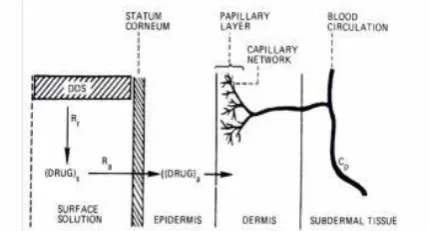
Transdermal permeation of a drug moiety involves the following steps:
i. Sorption by stratum corneum.
ii. Permeation of drug through viable epidermis. iii. Uptake of the drug moiety by the capillary network
in the dermal papillary layer.
iv. The drug must possess some physicochemical properties to reach target site via systemically through stratum corneum. The rate of permeation of drug moiety across the skin is governed by following equation:
dQ/dT = Ps( Cd –Cr)
Where, Cd= concentration of penetrate in the donor phase (on the surface of skin);
Cr= concentration of penetrate in the receptor phase (body); and Psis the overall permeability coefficient of the skin which is defined as:
Ps = Ks Dss/hs
Where, K = Partition coefficient of the penetrant; Dss=Apparent diffusivity of penetrant
hs= Thickness of skin.
Fig. 2: Different route of skin penetration.
A constant rate of drug permeation achieved, if Cd >Cr then the equation reduced as:
dQ/dT=Ps.Cd
The rate of skin permeation (dQ/dt) becomes a constant, if the Cvalue remains fairly constant throughout the course of skin permeation Tomaintain the Cdat a constant value, it is critical tomake the drug to be released at a rate (Rr) which is always greater than the rate of skin uptake (Ra), i. e., Rr>>Ra as shown in figure.8.
www.wjpmr.com 65 By doing so, the drug concentration on the skin surface
(Cd) is maintained at a level which is always greater than the equilibrium (or saturation) solubilityof the drug in the stratum corneum (Ces),i.e.,Cd>>C; and maximum rate of skin permeation(dQ/dt)m as expressed by equation.
[dQ/dT]m= PsCse
Apparently, the magnitude of (dQ/dt)mis determined by the skin permeability coefficient (Ps) of the drugand its equilibrium solubility in the stratum corneum(Cse).
A transdermal patch is defined as medicated adhesive patch which is placed above the skin to deliver a specific dose of medication through the skin with a predetermined rate of release to reach into the bloodstream. Today the most common transdermal system present in the market mainly based on semipermeable membranes which were called as patches.
Fig. 3: Transdermal patch showing its different components.
Types Of Transdermal Patches[12,5] Single-layer drug-in-adhesive
In this system drug and excipients is inclusive with skin adhesive which serve as formulation foundation as a single breaking layer. The rate of release of drug through diffusion phenomenon.
Fig. 4: Single layer drug in adhesive patch and its different component.[12,5]
The rate of release of drug is expressed as: dQ/dT =Cr/1/Pm + 1/Pa
Where Cr = drug concentration in reservoir compartment;
Pa = Permeability coefficient of adhesive layer;
Pm = Permeability coefficient of rate controlling membrane.
Multi-layer drug-in-adhesive
In this system drug and excipients incorporated with adhesive but both layer of adhesive separated by single
layer membrane. The released of drug occurred through diffusion phenomenon.
Fig. 5: Multi-layer drug in adhesive patch and its different component.[12,5]
The rate of release of drug is governed by following equation:
dQ/dT = Ka/r .Da/ha. Cr
Where Ka/r = partition coefficient for theinterfacial partitioning of the drug from the reservoirlayer to adhesive layer
Drug reservoir-in-adhesive
In the reservoir system,inclusion of liquid compartment containing drug solution/suspension between baking layer and semipermeable membrane followed by adhesive layer and release liner.
Fig. 6: Drug reservoir in adhesive patch and its different component[12,5]
The rate of drug release from this drug reservoir system is given by
dQ/dT = Ka/r .Da/ha(t).A(ha)
Where ha = thickness of adhesive layer; A = thickness of diffusional path.
Drug matrix-in-adhesive
This system is designed by inclusion of semisolid matrix having drug in solution or suspension form which is in direct contact with the release liner.
Fig. 7: Single layer drug in adhesive patch with its different component.[12,5]
The rate of release of drug is goverened by following equation:
dQ/dT = ACpDp1/2/ 2t
Where A = the initial drug loading dose dispersed in the polymer matrix;
Cp = solubility of the drug;
www.wjpmr.com 66 Basic components of TDDS
Polymer matrix/drug reservoir
Drug
Permeation enhancer
Adhesive
Backing film
Liner
Plasticizer
Polymer matrix/Drug Reservoir[13,14,15]
It is the very important component in TDDS and control the release of drug from patch.The polymers used in
TDDS should be stable. They should not produce any toxic effect either alone (or) with other excipients in TDDS formulation. They shouldn’t expensive one and it should be easily manufactured. They should have good stability and more compatibility with drugs and other components of System.The cross linked poly ethylene glycol, eudragit, ethyl cellulose, poly vinyl pyrolidine and hydroxyl propyl methyl cellulose are used as matrix formers in TDDS.
The polymers like EVA, poly urethane and silicone rubber are used as rate controlling membrane.
Table 1: List of Polymers Used In TDDS.
Polymers used in TDDS
Natural polymers Synthetic polymer Synthetic elastomer Cellulose derivative
Gelatin Shellac Starch Waxes Gums
Natural rubber Chitosan etc.
PVA
Poly vinyl chloride Polyethylene PVP
Poly acrylate etc
Poly butadiene Hydrin rubber Poly iso butylenes Silicon Rubber Nitrile
Acronitryle Neoprene Butyl rubber etc
Drug[16,17]
The selection of drug is based on its properties like physiochemical as well as biological properties.
Drug should have higher first pass metabolism. Drugs having narrow therapeutic window. Drugs with short half-life.
Drugs with frequent dosing.
Low molecular weight moieties (<1000 Dalton) Drugs with low dose (mg/day).
Low melting point substances (<200⁰C)
Drugs having affinity with both lipophilic and hydrophilic phases.
Drugs without any dermatological effect are suitable for formulation as transdermal
Permeation Enhancers
These are the substances which are reversibly changes the structure of stratum cornium and increase the permeation of drug from skin to blood stream. They are two types
1) Chemical enhancers [accelerants, absorption promoters (or) permeation enhancers][18,19]
They act by Increasing drug permeability by reversible damage to stratum cornium.
To consider the stratum cornium. To increase partition coefficient of drug.
Table 2: List of Chemical Enhancers Used In TDDS. Chemical
Enhancers Examples
Solvents
Water, Methanol, Ethanol,
Propylene Glycol Di-Methyl Acetamide
Fatty Acids & Esters
Oleic Acid, Linoleic Acid Lauric Acid, Capric Acid
Terpenes
Menthol, Cardamom Oil Cinnamon Oil, 18-Cineol Carvone
Surfactants
Anionic
SLS,Decodecyl Methyl Sulfomide Non-Ionic:
Pluronic F127, Pluronic F68 Bile Salts:Sodium Taurocholate Sodium Deoxy Cholatte
Pyrolidine N-Methyl 2-Pyrolidine
Axone Amides
Dimethyl Acetamide Dimethyl Formamide
Sulfoxides DMS
Didecyl Sulfoxides Miscellaneous
www.wjpmr.com 67 2) Physical Enhancers[18]
The following physical techniques have been used for enhancing the permeability of drug through skin.
Ionotophoresis
Electrophoresis
Sonophoresis
By using micro needles
Magnetophoresis
By using laser radiation
The combination of chemical enhancer and the magnetophoresis having greater enhancing power which is recently detected in lidocaine hydrochloride patch.
Adhesive[20]
It is used to affix the patch on the skin. It should be adhere on the skin with light pressure applied by finger. It should be easily removed from the skin surface without leaving any residue. It should not produce any irritation. It should have excellent contact with the skin. Should compatible with other components in formulation. It should be allow permeating the drug freely from the patch.
E.g. polyacrylates, poly iso butylenes, silicone derivatives.
Backing Laminate[21,22]
It is used to protect the patch from outer environment. They must be chemically resistant. They won’t allow to permeation of components in the patches. They have optimal elasticity, flexibility and tensile strength. It should have low water vapour transition rate. If a drug incorporated into a liquid (or) gel in the formulation, the backing material should be heat stable to allow fluid tight packing of drug reservoir (form-fill seal process). E.g. vinyl, poly ethylene and poly ester film
Liner[22]
It is used to protect the patch during the storage. It is removed during application of patch on skin. It should be chemically inert.It consists of two layers, one is base layer and other isrelease coating layer. The base layer may be occlusive (E.g. poly ethylene, poly vinyl chloride).
The release coat layer made up of silicon (or) Teflon. The polyester foil and metallized laminate are also used as release liner.
Plasticizer[23]
They are used to provide plasticity to transdermal patch. This also chemically inert and compatible will all other ingredients in the formulation.
E.g. PEG, PG, tri-ethyl citrate, di-butyl phthalate.
Some of the plasticizer also act as a permeation enhancer E.g. propylene glycol.
Table 3: Ideal properties of drugs for TDDS.[23-25] Parameters Properties
Dose Should be low (low than 20 mg/day)
Half-life 10 or less (h) Molecular weight <400 Da
Partition Log P (octanol-water) between 1.0 and
Coefficient 4.0
Skin permeability >0.5 x 10⁻³ cm/h Coefficient
Liophilicity 10 < Ko/w < 1000
Oral bioavailability Low Therapeutic index Low Melting point <2000C
pH Between 5.0 and -9.0
Various methods for preparation TDDS a. Asymmetric TPX membrane method
A prototype patch can be fabricated for this a heat sealable polyester film (type 1009, 3m) with a concave of 1cm diameter will be used as the backing membrane. Drug sample is dispensed into the concave membrane, covered by a TPX {poly (4-methyl-1-pentene)} asymmetric membrane, and sealed by an adhesive. [(Asymmetric TPX membrane preparation): These are fabricated by using the dry/wet inversion process. TPX is dissolved in a mixture of solvent (cyclohexane) and nonsolvent additives at 60°c to form a polymer solution.
The polymer solution is kept at 40°C for 24 hrs and cast on a glass plate to a pre-determined thickness with a gardner knife. After that the casting film is evaporated at 50°C for 30 sec, then the glass plate is to be immersed immediately in coagulation bath [maintained the temperature at 25°C]. After 10 minutes of immersion, the membrane can be removed, air dry in a circulation oven at 50°C for 12 hrs].[27]
b. Circular teflon mould method
www.wjpmr.com 68 c. Mercury substrate method
In this method drug is dissolved in polymer solution along with plasticizer. The above solution is to be stirred for 10- 15 minutes to produce a homogenous dispersion and poured in to a leveled mercury surface, covered withinverted funnel to control solvent evaporation.[29]
d. By using “IPM membranes” method
In this method drug is dispersed in a mixture of water and propylene glycol containing carbomer 940 polymer and stirred for 12 hrs in magnetic stirrer. The dispersion is to be neutralized and made viscous by the addition of triethanolamine. Buffer pH 7.4 can be used in order to obtain solution gel, if the drug solubility in aqueous solution is very poor. The formed gel will be incorporated in the IPM membrane.[30]
e. By using “EVAC membranes” method
In order to prepare the target transdermal therapeutic system, 1% carbopol reservoir gel, polyethelene (PE), ethylene vinyl acetate copolymer (EVAC) membranes can be used as rate control membranes. If the drug is not soluble in water, propylene glycol is us+ed for the preparation of gel. Drug is dissolved in propylene glycol, carbopol resin will be added to the above solution and neutralized by using 5% w/w sodium hydroxide solution. The drug (in gel form) is placed on a sheet of backing layer covering the specified area. A rate controlling membrane will be placed over the gel and the edges will be sealed by heat to obtain a leak proof device.[31]
f. Aluminium backed adhesive film method
Transdermal drug delivery system may produce unstable matrices if the loading dose is greater than 10 mg. Aluminium backed adhesive film method is a suitable one. For preparation of same, chloroform is choice of solvent, because most of the drugs as well as adhesive are soluble in chloroform. The drug is dissolved in chloroform and adhesive material will be added to the drug solution and dissolved. A custammade aluminium former is lined with aluminium foil and the ends blanked off with tightly fitting cork blocks.[32]
g. Preparation of TDDS by using Proliposomes The proliposomes are prepared by carrier method using film deposition technique. From the earlier reference drug and lecithin in the ratio of 0.1:2.0 can be used as an optimized one. The proliposomes are prepared by taking 5mg of mannitol powder in a 100 ml round bottom flask which is kept at 60-70°c temperature and the flask is rotated at 80-90 rpm and dried the mannitol at vacuum for 30 minutes. After drying, the temperature of the water bath is adjusted to 20-30°C. Drug and lecithin are dissolved in a suitable organic solvent mixture, a 0.5ml aliquot of the organic solution is introduced into the round bottomed flask at 37°C, after complete drying second aliquots (0.5ml) of the solution is to be added. After the last loading, the flask containing proliposomes are connected in a lyophilizer and subsequently drug loaded mannitol powders (proliposomes) are placed in a
desiccator over night and then sieved through 100 mesh. The collected powder is transferred into a glass bottle and stored at the freeze temperature until characterization.[33,34]
h. By using free film method
Free film of cellulose acetate is prepared by casting on mercury surface. A polymer solution 2% w/w is to be prepared by using chloroform. Plasticizers are to be incorporated at a concentration of 40% w/w of polymer weight. Five ml of polymer solution was poured in a glass ring which is placed over the mercury surface in a glass petri dish. The rate of evaporation of the solvent controlled by placing an inverted funnel over the petri dish. The film formation is noted by observing the mercury surface after complete evaporation of the solvent. The dry film will be separated out and stored between the sheets of wax paper in a desiccator until use. Free films of different thickness can be prepared by changing the volume of the polymer solution.[35]
Evaluation
1) Physical Appearance[39]
All the formulated patches were visually inspected for colour, clarity, opaque, transparency, flexibility & smoothness.
2) Interaction Studies[39,40]
Not only in TDDS almost all the dosage forms contain the exipients.These excipients must be compatible with the drug to avoid a loss of stabiliaty and reduce in bioavailability. The interaction studies are commonly carried out in thermal analysis, FT-IR, UV and chromatographic techniques by comparing their physiochemical properties of drug excipients.
3) Thickness of Patch[41]
The thickness of patch is measured in a different points of the formulated patches by different points of formulated patches by using digital micrometer/micrometer screw gauge/ travelling microscope/vernier callipers. Determine the average thickness and standard deviation for the same ensure the thickness of the formulated patch.
4) Weight Uniformity[41]
Before done the weight uniformity test the formulated patches were dried at 60⁰c for 4 hours. A specified area of the patch is to be cut in different parts of patch and it is weighed in digital balance. The average weight and standard deviation values are to be calculated from individual weights.
5) Folding Endurance[41]
www.wjpmr.com 69 6) Percentage Moisture Loss[41]
The formulated patches are weighed individually and kept in a desiccators containing anhydrous calcium chloride at room temperature for 24 hours. After the 24 hours the patches are weighed at a specific time interval until the constant weight is obtained. The percentage moisture loss is calculated by using following formulae:
Percentage moisture loss= (Initial wt - final wt)/initial wt) X 100
7) Percentage Moisture Uptake[41]
Formulated patches are weighed individually and kept in a desiccators containing saturated potassium chloride or ammonium chloride. The RH is maintained as 84%. After 24 hours the patches are reweighed at a specific time intervals till the constant weight is attained.
Percentage moisture uptake = (final wt - initial wt)/initial wt) X 100
8) Water Vapour Permeability Evaluation (WVP)[42] It is determined by natural air circulation over. It can be determined by following formulae;
WVP=W/A
WVP is expressed in g/m2 per 24 hours.
Where, W = amount of vapour permeated through the patch (gm/24 hour)
A = surface area of the exposure samples (m2)
9) Drug Content Analysis[42]
An accurately weighed portion of formulated patches is dissolved in a suitable solvent in which drug is soluble and then the solution is shaken continuously for 24 hours by using shaker incubator. Then the solution is sonicated and it is filtered. Then the filtrate is analysed by using suitable techniques such as UV (or) HPLC etc., with proper dilution.
10) Uniformity Of Dosage Unit[43]
An accurately weighed portion of formulated patches are cut in small pieces which are transferred in to a specific volume in volumetric flask. Dissolve it in a suitable solvent and sonicate for complete extraction of drug from patch and volume make up with solvent. The solution is allowed to settle down for an hour and the supernatant liquid was collected and performs a proper dilution to give desired concentration. It is filtered using 0.2 μm membrane filter and analysed by using suitable analytical techniques like UV, HPLC etc.
11) Percentage Elongation Break Test[45]
It is determined by calculating the length of the patch just before the break point.
Percentage elongation = (Final length-initial length)/initial lengthx100
12) Flatness[44]
A transdermal patch should posses a smooth surface which not constrict with time. It can be studied by flatness test. In this test, one strip is cut from centre and two strips are cutted from right and left sides. The length of each strip is measured. The variation in length is measured by percentage constriction. If the percentage constriction is 0%, it indicates 100% flatness.
% construction = (initial length -final length)/initial length x100
13) Thumb Tack Test[43]
It is one of the qualitative test applied for the determination of tack property of adhesives. Simply the thumb is pressed over the adhesive layer and the relative tack property is determined.
14) Rolling Ball Tack Test[46]
In this evaluation, the distance that stainless steel ball travels along an upward facing adhesive is measured. If the further travelling of ball, it indicates the adhesive is less tacky.
15) Quick Stick (Or) Peel Tack Test[46]
It is used for the measurement of the peel force required to break the bond between the adhesive and the substrate by pulling the tape (adhesive layer) away from substrate (stainless steel plate) at the speed of 12 inch/minute.
16) Probe Tack Test[46]
The measurement of the force which is required to pull the probe away from the adhesive lower at fixed rate. It is expressed in grams.
17) Polariscope Examination[43]
The specific surface area of pieces from the patch is cutted and placed on the objective slide to observe the drugs crystals. It is used to find out the drug whether in crystal form (or) amorphous form in the patch.
18) Shear Adhesion Test[43]
It is used to measure the cohesive strength of the adhesive polymer. Adhesive film is placed over a stainless steel and a specified amount is hung from the tape to affect it pulling in direction parallel to the plate. Shear adhesion strength is measured by calculating the it takes to pull the tape of the plate. If the longer time take for removed, the shear strength is greater.
19) Peel Adhesion Properties[43]
The peel adhesion is known the force required to remove the adhesive film from the substrate. The force required to pull a single coated tape is measured in this test. The coat is must applied to a substrate at 180⁰C.
20) In-vitro Drug Release Studies[39]
www.wjpmr.com 70 size and the shape and it is weighed accurately. Then the
piece of cutted patch is affixed in a glass plate by using adhesive. Then the plates are immersed in a 500ml of dissolution medium placed in the cylindrical vessel. The temperature is maintained at 30⁰ + 5⁰ C and the paddle was set at a distance of 2.5cm from the glass plate at the bottom. RPM is fixed as 50. The samples are withdrawn at appropriate time intervals up to 24 hours, fresh medium is replaced during each sampling. Then the samples are analysed by UV (or) HPLC to detect the drug release.
21) In-Vitro Drug Permeation Studies[39]
It is done by using Franz diffusion cell. Abdominal skin with full thickness of male wistar rats (200-250 gm weight) is act as a semi permeable membrane. The membrane (abdominal skin) was isolated from rat abdomen and it is cleared properly, the tissues and the blood vessels present over the skin also removed. Then the skin is equilibrated in medium for 1 hour before starting the experiments and was placed on a magnetic stirrer with a small magnetic needle for uniform distribution of diffustant. The temperature of the cell was maintained at 32⁰ + 5⁰ C using thermostatically controlled heater. The isolated rate spin is mounted between the donor receptor compartments of the cell, with the epidermis facing upward in to the compartment. The specified volume is taken out from the receptor compartment and it is repeated with fresh medium. Then the samples are filtered and analysed by UV (or) HPLC.
22) Skin Irritation Test[43]
Skin permeation and sensitization testing is performed by using healthy rabbits. The formulated patches are applied on the dorsal surface of the skin rabbits. Before affixing the patch the hair is removed from the skin of the rabbits. After 24 hours the skin is to be observed.
23) Stability Studies[39]
It is carried out according to ICH guidelines. The formulated transdermal patches are stored at 40⁰ + 0.5⁰ C and 75 + 5% RH for six months. The samples were withdrawn at 0,30, 60, 90 and 180 days and it analyse suitably for drug content.
REFERENCE
1. Jain, Controlled and Novel Drug Delivery, CBS Publishers, and Distributors, 2002; 107.
2. Chien, YW, Novel drug delivery systems, Drugs and the Pharmaceutical Sciences, Marcel Dekker, New York, NY, 50: 1992; 797.
3. Hadgraft J, Guy R. H. Transdermal Drug Delivery. 2nd ed. New York: Marcel Dekker, 35: 14-16. 4. Ashok K, Nikhila P, Lakshmana S, Gopal V.
Transdermal drug delivery system an overview: Int. J. pharm. Sci. rev. res. July- August, 2010; 3(2): 9-52.
5. Arunachalam, A., Karthikeyan, M., Kumar,V. D., Prathap, M., Sethuraman, S., Ashutoshkumar, S., Manidipa, S., Transdermal Drug Delivery System: A
Review, Current Pharma Research, 2010; 1(1): 70-81.
6. Kapoor D., Patel, M. and Singhal M., Innovations in Transdermal drug delivery system, International Pharmaceutica Sciencia, 2011; 1(1), 54-61.
7. Keleb, E., Sharma, R.K., Mosa Esmaeil, B., Abdalkadar Z aljahwi, Review on Transdermal Drug Delivery System- Design and Evaluation, International Journal of Advances in Pharmaceutical Sciences, 2010; 1: 201-211.
8. Sharma, N., Agarwal, G., Rana, A.C., Ali Bha,tZ.,Kumar ,D., A Review: Transdermal, Drug Delivery System: A Tool For Novel Drug Delivery System, International Journal of Drug Development & Research, 2011; 3(3): 70-84.
9. Alexander,A.,Dwivedi,S.,Ajazuddin,giri,T.K.,Saraf, S.,Saraf,S.,Tripathi,D.K., Approaches for breaking the barriers of drug permeation through transdermal drug delivery, Journal of Controlled Release, 2012; 164: 26-40.
10. Mathur, V., Satrawala ,Y., Rajput, M. S., Physical and chemical penetration enhancers in transdermal drug delivery system Asian Journal Of Pharmacy, 2010; 4(3): 173-183.
11. Jain N.K., Introduction To Novel Drug Delivbery Systems, Transdermal Drug Delivery, 97-117. 12. Patel, D., Sunita, A., Parmar, B., Bhura, N.
Transdermal Drug Delivery System: A Review, The Pharma Innovation, 2012; 1(4): 66-75.
13. Keith AD. Polymer matrix considerations for transdermal devices. Drug Dev Ind Pharm, 1983; 9: 605.
14. Bromberg L. Cross linked polyethylene glycol networks as reservoirs for protein delivery. J Apply Poly Sci., 1996; 59: 459-66.
15. Verma PRP and Iyer SS. Transdermal delivery of propranolol using mixed grades of eudragit: Design and in vitro and in vivo evaluation. Drug Dev Ind Pharm, 2000; 26: 471-6.
16. Boretos JW, Detmer DE and Donachy JH. Segmented polyurethane: a polyether polymer. J Biomed Mat Res., 1971; 5: 373.
17. Chung SJ. Future drug delivery research in south korea. J Controlled. Release, 1999; 62: 73-9.
18. Gordon RA and Peterson TA. Four myths about transdermal drug delivery. Drug Delivery Technology, 2003; 3: 1-7.
19. Williams AC and Barry BW. Penetration enhancers, Advanced drug delivery reviews, 2004; 56: 603-18. 20. Karande P, Jain A, Ergun K, Kispersky V and
Mitragotri S. Design principles of chemical penetration enhancers for transdermal drug delivery, Proceedings of the national academy of sciences of the United States of America, 2005; 102: 4688-93. 21. Walters KA. Transdermal drug delivery systems In:
www.wjpmr.com 71 22. Godbey KJ. Improving patient comfort with non
occlusive transdermal backings. American Association ofPharmaceutical Scientists, 1996; 1-2. 23. Foco A, Hadziabdic J and Becic F. Transdermal
drug delivery systems. Med Arch, 2004; 58: 230-4. 24. Rao PR and Diwan PY. Permeability studies of
cellulose acetate free films for transdermal use: Influence of plasticizers. Pharm Acta Helv, 1997; 72: 47-51.
25. Patel RP, Baria AH. Formulation and evaluation consideration of transdermal drug delivery system. Int J Pharm Res, 2011; 3: 1-9. Naik A, Kalia YN, Guy RH. Transdermal drug delivery: Overcoming the skin’s barrier function. Pharm Sci Technol Today 2009; 3:318-26. 23. Keleb E, Sharma RK, Mosa EB, Aljahwi A. Transdermal drug delivery system and evaluation. Int J Adv Pharm Sci, 2010; 1: 201-11.
26. Keleb E, Sharma RK, Mosa EB, Aljahwi A. Transdermal drug delivery system and evaluation. Int J Adv Pharm Sci, 2010; 1: 201-11.
27. Baker W and Heller J.” Material Selection for Transdermal Delivery Systems”, In Transdermal Drug Delivery: Developmental Issues and Research Initiatives, J.Hadgraft and R.H.Guys, Eds. Marcel Dekker, Inc., New york, 1989; 293-311.
28. Wiechers J. Use of chemical penetration enhancers in Transdermal drug delivery-possibilities and difficulties. Acta pharm, 1992; 4: 123.
29. Yamamoto T, Katakabe k, Akiyoshi K, Kan K and Asano T. Topical application of glibenclamide lowers blood glucose levels in rats. Diabetes res. Clin. Pract, 1990; 8: 19-22.
30. Al- Khamis K, Davis S.S and Hadgraft J. Microviscosity and drug release from topical gel formulations. Pharm. Res., 1986; 3: 214-217. 31. Anon. Transdermal delivery systems-general drug
release standards. Pharmacopeial Forum, 1980; 14: 3860-3865.
32. Mayorga P, Puisieux F and Couarraze G. Formulation study of a Transdermal delivery system of primaquine. Int. J. pharm, 1996; 132: 71-79. 33. Deo M.R, Sant V.P,Parekh S.R, Khopade A.J and
Banakar U.V. Proliposome-based Transdermal delivery of levonorgestrel. Jour. Biomat. Appl., 1997; 12: 77-88.
34. Yan-yu X, Yun- mei S, Zhi-Peng C and Qi-nerg P. Preparation of silymarin proliposomes; A new way to increase oral bioavailability of silymarin in beagle dogs. Int. pharm, 2006; 319: 162-168.
35. Crawford R.R and Esmerian O.K. Effect of plasticizers on some physical properties of cellulose acetate phthalate films. J. Pharm. Sci., 1997; 60: 312-314.
36. Vyas S, Khar RK. Controlled Drug Delivery - Concept and Advances. Vallab Prakash, 2002; 418-22.
37. Foco A, Hadziabdic J, Becic F. “Transdermal drug delivery systems”.Med Arch, 2004; 58: 230-234.
38. Patel M. Dipen, et al; “A review article on formulation and evaluation aspect of transdermal drug delivery system” International Journal of Pharmaceutical Sciences Review and Research”, 2011; 6(2): 83-90.
39. Singh J, Tripathi KT and Sakia TR. Effect of penetration enhancers on the invitro transport of ephedrine through rate skin and human epidermis from matrix based Transdermal formulations. Drug Dev Ind Pharm, 1993; 19: 1623-1628.
40. Wade A and Weller PJ. Handbook of pharmaceutical Excipients. Washington, DC: American Pharmaceutical Publishing Association, 1994; 362-366.
41. Rhaghuram reddy K, Muttalik S and Reddy S. Once daily sustained- release matrix tablets of nicorandil: formulation and invitro evaluation. AAPS Pharm Sci Tech., 2003; 4: 4.
42. Shaila L, Pandey S and Udupa N. Design and evaluation of matrix type membrane controlled Transdermal drug delivery system of nicotin suitable for use in smoking cessation. Indian Journ. Pharm Sci., 2006; 68: 179-184.
43. Aarti N, Louk ARMP, Russsel OP and Richard HG. Mechanism of oleic acid induced skin permeation enhancement in vivo in humans. Jour control Release, 1995; 37: 299-306.
44. Wade A and Weller PJ. Handbook of pharmaceutical Excipients. Washington, DC: American Pharmaceutical Publishing Association, 1994; 362-366.
45. Lec ST, Yac SH, Kim SW and Berner B. One way membrane for Transdermal drug delivery systems / system optimization. Int J Pharm, 1991; 77: 231 -237.
![Fig. 5: Multi-layer drug in adhesive patch and its different component.[12,5]](https://thumb-us.123doks.com/thumbv2/123dok_us/8360105.1670816/3.595.318.482.565.622/fig-multi-layer-drug-adhesive-patch-different-component.webp)

![Table 3: Ideal properties of drugs for TDDS.[23-25]](https://thumb-us.123doks.com/thumbv2/123dok_us/8360105.1670816/5.595.311.535.88.294/table-ideal-properties-drugs-tdds.webp)
