CLINICAL REPORT
Neonatal Drug Withdrawal
abstract
Maternal use of certain drugs during pregnancy can result in transient neonatal signs consistent with withdrawal or acute toxicity or cause sustained signs consistent with a lasting drug effect. In addition, hos-pitalized infants who are treated with opioids or benzodiazepines to provide analgesia or sedation may be at risk for manifesting signs of withdrawal. This statement updates information about the clinical presentation of infants exposed to intrauterine drugs and the thera-peutic options for treatment of withdrawal and is expanded to include evidence-based approaches to the management of the hospitalized in-fant who requires weaning from analgesics or sedatives. Pediatrics 2012;129:e540–e560
INTRODUCTION
Use and abuse of drugs, alcohol, and tobacco contribute significantly to the health burden of society. The 2009 National Survey on Drug Use and Health reported that recent (within the past month) use of illicit drugs, binge or heavy alcohol ingestion, and use of tobacco products occurred in 8.7%, 23.7%, and 27.7%, respectively, of the population 12 years or older.1Numerous case reports have documented the use of a variety of drugs by women of childbearing age (Table 1). In-trauterine exposure to certain drugs may cause congenital anomalies and/or fetal growth restriction, increase the risk of preterm birth, produce signs of withdrawal or toxicity in the neonate, or impair normal neurodevelopment.2 Fetal exposure to marijuana, the illicit drug most commonly used by pregnant women, does not cause clinically important neonatal withdrawal signs but may have subtle effects on long-term neurobehavioral outcomes.3 With the use of computer-assisted interviewing techniques that preserved confi den-tiality, the 2009 National Survey on Drug Use and Health noted that 4.5% of pregnant women 15 to 44 years of age reported recent use of illicit drugs (eg, marijuana, cocaine, hallucinogens, heroin, meth-amphetamines, and nonmedical use of prescription drugs). Binge or heavy drinking in the first trimester was reported by 11.9%, and recent tobacco use was reported by 15.3%. Rates of recent illicit drug use and smoking were lower among pregnant compared with non-pregnant women across all age groups, except for those 15 to 17 years of age. In the latter age group, the rates of illicit drug use and smoking were higher among those who were pregnant compared with those who were not pregnant (15.8% vs 13.0% and 20.6% vs 13.9%, respectively). The reported rates of illicit drug use most likely underestimate true rates, because the percentage of pregnant women who report the recent use of illicit drugs on screening interviews can
Mark L. Hudak, MD, Rosemarie C. Tan, MD,, PhD, THE COMMITTEE ON DRUGS, and THE COMMITTEE ON FETUS AND NEWBORN
KEY WORDS
opioid, methadone, heroin, fentanyl, benzodiazepine, cocaine, methamphetamine, SSRI, drug withdrawal, neonate, abstinence syndrome
ABBREVIATIONS
CNS—central nervous system DTO—diluted tincture of opium
ECMO—extracorporeal membrane oxygenation FDA—Food and Drug Administration 5-HIAA—5-hydroxyindoleacetic acid
ICD-9—International Classification of Diseases, Ninth Revision NAS—neonatal abstinence syndrome
SSRI—selective serotonin reuptake inhibitor
This document is copyrighted and is property of the American Academy of Pediatrics and its Board of Directors. All authors havefiled conflict of interest statements with the American Academy of Pediatrics. Any conflicts have been resolved through a process approved by the Board of Directors. The American Academy of Pediatrics has neither solicited nor accepted any commercial involvement in the development of the content of this publication.
The guidance in this report does not indicate an exclusive course of treatment or serve as a standard of medical care. Variations, taking into account individual circumstances, may be appropriate.
www.pediatrics.org/cgi/doi/10.1542/peds.2011-3212
doi:10.1542/peds.2011-3212
All clinical reports from the American Academy of Pediatrics automatically expire 5 years after publication unless reaffirmed, revised, or retired at or before that time.
PEDIATRICS (ISSN Numbers: Print, 0031-4005; Online, 1098-4275).
Copyright © 2012 by the American Academy of Pediatrics
be substantially lower than that de-termined by drug screening using bi-ological samples. For infants, the use of International Classification of Dis-eases, Ninth Revision(ICD-9)-based hos-pital discharge databases to determine the incidence of neonatal drug with-drawal secondary to intrauterine expo-sure has in the past underestimated the incidence of this condition.4 Data compiled by the Agency for Health-care Research and Quality and by the Florida Department of Health attest to an increased incidence and/or recog-nition of neonatal withdrawal syn-drome (ICD-9 code 779.5). Nationally, the number of infants coded at discharge with neonatal withdrawal increased from 7653 in 1995 to 11 937 in 2008. In Florida, the number of newborns dis-charged with ICD-9 code 779.5 climbed by more than 10-fold, from 0.4 to 4.4 discharges per 1000 live births, from 1995 to 2009. An indeterminate part of these observed increases has resulted from more liberal use of prescription opiates in pregnant women to palliate a wide variety of etiologies of acute
or chronic pain. In a recent report, chronic use of narcotic prescriptions (use for≥1 intrapartum month) among pregnant women cared for at a single clinic increasedfivefold from 1998 to 2008, and 5.6% of infants delivered to these women manifested signs of neonatal withdrawal.5
Signs characteristic of neonatal with-drawal have been attributed to intra-uterine exposure to a variety of drugs (Table 2). Other drugs cause signs in neonates because of acute toxicity. Chronic in utero exposure to a drug (eg, alcohol) can lead to permanent phe-notypical and/or neurodevelopmental-behavioral abnormalities consistent with drug effect. Signs and symptoms of withdrawal worsen as drug levels decrease, whereas signs and symp-toms of acute toxicity abate with drug elimination. Clinically important neo-natal withdrawal most commonly re-sults from intrauterine opioid exposure. The constellation of clinical findings associated with opioid withdrawal has been termed the neonatal abstinence
syndrome (NAS). Among neonates ex-posed to opioids in utero, withdrawal signs will develop in 55% to 94%.6–9 Neonatal withdrawal signs have also been described in infants exposed an-tenatally to benzodiazepines,10,11 bar-biturates,12,13and alcohol.14,15
COCAINE AND OTHER STIMULANTS
An abstinence syndrome after intra-uterine exposure to central nervous system (CNS) stimulants such as co-caine and amphetamine has not been clearly defined. Many studies that have assessed behavior and neurologic signs in cocaine-exposed infants have used scoring systems that were designed to evaluate opioid withdrawal. Neuro-behavioral abnormalities16,17frequently occur in neonates with intrauterine cocaine exposure, most frequently on the second or third postnatal days.18 These abnormalities may include ir-ritability, hyperactivity, tremors, high-pitched cry, and excessive sucking. Because cocaine or its metabolites may be detected in neonatal urine
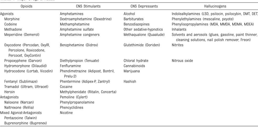
TABLE 1 Major Drugs of Abusea
Opioids CNS Stimulants CNS Depressants Hallucinogens
Agonists Amphetamines Alcohol Indolealkylamines (LSD, psilocin, psilocybin, DMT, DET) Morphine Dextroamphetamine (Dexedrine) Barbiturates Phenylethylamines (mescaline, peyote)
Codeine Methamphetamine Benzodiazepines Phenylisopropylamines (MDA, MMDA, MDMA, MDEA) Methadone Amphetamine sulfate Other sedative-hypnotics Inhalants
Meperidine (Demerol) Amphetamine congeners Methaqualone (Quaalude) Solvents and aerosols (glues, gasoline, paint thinner, cleaning solutions, nail polish remover, Freon) Oxycodone (Percodan, OxyIR,
Percolone, Roxicodone, Percocet, OxyContin)
Benzphetamine (Didrex) Glutethimide (Doriden) Nitrites
Propoxyphene (Darvon) Diethylpropion (Tenuate) Chloral hydrate Nitrous oxide Hydromorphone (Dilaudid) Fenfluramine Cannabinoids
Hydrocodone (Lortab, Vicodin) Phendimetrazine (Adipost, Bontril, Prelu-2)
Marijuana
Fentanyl (Sublimaze) Phentermine (Adipex-P, Zantryl) Hashish Tramadol (Ultram, Ultracet) Cocaine
Heroin Methylphenidate (Ritalin, Concerta) Antagonists Pemoline (Cylert)
Naloxone (Narcan) Phenylpropanolamine Naltrexone (ReVia) Phencyclidines Mixed Agonist-Antagonists Nicotine
Pentazocine (Talwin) Buprenorphine (Buprenex)
DET, diethyltryptamine; DMT, dimethyltryptamine; LSD, lysergic acid diethylamide; MDA, methylenedioxyamphetamine; MDEA, 3,4-methylenedioxyethamphetamine; MDMA, 3,4-methylene-dioxymethamphetamine (ecstasy); and MMDA, 3-methoxy-4,5-methylenedioxyamphetamine.
for as long as 7 days after delivery,18 observed abnormalities in exposed in-fants may reflect drug effect rather than withdrawal. In an unmasked study, 6%, 14%, and 35% of infants exposed to cocaine only, heroin only, or cocaine plus heroin, respectively, qualified for treatment on the basis of scoring.19 Several studies that used masked eval-uators found that cocaine-exposed in-fants had either no20,21or minimal22 withdrawal signs compared with cocaine-naïve infants (ie, those never exposed). Eyler et al16 conducted a prospective controlled study of 3 groups of infants: 1 group had no documented exposure to cocaine by history or by maternal and infant urine testing; a second group was cocaine exposed but had negative urine screening at birth; and a third group had cocaine metabolites de-tected in neonatal urine. Observers masked to infant status performed as-sessments using the Brazelton Neo-natal Behavioral Assessment Scale.23 Infants who were positive for cocaine metabolites did not differ significantly
from metabolite-negative infants with a history of exposure nor from cocaine-naïve infants. Thesefindings supported neither a withdrawal nor a drug tox-icity syndrome. Cocaine-exposed infants have been described as having a higher incidence of abnormal auditory brain-stem responses and EEGs, compared with nonexposed infants.24,25In another study, infants with heavy exposure to cocaine had similar Brazeltonfindings at 2 to 3 days of age as did infants with light or no exposure; however, by 17 days of age, heavily exposed infants were more excitable and demonstrated poorer state regulation.26No published studies have carefully evaluated phar-macologic treatment of infants with signs attributable to prenatal cocaine exposure.
Methamphetamine abuse has been reported among pregnant women,27 although overall rates are low com-pared with cocaine and appear to have decreased in the general popu-lation between 2006 and 2008.1 Meth-amphetamine is an extremely potent
sympathomimetic agent that induces euphoria and increases alertness and self-confidence, because it produces a massive efflux of dopamine in the CNS. Pregnant women who abuse metham-phetamine are at increased risk of pre-term birth, placental abruption, fetal distress, and intrauterine growth re-striction at rates similar to those for pregnant women who use cocaine. In 1 study, only 4% of infants exposed to methamphetamine were treated for drug withdrawal, but it was not pos-sible to exclude concomitant abuse of other drugs as contributory in all cases.27There are reports of long-term adverse neurotoxic effects of in utero methamphetamine exposure on behav-ior, cognitive skills, and physical dex-terity.28,29
SELECTIVE SEROTONIN REUPTAKE INHIBITORS
Selective serotonin reuptake inhibitors (SSRIs) are a class of antidepressant medications that became available for widespread clinical use in 1988. SSRIs
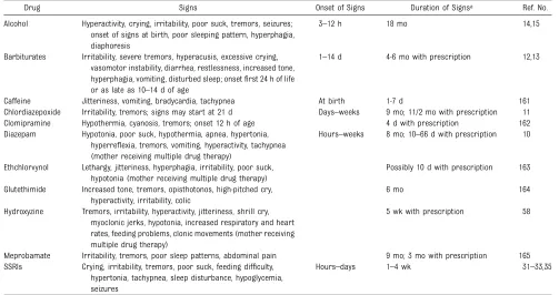
TABLE 2 Maternal Nonnarcotic Drugs That Cause Neonatal Psychomotor Behavior Consistent With Withdrawal
Drug Signs Onset of Signs Duration of Signsa Ref. No.
Alcohol Hyperactivity, crying, irritability, poor suck, tremors, seizures; onset of signs at birth, poor sleeping pattern, hyperphagia, diaphoresis
3–12 h 18 mo 14,15
Barbiturates Irritability, severe tremors, hyperacusis, excessive crying, vasomotor instability, diarrhea, restlessness, increased tone, hyperphagia, vomiting, disturbed sleep; onsetfirst 24 h of life or as late as 10–14 d of age
1–14 d 4-6 mo with prescription 12,13
Caffeine Jitteriness, vomiting, bradycardia, tachypnea At birth 1-7 d 161 Chlordiazepoxide Irritability, tremors; signs may start at 21 d Days–weeks 9 mo; 11/2 mo with prescription 11 Clomipramine Hypothermia, cyanosis, tremors; onset 12 h of age 4 d with prescription 162 Diazepam Hypotonia, poor suck, hypothermia, apnea, hypertonia,
hyperreflexia, tremors, vomiting, hyperactivity, tachypnea (mother receiving multiple drug therapy)
Hours–weeks 8 mo; 10–66 d with prescription 10
Ethchlorvynol Lethargy, jitteriness, hyperphagia, irritability, poor suck, hypotonia (mother receiving multiple drug therapy)
Possibly 10 d with prescription 163
Glutethimide Increased tone, tremors, opisthotonos, high-pitched cry, hyperactivity, irritability, colic
6 mo 164
Hydroxyzine Tremors, irritability, hyperactivity, jitteriness, shrill cry, myoclonic jerks, hypotonia, increased respiratory and heart rates, feeding problems, clonic movements (mother receiving multiple drug therapy)
5 wk with prescription 58
Meprobamate Irritability, tremors, poor sleep patterns, abdominal pain 9 mo; 3 mo with prescription 165 SSRIs Crying, irritability, tremors, poor suck, feeding difficulty,
hypertonia, tachypnea, sleep disturbance, hypoglycemia, seizures
Hours–days 1–4 wk 31–33,35
(eg, fluoxetine [Prozac], paroxetine [Paxil], sertraline [Zoloft], citalopram [Celexa], escitalopram [Lexapro], and fluvoxamine [Luvox]) are now the most frequently used drugs to treat depres-sion both in the general population and in pregnant women.30 Case reports,31 adverse drug reaction reports,32 and prospective studies33,34 linked third-trimester use of SSRIs in pregnant women to a constellation of neonatal signs that include continuous crying, ir-ritability, jitteriness, and/or restlessness; shivering; fever; tremors; hypertonia or rigidity; tachypnea or respiratory dis-tress; feeding difficulty; sleep distur-bance; hypoglycemia; and seizures.35 The onset of these signs ranged from several hours to several days after birth and usually resolved within 1 to 2 weeks. In 1 infant exposed to parox-etine, signs persisted through 4 weeks of age.36In severely affected infants, a short-term course of chlorpromazine provided measurable relief of symp-toms.36
Several authors have discussed whether these signs are better explained by se-rotonin syndrome (attributable to in-creased serotonin concentration in the intersynaptic cleft) or by SSRI with-drawal (attributable to a relative hypo-serotonergic state).30,32,35,37–40In adults, treatment with a single SSRI may cause mild to moderate serotonin syn-drome, but severe signs are more likely to occur when 2 or more drugs that increase serotonin concentration by different mechanisms are pre-scribed.35 In adults, serotonin syn-drome is characterized by the following triad of clinical signs: changes in men-tal status (agitation, confusion); auto-nomic hyperactivity (fever, tachycardia, tachypnea, diaphoresis, mydriasis); and neuromuscular abnormalities (tremor, clonus, hyperreflexia, hypertonia). On the other hand, serotonin withdrawal in adults manifests with subjective symp-toms that include anxiety, headache,
nausea, fatigue, low mood, and, rarely, extrapyramidal signs such as dysto-nia. Hence, in most cases, the clinical syndrome reported among neonates born to mothers on SSRI treatment is consistent with a gradual resolution of a hyperserotonergic condition rather than with the evolution of a hyposer-otonergic state. Still, in a few cases, drug withdrawal may be a better ex-planation.35
Biochemical studies that correlate serial serum SSRI (or active metabo-lite) concentrations and markers of CNS serotonin activity (eg, 5-hydrox-yindoleacetic acid [5-HIAA], a metabo-lite of serotonin) with changes in clinical signs could be helpful in dif-ferentiating toxicity from withdrawal. In adults, cerebrospinal fluid concen-trations of 5-HIAA (but not serum concentrations of serotonin) correlate inversely with increased CNS serotonin activity that results from SSRI treat-ment. One prospective study compared concentrations of SSRI and active metabolites at birth, 2 days of life, and 2 weeks of life; cord blood monoamine and metabolite; and serial serotoner-gic scores in infants born to mothers on treatment with SSRIs and those of SSRI-naïve control infants.39 The in-fants born to mothers on SSRIs had an average serotonergic score four-fold greater than SSRI-naïve infants. Cord blood 5-HIAA concentrations were inversely related to the initial serotonergic score, and the resolution of neonatal signs correlated with rapid declines in serially measured serum SSRI and metabolite concen-trations.39 These results do support drug toxicity rather than drug with-drawal as the cause of clinical signs. Recent authors have suggested the terms “serotonin discontinuation syn-drome”34 or “prenatal antidepressant exposure syndrome.”41
Although 1 study reported decreased pain reactivity at 2 months of age
benefits of breastfeeding as well as the potential risk that her infant may continue to be exposed to a measure-able level of the SSRI with unknown long-term effects.
OPIOIDS
Opioids are a class of natural, en-dogenous, and synthetic compounds that activate primarily µ-opioid (but alsoκ- andδ-opioid) receptors in the CNS to produce supraspinal analgesia. Other acute effects include sedation, euphoria, miosis, respiratory depres-sion, and decreased gastrointestinal motility. Prolonged use results in phys-ical and psychologphys-ical dependence. As a class, opioids demonstrate a nar-row therapeutic index. On the other hand, the interpatient range of dose necessary to achieve a similar thera-peutic effect is fairly wide because of genetic differences in pharmacoki-netics and pharmacodynamics.47 Mor-phine is 1 of many natural opioids that can be extracted from the opium poppy. Codeine, heroin (diacetylmor-phine), hydromorphone (Dilaudid), fen-tanyl (Sublimaze), and methadone are examples of synthetic opioids. Endog-enous opioids include enkephalins, endorphins, and endomorphins. The term opiate refers to a subclass of alkaloid opioids. Methadone exerts secondary effects by acting as an N-methyl-D-aspartate receptor
antago-nist, blocking the actions of glutamate, the primary excitatory neurotrans-mitter in the CNS. Opioids acutely in-hibit the release of noradrenaline at synaptic terminals. With chronic opi-oid exposure, tolerance develops as the rate of noradrenaline release over time increases toward normal. Abrupt discontinuation of exogenous opioids results in supranormal re-lease of noradrenaline and produces the autonomic and behavioral signs and symptoms characteristic of with-drawal.
Opioid abuse in pregnant women presents additional risks for the fetus and newborn. Opioids are small lipo-philic molecular weight compounds that cross placental and blood-brain barriers. Active or passive maternal detoxification is associated with in-creased risk of fetal distress and fe-tal loss. Maintenance programs with methadone (a full µ-opioid agonist and a Food and Drug Administration [FDA] schedule II controlled substance) for pregnant women can sustain opi-oid concentrations in the mother and fetus in ranges that minimize opioid craving, suppress abstinence symp-tomatology, block heroin-induced eu-phoria, and prevent fetal stress. Other benefits from this once controversial treatment are optimization of prenatal care and general maternal physical and mental health, as well as antici-pation of potential withdrawal signs in the newborn infant. Disadvantages of methadone include the extremely un-likely achievement of successful de-toxification after delivery and a more severe and prolonged course of NAS compared with heroin exposure. These issues have encouraged the develop-ment of other synthetic opioids as al-ternative treatments to methadone.
Subsequent to the Drug Addiction Treatment Act of 2000 that allowed office-based treatment of addiction by using FDA schedule III to V drugs, the synthetic opioid buprenorphine (a partial µ-opioid agonist) was ap-proved by the FDA in 2002 as a sched-ule III controlled substance for the treatment of opioid dependence. Nei-ther methadone nor buprenorphine is approved for use in pregnant women, and both are categorized by the FDA as class C pregnancy drugs. None-theless, buprenorphine, either alone (Subutex) or in combination with nal-oxone (Subnal-oxone), has been used both as a first-line treatment of heroin ad-diction and as a replacement drug for
methadone. Recent results from the Maternal Opioid Treatment: Human Experimental Research study suggest that buprenorphine has some advan-tages to methadone as a treatment of opioid addiction in pregnant women. Infants born to mothers treated with buprenorphine had shorter hospital stays (10 vs 17.5 days), had shorter treatment durations for NAS (4.1 vs 9.9 days), and required a lower cumu-lative dose of morphine (1.1 vs 10.4 mg) compared with infants born to moth-ers on methadone maintenance.48
CLINICAL PRESENTATION OF OPIOID WITHDRAWAL
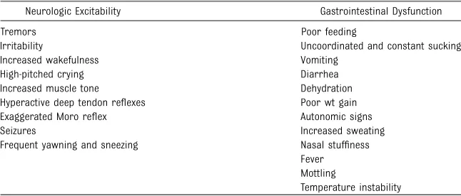
The clinical presentation of NAS varies with the opioid, the maternal drug history (including timing of the most recent use of drug before delivery), maternal metabolism, net transfer of drug across the placenta, placental metabolism (W. Snodgrass, MD, PhD, personal communication, 2008), infant metabolism and excretion, and other factors. In addition, maternal use of other drugs and substances such as cocaine, barbiturates, hypnotics-sedatives, and cigarettes may infl u-ence the severity and duration of NAS. Because opioid receptors are concentrated in the CNS and the gastrointestinal tract, the predom-inant signs and symptoms of pure opioid withdrawal reflect CNS irrita-bility, autonomic overreactivity, and gas-trointestinal tract dysfunction (Table 3). Excess environmental stimuli and hun-ger will exacerbate the perceived se-verity of NAS.
infants exposed to buprenorphine, 1 study found that onset of withdrawal peaked at 40 hours and that signs were most severe at 70 hours of age.51 The different time courses re-flect variations in the half-lives of drug elimination. However, if 1 week or longer has elapsed between the last maternal opioid use and delivery of the infant, the incidence of neona-tal withdrawal is relatively low.52 The incidence and severity of NAS are greater in infants exposed to metha-done compared with those exposed to buprenorphine48 or heroin. Still, se-vere withdrawal has been described in 0 to 50% of buprenorphine-exposed infants.53–55 In the acute phase, seiz-ures have occurred in 2% to 11% of infants withdrawing from opioids49,50,56; however, abnormal EEG results with-out overt seizure activity have been reported in >30% of neonates.57,58 Subacute signs of opioid withdrawal may last up to 6 months.59
Seizures also may be associated with withdrawal from a variety of non-narcotic drugs (eg, barbiturates,12,14 alcohol,14 and sedative-hypnotics60,61). The mechanism and significance of seizures associated with withdrawal are unclear. Withdrawal from ethanol begins early, in general, during the first 3 to 12 hours after delivery.12,15 Diagnosis of sedative withdrawal is more difficult, because classically it appears after the first few days of
life. Barbiturate withdrawal has a me-dian onset of 4 to 7 days, but a wide range from days 1 through 14.12,13 Other sedative-hypnotics have exhibi-ted even later onset, including as late as day 12 for diazepam10 and day 21 for chlordiazepoxide.11
Studies of the relationship between maternal methadone dose and the incidence and severity of NAS have provided contradictoryfindings. Some studies demonstrated that larger maternal methadone dosages in late pregnancy were associated with greater neonatal concentrations and increased risk of withdrawal,8,9,62–68 but others refuted a correlation.69–74This lack of consensus is explained in part by dif-ferent approaches to the management of antenatal methadone maintenance therapy. There were substantial var-iations in the mean and range of daily methadone dose in the populations studied. Studies that found no corre-lation tended to enroll infants born to mothers who had been prescribed higher doses of methadone (50–200 mg/day), whereas those that did note a relationship between maternal dose and NAS sequelae reported lower maternal doses (eg, <50 mg/day) or included women undergoing partial detoxification.67 Another potential ex-planatory factor is the significant in-terindividual variability in maternal methadone metabolism.75 As a re-sult, cumulative fetal exposure can
be expected to vary among infants born to mothers on equivalent metha-done regimens.
Methadone concentrations in cord blood and at 48 hours of age,72as well as the rate of decline in neonatal se-rum concentration,65 appear to corre-late with NAS signs. Kuschel et al72 found that infants who required rescue treatment had lower cord blood meth-adone concentrations and that, in all but 1 infant, methadone concentrations were undetectable in the serum at 48 hours. Doberczak65noted that faster declines in postnatal blood methadone concentrations were associated with more severe CNS withdrawal.
Preterm Infants
Preterm infants have been described as being at lower risk of drug with-drawal with less severe and/or pro-longed courses. Infants born at <35 weeks’ gestation whose mothers re-ceived methadone maintenance had significantly lower total and CNS ab-stinence scores than did term infants of mothers receiving similar metha-done dosages.64 In a more recent study, lower gestational age corre-lated with a lower risk of neonatal withdrawal.68The apparent decreased severity of signs in preterm infants may relate to developmental immatu-rity of the CNS, differences in total drug exposure, or lower fat depots of drug. Alternatively, the clinical evaluation of the severity of absti-nence may be more difficult in pre-term infants, because scoring tools to describe withdrawal were largely developed in term or late preterm infants.76,77 In a retrospective study, Dysart et al78compared the length of hospital stay, duration of medication, and cumulative medication exposure for preterm and term infants born to mothers enrolled in a methadone maintenance program. Infants were evaluated by using an abstinence scoring system77and treated uniformly
TABLE 3 Clinical Features of the Neonatal Narcotic Abstinence Syndrome
Neurologic Excitability Gastrointestinal Dysfunction
Tremors Irritability
Increased wakefulness High-pitched crying Increased muscle tone Hyperactive deep tendon reflexes Exaggerated Moro reflex Seizures
Frequent yawning and sneezing
Poor feeding
Uncoordinated and constant sucking Vomiting
Diarrhea Dehydration Poor wt gain Autonomic signs Increased sweating Nasal stuffiness Fever Mottling
with a neonatal opiate solution. All adverse outcomes were reduced in the preterm cohort.
Abuse of Multiple Drugs
The abuse of multiple drugs during pregnancy is not uncommon,79but its effect on the occurrence and severity of neonatal abstinence is controver-sial. In 1 study, abstinence scores of infants whose mothers abused co-caine and methadone were similar to the scores of infants whose mothers received high-dose maintenance meth-adone.64 In another study, the neuro-behavioral scores of infants exposed to intrauterine cocaine were similar to those of infants exposed to both cocaine and methadone.80Conversely, an unmasked study reported higher abstinence scores in infants exposed to both cocaine and heroin in compar-ison with those exposed to heroin or cocaine alone.19 Infants born to moth-ers maintained on methadone who were also heavy smokers (>20 ciga-rettes per day) demonstrated higher withdrawal scores that peaked later than infants born to light smokers.81
A 1989 case report linked the adminis-tration of naloxone for the treatment of apnea in a baby born to a mother with recent methadone ingestion to the onset of seizures. The seizures resolved after morphine treatment but did not respond to administration of phenobarbital or diazepam.82 For this reason, maternal use of opiates during pregnancy has remained a relative contraindication to the use of naloxone for the treatment of apnea or hypoventilation during the transition period after birth.
DIFFERENTIAL DIAGNOSIS
The presence of maternal character-istics known to be associated with drug abuse during pregnancy can be considered an indication to screen for intrauterine drug exposure. These characteristics include absent, late, or
inadequate prenatal care; a previously documented or admitted history of drug abuse; a previous unexplained late fetal demise; precipitous labor; abruptio placentae; hypertensive episodes; se-vere mood swings; cerebrovascular accidents; myocardial infarction; and repeated spontaneous abortions.80,83–88 The legal implications of testing and the need for consent from the mother may vary among the states.89Each hospital should consider adopting a policy for maternal and newborn screening to avoid discriminatory practices and to comply with local laws.
Withdrawal signs in the newborn may mimic other conditions, such as in-fection, hypoglycemia, hypocalcemia, hyperthyroidism, intracranial hemor-rhage, hypoxic-ischemic encephalopa-thy, and hyperviscosity.90 If none of these diagnoses is readily apparent, a detailed maternal drug history should be obtained that includes interviewing the mother about drug use and abuse by her partner, friends, and parents, in addition to queries about the mother’s prescription and nonprescription drug use.90,91Because maternal self-reporting underestimates drug exposure and maternal urine screening during pregnancy fails to identify many cases of drug use,83 appropriate neonatal drug screening should be performed. Conversely, no clinical signs should be attributed solely to drug withdrawal on the basis of a positive maternal history without a careful assessment to exclude other causes.
Screening is most commonly accom-plished by using neonatal urine speci-mens. A urine sample must be collected as soon as possible after birth, be-cause many drugs are rapidly me-tabolized and eliminated.90,92,93 Even so, a positive urine screening result may only reflect recent drug use. Al-cohol is detectable in neonatal urine for 6 to 16 hours after the last
ma-ternal ingestion. Amphetamines, ben-zodiazepines, cocaine metabolites, and opioids are usually cleared within 1 to 3 days after birth. Marijuana and co-caine metabolites may be detectable for weeks, depending on maternal usage.94
Drugs that are excreted in the hep-atobiliary system as well as drugs excreted by the fetal kidneys into the amniotic fluid are concentrated in meconium. Hence, meconium analysis is most useful when the history and clinical presentation strongly suggest neonatal withdrawal, but the maternal and neonatal urine screening results are negative. Drawbacks of testing for drugs in meconium are that it is not typically performed by hospitals and that results are often not available for days to weeks. Meconium must be collected before it is contaminated by transitional, human milk, or formula stools—otherwise, the assay may not be valid or the reference laboratory may reject the sample. Assay of me-conium, although not conclusive if the results are negative, is more likely to identify infants of drug-abusing mothers than is the testing of infant or maternal urine.95,96 Other speci-mens that have been tested in re-search laboratories are maternal and neonatal hair.97,98 Recently, testing of umbilical cord tissue by using drug class-specific immunoassays was shown to be in concordance with testing of paired meconium specimens at rates of 97%, 95%, 99%, and 91% for the detection of amphetamines, opiates, co-caine, and cannabinoids, respectively.99 The availability of this tissue from the moment of birth (in contrast to the inherent delay in collecting urine or meconium) may foster the adoption of this method of testing.
ASSESSMENT AND
NONPHARMACOLOGIC TREATMENT
neonatal withdrawal signs. Clinicians have used discrete or serial scores to assist with therapeutic decisions. The Lipsitz tool, also known as the Neonatal Drug Withdrawal Scoring System,76 was recommended in the 1998 Amer-ican Academy of Pediatrics statement “Neonatal Drug Withdrawal,”100 prob-ably because it is a relatively simple metric with good sensitivity for iden-tifying clinically important withdrawal. The modified Neonatal Abstinence Scoring System (Fig 1),101 is the pre-dominant tool used in the United States.102 This more comprehensive instrument assigns a cumulative score based on the interval observation of
21 items relating to signs of neonatal withdrawal.103 In 1 study,
administra-tion of this scoring system with
in-fants verified not to have been exposed to prenatal opiates by meconium
anal-ysis resulted in a stable median score
of 2 during each of thefirst 3 days of life, with 95th percentile scores of 5.5
and 7 on days 1 and 2, respectively.104
Infants at risk for NAS should be carefully monitored in the hospital for the development of signs consis-tent with withdrawal. The appropriate duration of hospital observation is variable and depends on a careful assessment of the maternal drug
history. An infant born to a mother on a low-dose prescription opiate with a short half-life (eg, hydrocodone; av-erage half-life, 4 hours) may be safely discharged if there are no signs of withdrawal by 3 days of age, whereas an infant born to a mother on an opiate with a prolonged half-life (eg, metha-done) should be observed for a mini-mum of 5 to 7 days. Initial treatment of infants who develop early signs of withdrawal is directed at minimiz-ing environmental stimuli (both light and sound) by placing the infant in a dark, quiet environment; avoiding auto-stimulation by careful swaddling; re-sponding early to an infant’s signals;
FIGURE 1
adopting appropriate infant position-ing and comfortposition-ing techniques (sway-ing, rocking); and providing frequent small volumes of hypercaloric formula or human milk to minimize hunger and allow for adequate growth. Caloric needs may be as high as 150 to 250 cal/kg per day because of increased energy expenditure and loss of calories from regurgitation, vomiting, and/or loose stools.105,106 The infant needs to be carefully observed to recognize fever, dehydration, or weight loss promptly. The goals of therapy are to ensure that the infant achieves ade-quate sleep and nutrition to establish a consistent pattern of weight gain and begins to integrate into a social environment. Maternal screening for comorbidities, such as HIV or hepatitis C virus infections and polydrug abuse, needs to be performed. Additional supportive care in the form of intra-venous fluids, replacement electro-lytes, and gavage feedings may be necessary to stabilize the infant’s con-dition in the acute phase and obviate the need for pharmacologic interven-tion. When possible, and if not other-wise contraindicated, mothers who adhere to a supervised drug treat-ment program should be encouraged to breastfeed so long as the infant con-tinues to gain weight. Breastfeeding or the feeding of human milk has been associated with less severe NAS that presents later and less frequently re-quires pharmacologic intervention.107,108 Methadone is present in very low con-centrations in human milk. Cumula-tive daily intake of methadone in fully breastfed infants has been estimated to range from 0.01 to 0.15 mg/day in thefirst 30 days of life109 and 0.15 to 0.30 mg/day between 30 and 180 days of age.110 Similarly, the amount of buprenorphine excreted in human milk is small. Although more informa-tion is needed to evaluate long-term neurodevelopmental outcome of in-fants exposed to small quantities of
buprenorphine, there is no clear rea-son to discourage breastfeeding in mothers who adhere to methadone or buprenorphine maintenance treat-ment.111
Each nursery should adopt a protocol for the evaluation and management of neonatal withdrawal, and staff should be trained in the correct use of an abstinence assessment tool. In a re-cent survey of accredited US neo-natology fellowship programs, only 55% had implemented a written NAS protocol, and only 69% used a pub-lished abstinence scoring system.102
RATIONALE AND COMPARATIVE EVIDENCE FOR PHARMACOLOGIC TREATMENT
Drug therapy is indicated to relieve moderate to severe signs of NAS and to prevent complications such as fever, weight loss, and seizures if an infant does not respond to a committed program of nonpharmacologic sup-port. Since the introduction of the abstinence scales in 1975, published reports have documented that the decision to initiate pharmacologic treatment has been based on single or serial withdrawal scores. However, no studies to date have compared the use of different withdrawal score thresh-olds for initiating pharmacologic in-tervention on short-term outcomes (eg, severity and duration of with-drawal signs, weight gain, duration of hospitalization, need for pharmaco-logic treatment, or cumulative drug exposure). Withdrawal from opioids or sedative-hypnotic drugs may be life-threatening, but ultimately, drug withdrawal is a self-limited process. Unnecessary pharmacologic treatment will prolong drug exposure and the duration of hospitalization to the pos-sible detriment of maternal-infant bond-ing. The only clearly defined benefit of pharmacologic treatment is the short-term amelioration of clinical signs.
Studies have not addressed whether long-term morbidity related to neona-tal drug withdrawal is decreased by pharmacologic management of affected infants, or whether continued postnatal drug exposure augments the risk of neurobehavioral and other morbidities. It is possible that pharmacologic ther-apy of the infant may introduce or re-inforce a maternal disposition to rely on drugs for the treatment of infant discomfort or annoying behavior.112
Clinicians have treated NAS with a va-riety of drug preparations, including opioids (tincture of opium, neonatal morphine solution, methadone, and paregoric), barbiturates (phenobar-bital), benzodiazepines (diazepam, lorazepam), clonidine, and phenothia-zines (chlorpromazine). Information pertinent to the use of these drug preparations in infants is well sum-marized in the previous American Academy of Pediatrics statement.100 Recent surveys have documented that, in accord with the recommendations of that statement, 94% of UK and 83% of US clinicians use an opioid (mor-phine or methadone) as the drug of first choice. The majority of practi-tioners use phenobarbital as a second drug if the opiate does not adequately control withdrawal signs.102,113 Daily doses of morphine ranged from 0.24 mg/kg per day to 1.3 mg/kg per day.113 Paregoric is no longer used, because it contains variable concentrations of other opioids, as well as toxic ingre-dients such as camphor, anise oil, al-cohol, and benzoic acid.100The use of diazepam has also fallen into disfavor because of a documented lack of effi -cacy compared with other agents and because of its adverse effects on infant suck and swallow reflexes.114–116
was compared with a control treatment that could include a nonpharmacologic intervention, a placebo treatment, or another opioid and/or sedative drug. The authors prospectively designated 4 primary outcomes (failure of treatment to control withdrawal signs; incidence of seizures; survival; and neurodevel-opmental outcome) for meta-analysis. Treatment failure was defined vari-ously as the inability of the treatment to maintain abstinence scores within a preset“safe” level and/or the need to add another drug therapy. Some studies did not report primary out-comes and instead quantified second-ary outcomes (eg, duration of treatment, duration of hospitalization, rate of weight gain, etc).
Seven studies of opioid treatment that enrolled a total of 585 infants were identified between 1983 and 2004. Methodologicflaws were common and included quasirandom patient alloca-tion; substantial and often unexplained differences in allocation of patients to treatment groups; imbalances in group characteristics after randomi-zation; failure to mask study treat-ments; and failure to mask outcome measurements. In the single study that assessed oral morphine treatment versus supportive therapy only, 3 con-secutive Finnegan scores ≥8 promp-ted institution of the intervention.119 No significant effect of morphine was found on the rate of treatment failure. Oral morphine significantly increased the duration of treatment and the length of hospital stay, but it did re-duce the number of days required to regain birth weight and duration of supportive care. Four studies com-pared treatment failures of opioids (paregoric, oral morphine, or metha-done) with phenobarbitone.8,119–121 Neither the meta-analysis nor any in-dividual study identified a significant difference in treatment failure. One study reported a lower incidence of
seizures in the opioid (paregoric) treatment group.122 No consistent trends in secondary outcomes were observed, although 1 study reported a shorter duration of therapy in the phenobarbitone compared with the paregoric treatment group,123 and another made the opposite observa-tion when the opioid used was oral morphine.121Three studies individually and in combination reported signifi -cantly lower rates of treatment failure in infants assigned to opioid (pare-goric or methadone) compared with diazepam therapy8,114,120 but did not define differences in secondary out-comes. No studies reported mortality or neurodevelopmental outcomes.
A second Cochrane review analyzed 6 trials involving 305 infants published between 1969 and 2002 in which sed-ative treatment of NAS was compared with a nonopioid therapy. Methodologic concerns were similar to the opioid treatment trials. In the sole study of phenobarbitone versus supportive care, no difference in treatment failure was found, but treatment significantly in-creased the duration of therapy and hospital stay.119 A small study that allocated infants already treated with diluted tincture of opium (DTO) to phe-nobarbitone as a second drug versus no additional treatment identified no infants in either group with treatment failure but observed significant reduc-tions in the duration of hospitalization (38 vs 79 days) and the maximal daily dose of opioid in the phenobarbitone-treated infants.124 Infants were dis-charged from the hospital once they were no longer taking opioids. How-ever, the mean duration of phenobar-bitone treatment was 3.5 months. Of 3 studies that compared phenobarbi-tone and diazepam treatment, 1 found a significantly lower rate of treat-ment failure in the phenobarbitone group.8,114,120 One study of phenobar-bitone versus chlorpromazine125found
no differences in primary or second-ary outcomes.
Since 2004, a number of small studies of varying methodologic quality have compared pharmacologic treatments. In a prospective randomized double-masked study, Langenfeld et al126 could not identify differences in du-ration of treatment, dudu-ration of hos-pitalization, or in weight gain (g/day) in infants treated with either DTO or oral morphine drops. A retrospective study found no difference in length of hospitalization in infants with NAS who were treated with methadone or oral morphine solution, but did cor-relate higher maternal methadone doses with longer lengths of stay.127 Ebner et al128examined the incidence of NAS in infants born to mothers maintained with methadone, morphine, or buprenorphine and compared phe-nobarbital and oral morphine treat-ments in affected infants. Sixty-eight percent of infants born to mothers maintained on methadone required pharmacologic treatment at a mean age of 58 hours, compared with 82% of infants at a mean age of 33 hours in the morphine group and 21% of infants at a mean age of 34 hours in the buprenorphine group. The dura-tion of treatment was significantly shorter for infants who received mor-phine compared with infants who were treated with phenobarbital. A random-ized comparison trial of sublingual buprenorphine versus neonatal opium solution for the treatment of NAS showed a nonsignificant reduction in length of treatment and duration of hospitalization in the buprenorphine group.129 Buprenorphine therapy was well tolerated.
Clonidine is anα2-adrenergic receptor
reduces CNS sympathetic outflow and palliates symptoms of autonomic over-activity such as tachycardia, hyper-tension, diaphoresis, restlessness, and diarrhea. Cessation of clonidine treat-ment can result in a rebound of auto-nomic activity. Reported experience with clonidine as a primary or ad-junctive treatment of NAS is limited but promising. In a small case series, 6 of 7 infants with NAS showed signif-icant resolution of signs when treated with oral clonidine.132In a randomized double-masked controlled trial, Agthe et al133 compared the efficacy and safety of treating NAS with DTO plus oral clonidine (1µg/kg every 3 hours) versus DTO plus placebo in 80 infants with prenatal exposure to methadone and/or heroin. The combination ther-apy significantly reduced the median length of treatment of all infants and for infants exposed to methadone, but more infants in the DTO/clonidine group required resumption of DTO after initial discontinuation. The mean total dose of morphine over the treat-ment course was ∼60% lower in the combination therapy group. No clini-cally significant differences in feeding, weight gain or loss, heart rate, or blood pressure were observed. In another case series, oral clonidine was ad-ministered either as a primary or adjunctive therapy for the prevention or treatment of narcotic withdrawal in infants on intravenous fentanyl or infants with antenatal exposure to opiates.134In all cases, treatment was successful and clonidine was discon-tinued without sequelae after a mean duration of 7 days. In a retrospective case series, infants who had evidence of NAS attributable to antenatal meth-adone exposure had lower severity scores and required fewer days of drug therapy and hospitalization if they had been treated with a combination of clonidine and chloral hydrate rather than a combination of morphine and phenobarbital.135
A recently published case series from France that used a historical cohort for a comparison has suggested that the treatment of NAS with the pheno-thiazine, chlorpromazine, as a single drug may be more effective than treat-ment with morphine.136Infants treated with oral morphine had significantly longer median durations of treatment and hospitalization in comparison with infants treated with chlorpromazine. No adverse affects were reported.
OUTCOME
Assessment of potential long-term morbidity specifically attributable to neonatal drug withdrawal and its treatment is difficult to evaluate. Few studies have followed drug-exposed children beyond the first few years of life. Confounding variables, such as environment and dysfunctional care-givers, complicates the interpretation of outcomes. In a small study, devel-opmental scores on the mental index on the Bayley Scales of Infant Devel-opment were not affected by the se-verity of withdrawal or the treatment chosen.114 Mean scores on the Bayley Scales of Infant Development were similar for all infants treated for withdrawal, including those receiving phenobarbital, paregoric, or a combina-tion therapy. Scores of infants whose withdrawal was too mild to qualify for pharmacologic intervention were also similar.
Fourteen drug-exposed infants with withdrawal-associated seizures were reported by Doberczak et al.25 The abstinence scores for 5 of these infants were <7 (the cutoff for treat-ment); hence, they received no phar-macologic therapy before the onset of seizures. Thirteen of the 14 infants were offspring of mothers enrolled in a methadone treatment program; how-ever, the success of maternal treat-ment was not described. Of the 14 infants with seizures, 12 were available
for evaluation at 1 year of age; results of neurologic examinations were nor-mal in 9 of the 12 infants evaluated. EEG results were abnormal in 9 neo-nates; however, subsequent EEGs for 7 of 8 of these infants normalized during follow-up. Mean scores on the Bayley Scales of Infant Development were also normal by 1 year of age, similar to matched controls that were drug exposed, but in whom withdrawal-associated seizures did not develop.24 Withdrawal-associated seizures seem to be primarily myoclonic, to respond to opiates, and to carry no increased risk of poor outcome. Withdrawal-associated seizures in neonates are different from those associated with other causes. Based on the depression of norepinephrine and dopamine ob-served with methadone exposure in an-imal models, withdrawal seizures are speculated to be attributable to low-ered levels of neurotransmitters.137,138 The normalization of the EEG and nor-mal neurologic development are be-lieved to reflect recovery of normal neurotransmitter concentrations dur-ing early infancy. Bandstra et al139 have comprehensively reviewed out-comes of infants and toddlers who were exposed prenatally to opioids and cocaine.
MANAGEMENT OF ACQUIRED OPIOID AND BENZODIAZEPINE DEPENDENCY
and symptoms of withdrawal on acute dosage reduction or cessation of therapy. Infants who undergo complex surgery, who require prolonged med-ical intensive care for conditions such as respiratory failure or persistent pulmonary hypertension, or who are supported with extracorporeal mem-brane oxygenation (ECMO) therapy are among those at greatest risk of ac-quired drug dependency.
Extended treatment with opioids via continuous intravenous infusion re-sults in drug tolerance. Even short-term opioid exposure alters the number and affinity of receptors in key neuronal centers so that an escala-tion of the opioid infusion rate (which produces an increase in opioid plasma concentrations) becomes necessary to achieve the same physiologic effect.140 By itself, the development of tolerance does not predict physical dependency or withdrawal.141 Cumulative expo-sure to fentanyl, quantified by the to-tal dose in milligrams per kilogram or the number of consecutive days of treatment, correlated with the likeli-hood of withdrawal.140,142,143By using a multiple logistic regression analysis, Arnold et al140found that the duration of ECMO therapy was an even more powerful predictor of withdrawal than was cumulative fentanyl exposure. Katz et al142 reported that among 23 mechanically ventilated children aged 1 week to 22 months (mean, 6 months) who were treated for>24 hours with a continuous fentanyl infusion, 13 of 23 children (57%) developed withdrawal as defined by a Finnegan score≥8. In this prospective study, a cumulative fen-tanyl exposure in excess of 2.5 mg/kg or 9 days of therapy was 100% pre-dictive of withdrawal. More recently, in a prospective study of 19 neonates treated with fentanyl for a minimum of 24 hours, Dominquez et al143 docu-mented that a cumulative fentanyl dose
≥415µg/kg predicted withdrawal with
70% sensitivity and 78% specificity and that an infusion duration≥8 days was 90% sensitive and 67% specific for withdrawal. In adults, concomitant treatment with neuromuscular para-lytic agents or propofol for>24 hours also increased the likelihood of with-drawal.144 Signs and symptoms of with-drawal from fentanyl commence within 24 hours of cessation of therapy.
The refinement of pain management in children over the past 2 decades has witnessed an expansion of the use of opioids in the intensive care setting. As a result, more children have been treated for actual or potential with-drawal symptoms as a comorbidity of hospitalization. Fentanyl, a pureµ-opioid receptor antagonist, has become the opioid of choice because of its rapid onset of action, short duration of ef-fect (half-life of 0.5–1 hour), excellent potency, and minimal acute adverse effects. However, fentanyl has not been demonstrated to be safer or more ef-fective than morphine for the provision of long-term analgesia. Indeed, 1 study has reported that patients who were treated prospectively with a continu-ous morphine infusion during ECMO experienced a significantly lower need for supplemental analgesia, a lower rate of dependency, and a shorter hos-pital stay compared with a previous group of patients treated with fentanyl during ECMO.145
Practitioners have employed a variety of strategies to treat or, in high-risk patients, to prevent signs and symp-toms of opioid withdrawal in infants and children. Carr and Todres146 reported success with a gradual taper of the opioid infusion rate. Children who had received continuous opioid infusions for more than a week re-quired 2 to 3 weeks for complete weaning. One disadvantage of this approach was that intravenous access had to be maintained for the entire course of treatment. Tobias et al147
were among the first investigators to describe treatment of opioid with-drawal by conversion to enteral methadone. Methadone was chosen as the opioid of choice because of its excellent oral bioavailability (70%– 100%) and long half-life (19–41 hours), which allowed for long inter-vals between doses.148 In this initial report, 3 symptomatic patients who had been exposed to continuous or bolus opioids for up to 7 weeks were transitioned to a methadone regimen of 0.1 mg/kg, orally, every 12 hours. Dose reduction by 10% to 20% of the initial dose per week resulted in suc-cessful weaning in 4 to 6 weeks.
with 3 of the nonprotocol group, so that the decreased taper time on protocol was unlikely to have been confounded by other drug therapy. Weaning and discontinuation from benzodiazepines were successful dur-ing the methadone taper in all pro-tocol patients.
Meyer et al150described a protocol for rescue therapy in 29 patients 1 day to 20 years of age on admission who developed withdrawal during the course of nonstandardized tapers of prolonged continuous fentanyl in-fusion. Withdrawal was defined as the observation of 3 consecutive Finnegan scores ≥8 obtained at 2-hour inter-vals. The daily fentanyl dose for the period 24 to 48 hours before with-drawal symptoms was used to calcu-late an equipotent dose of morphine sulfate. Morphine was administered as a bolus dose every 4 hours and titrated to effect (Finnegan score consistently<8) over 12 to 24 hours. An equipotent amount of methadone was then determined by using the effective morphine dose. Three load-ing doses of methadone at 12-hour intervals were administered. After-ward, doses were given every 24 hours and weaned by 10% per day. Ten patients were receiving concomi-tant treatment with a benzodiazepine or chloral hydrate, but these medi-cations were not weaned during the methadone taper. Twenty-five of 29 patients successfully completed this taper over 10 days. Three patients required 21 days, and 1 patient died of sepsis. Sixteen of the patients were discharged from the hospital and completed methadone tapers on an outpatient basis. Nine of the patients had been started on clonidine during the phase of nonstandardized opioid weaning in unsuccessful attempts to prevent withdrawal. A subsequent ran-domized double-blind follow-up study by the same group of investigators151
found that in a group of 37 fentanyl-treated patients, a 5-day methadone taper was as successful as the longer 10-day course (13 of 16 vs 17 of 21 [not significant]) in discontinuing opi-oid infusions without causing with-drawal. In contrast to their previous study, a standardized taper of lor-azepam was allowed in 17 of the 37 patients while on the methadone protocol. Only 1 of these 17 patients who underwent dual tapers required rescue treatment with an increased dose of opioids.
Several factors potentially complicate the adoption of the protocols reported by Robertson, Meyer, and Berens (see Table 4) into routine neonatal clinical practices. Most obvious is that these studies were conducted in a PICU setting; few neonates were included, and their outcomes were not sepa-rately analyzed. Other investigators have emphasized that the Finnegan instrument common to all 3 studies has been validated only in term in-fants undergoing withdrawal secondary to in utero opioid exposure.152,153 Therefore, the use of this tool may have underestimated withdrawal symp-tomatology in an older pediatric pop-ulation. A third concern is that opioids and benzodiazepines are often used concurrently in the same patient, yet symptoms of opioid and benzodiaze-pine withdrawal overlap to a great ex-tent. Hence, current instruments will not reliably differentiate whether with-drawal symptoms stem from relative opioid or benzodiazepine abstinence.153 Other scales have been proposed for children and are in various stages of evaluation, including the Opioid and Benzodiazepine Withdrawal Scale,151 the Sedation Withdrawal Score,154and the Sophia Benzodiazepine and Opioid Withdrawal Checklist.155
At this time, no optimal pharmacologic regimen for the prevention or treat-ment of acquired opioid and/or
benzodiazepine dependency can be recommended, because the necessary comparative studies of safety and ef-ficacy are not available.156Hence, it is even more incumbent on the practi-tioner to prescribe pharmacologic interventions with the goal of achiev-ing the desired therapeutic effect by using the fewest drugs at the lowest doses and for the shortest durations possible.
Nonetheless, because many critically ill infants and children do receive treat-ment with prolonged courses of opioids and benzodiazepines, the following practices are reasonable based on the available evidence:
1. Each clinical unit can establish a threshold level of cumulative exposure to opioids and benzodia-zepines above which drug depen-dency can be expected to occur with a likelihood that justifies an-ticipatory initiation of a weaning protocol. For example, setting a threshold at a cumulative fentanyl exposure of>2 mg/kg or>7 days’ duration would predict a likeli-hood of dependency >50% but
<100%.141,142
2. Infants with a cumulative exposure to opioids or benzodiazepines be-low the thresholds for initiation of weaning protocols can undergo a rapid taper of these medications over a 24- to 48-hour period. Many such children will not subsequently exhibit drug dependency.
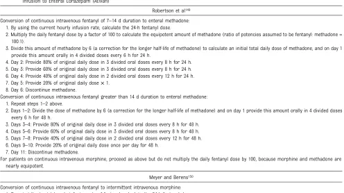
TABLE 4 Weaning Protocols by Using Conversion of Continuous Opioid Infusions to Enteral Methadone and for Conversion of Midazolam (Versed) Infusion to Enteral Lorazepam (Ativan)
Robertson et al149
Conversion of continuous intravenous fentanyl of 7–14 d duration to enteral methadone: 1. By using the current hourly infusion rate, calculate the 24-h fentanyl dose.
2. Multiply the daily fentanyl dose by a factor of 100 to calculate the equipotent amount of methadone (ratio of potencies assumed to be fentanyl: methadone = 100:1).
3. Divide this amount of methadone by 6 (a correction for the longer half-life of methadone) to calculate an initial total daily dose of methadone, and on day 1 provide this amount orally in 4 divided doses every 6 h for 24 h.
4. Day 2: Provide 80% of original daily dose in 3 divided oral doses every 8 h for 24 h. 5. Day 3: Provide 60% of original daily dose in 3 divided oral doses every 8 h for 24 h. 6. Day 4: Provide 40% of original daily dose in 2 divided oral doses every 12 h for 24 h. 7. Day 5: Provide 20% of original daily dose×1.
8. Day 6: Discontinue methadone.
Conversion of continuous intravenous fentanyl greater than 14 d duration to enteral methadone: 1. Repeat steps 1–2 above.
2. Days 1–2: Divide the dose of methadone by 6 (a correction for the longer half-life of methadone) and on day 1 provide this amount orally in 4 divided doses every 6 h for 48 h.
3. Days 3–4: Provide 80% of original daily dose in 3 divided oral doses every 8 h for 48 h. 4. Days 5–6: Provide 60% of original daily dose in 3 divided oral doses every 8 h for 48 h. 5. Days 7–8: Provide 40% of original daily dose in 2 divided oral doses every 12 h for 48 h. 6. Days 9–10: Provide 20% of original daily dose once per day for 48 h.
7. Day 11: Discontinue methadone.
For patients on continuous intravenous morphine, proceed as above but do not multiply the daily fentanyl dose by 100, because morphine and methadone are nearly equipotent.
Meyer and Berens150
Conversion of continuous intravenous fentanyl to intermittent intravenous morphine: 1. By using the target hourly infusion rate of fentanyl, calculate the 24-h fentanyl dose.
2. Multiply the daily fentanyl dose by a factor of 60 to calculate the equipotent dose of morphine (ratio of potencies assumed to be fentanyl: morphine = 60:1). 3. Divide the dose of morphine by 4 (correcting for the longer half-life of morphine) and on day 1 administer this amount intravenously in 6 divided doses every
4 h.
4. Titrate the morphine dose for adequate effect over 12 to 24 h. Conversion of intermittent intravenous morphine to enteral methadone:
1. Multiply the dose of morphine given every 4 h by 2 (ratio of potencies assumed to be morphine: methadone = 2:1) to determine an equipotent amount of methadone.
2. Provide this amount of methadone as an oral dose every 12 h for 3 doses.
3. Double this amount of methadone and provide as a single oral dose per day at bedtime.
4. Provide 90% of the initial dose on day 2, 80% on day 3, etc, so that the last dose of methadone (10% of the original dose) is given on day 10.
Protocols at Wolfson Children’s Hospital, Jacksonville, Florida
Conversion of continuous intravenous fentanyl>7 d duration to enteral methadone: 1. By using the current hourly infusion rate, calculate the 24-h fentanyl dose.
2. Multiply the daily fentanyl dose by a factor of 100 to calculate the equipotent amount of methadone (ratio of potencies assumed to be fentanyl: methadone = 100:1).
3. Divide this amount of methadone by 8–12 (a correction for the longer half-life of methadone) to calculate an initial total daily dose of methadone (not to exceed 40 mg/day).
4. Days 1–2: Provide the total daily dose of methadone orally in 4 divided doses every 6 h for 48 h. At the time of the second methadone dose, reduce the fentanyl infusion rate to 50%; at the time of the third dose, reduce the fentanyl infusion rate to 25%; and after the fourth methadone dose, discontinue the fentanyl infusion.
5. Days 3–4: Provide 80% of original daily dose in 3 divided oral doses every 8 h for 48 h. 6. Days 5–6: Provide 60% of original daily dose in 3 divided oral doses every 8 h for 48 h. 7. Days 7–8: Provide 40% of original daily dose in 2 divided oral doses every 12 h for 48 h. 8. Days 9–10: Provide 20% of original daily dose once per day for 48 h.
9. Day 11: Discontinue methadone.
Conversion of continuous intravenous midazolam>7 d duration to enteral lorazepam: 1. By using the current hourly infusion rate, calculate the 24-h midazolam dose.
2. Because lorazepam is twice as potent as midazolam and has a sixfold longer half-life, divide the 24 h midazolam dose by 12 to determine the daily lorazepam dose.
3. Divide the calculated lorazepam dose by 4 and initiate every 6 h oral treatments with the intravenous product or an aliquot of a crushed tablet. 4. Wean lorazepam by 10% to 20% per day. The dosage interval can also be increased gradually to every 8 h, then every 12 h, then every 24 h, and then every
calculated methadone equivalent to achieve a successful conversion. Also, the rate of weaning should be adjusted on the basis of careful con-tinuing clinical assessment. Eighty percent of children can be success-fully weaned from methadone com-pletely within 5 to 10 days.
4. Signs and symptoms of withdrawal from benzodiazepine therapy can be delayed. Intravenous benzodia-zepines can be converted to oral lorazepam (Table 4). The required time for weaning can be expected to be proportional to the dura-tion of intravenous benzodiazepine treatment.
5. Infants and children at risk for withdrawal are prudently observed in the hospital for signs and symp-toms. Each clinical unit can choose 1 assessment tool and train staff to minimize individual variability in scoring.
6. Discharge from the hospital for infants and very young children is prudently delayed until they are free of withdrawal signs and symp-toms for a period of 24 to 48 hours after complete cessation of opioids. Earlier discharge of an older child can be individualized in consider-ation of the child’s overall clinical status, the home environment, and the availability of adequate and prompt follow-up.
7. No clinical studies to date support the premise that initiation of cloni-dine, chloral hydrate, or continuous
intravenous low-dose naloxone157,158 during the course of continuous opioid infusions will reduce the likelihood or severity of opioid dependency.
CLINICAL HIGHLIGHTS
1) Each nursery that cares for infants with neonatal withdrawal should develop a protocol that defines indications and proce-dures for screening for maternal substance abuse. In addition, each nursery should develop and ad-here to a standardized plan for the evaluation and comprehensive treatment of infants at risk for or showing signs of withdrawal.
2) Screening for maternal substance abuse is best accomplished by us-ing multiple methods, includus-ing maternal history, maternal urine testing, and testing of newborn urine and/or meconium speci-mens that are in compliance with local laws. The screening of bio-logical samples is an adjunct to provide additional information helpful in the ongoing medical care of the infant. The duration of urinary excretion of most drugs is relatively short, and maternal or neonatal urinary screening only addresses drug exposure in the hours immediately before urine collection. Thus, false-negative urine results may occur in the pres-ence of significant intrauterine
drug exposure. Although new-born meconium screening also may yield false-negative results, the likelihood is lower than with urinary screening. The more recent availability of testing of umbilical cord samples may be considered a viable screening tool, because it appears to reflect in utero expo-sures comparable to meconium screening.
3) Drug withdrawal should be consid-ered in the differential diagnosis for infants in whom compatible signs develop. Physicians should be aware of other potential diag-noses that need to be evaluated and, if confirmed, treated appro-priately.
4) Nonpharmacologic supportive measures that include minimiz-ing environmental stimuli, pro-moting adequate rest and sleep, and providing sufficient caloric intake to establish weight gain should constitute the initial ap-proach to therapy.
5) Signs of drug withdrawal can be scored by using a published ab-stinence assessment tool. Infants with confirmed drug exposure who are unaffected or demonstrating minimal signs of withdrawal do not require pharmacologic ther-apy. Caution should be exercised before instituting pharmacologic therapy that could lengthen the duration of hospitalization and inter-fere with maternal-infant bonding.
TABLE 4 Continued
Robertson et al149
Summary of Conversion Of Intravenous Opioids to Enteral Methadone
1. Tobias et al147: Converted 2 patients on morphine (0.1–0.15 mg/kg q3h) and 1 patient on fentanyl (1–2µg/kg every 1–2 h) to methadone at a starting dose of
0.2 mg/kg per day.
2. Robertson et al149: 1µg/kg per h fentanyl = 0.4 mg/kg per day methadone.
3. Meyer and Berens150: 1µg/kg per h fentanyl = 0.24 mg/kg per day methadone.
Together with individualized clini-cal assessment, the serial and accurate use of a withdrawal as-sessment tool may facilitate a de-cision about the institution of pharmacologic therapy and there-after can provide a quantitative measurement that can be used to adjust drug dosing.
6) The optimal threshold score for the institution of pharmacologic therapy by using any of the pub-lished abstinence assessment in-struments is unknown.
7) Breastfeeding and the provision of expressed human milk should be encouraged if not contraindi-cated for other reasons.111,159
8) Pharmacologic therapy for withdrawal-associated seizures is indicated. Other causes of neo-natal seizures must also be eval-uated.
9) Vomiting, diarrhea, or both asso-ciated with dehydration and poor weight gain in the absence of other diagnoses are relative indi-cations for treatment, even in the absence of high total withdrawal scores.
10) The limited available evidence from controlled trials of neonatal opioid withdrawal supports the use of oral morphine solution and methadone when pharmaco-logic treatment is indicated. Growing evidence suggests that oral clonidine is also effective ei-ther as a primary or adjunctive therapy, but further prospective trials are warranted. Dosing regi-mens are listed in Table 5. With respect to other drug treatments
and clinical situations, a number of important caveats apply. Treat-ment with paregoric is contrain-dicated, because this preparation contains multiple opiates in addi-tion to morphine, as well as other potentially harmful compounds (alcohol, anise). Morphine pre-scriptions should be written as milligrams of morphine per kilo-gram and not as milliliters of DTO per kilogram. Tincture of opium contains a 25-fold higher concen-tration of morphine than do avail-able oral morphine solutions; hence, it increases the likelihood of drug error and morphine over-dose. The relative efficacy and safety of buprenorphine for the treatment of NAS require addi-tional comparative study. The op-timal pharmacologic treatment of infants who are withdrawing from sedatives or hypnotics is un-known. Finally, there is also insuf-ficient evidence to state whether an infant born to a mother with multiple drug abuse who meets criteria for pharmacologic ther-apy of withdrawal signs is best treated with an opioid, a barbitu-rate, a medication from another drug class, or a combination of drugs from different classes.
11) Physicians need to be aware that the severity of withdrawal signs, including seizures, has not been proven to be associated with dif-ferences in long-term outcome af-ter intrauaf-terine drug exposure. Furthermore, treatment of drug withdrawal may not alter the long-term outcome.
12) Given the natural history of with-drawal, it is reasonable for neo-nates with known antenatal exposure to opioids and benzo-diazepines to be observed in the hospital for 4 to 7 days. After discharge, outpatient follow-up should occur early and include reinforcement of the education of the caregiver about the risk of late withdrawal signs.
13) Neonates cared for in ICUs who have developed tolerance to opioids and benzodiazepines as a result of an extended duration of treat-ment can be converted to an equivalent regimen of oral meth-adone and lorazepam. Doses may be increased as necessary to achieve patient comfort. These med-ications can then be reduced by 10% to 20% of the initial dose every 1 to 2 days on the basis of clinical response and serial assessments by using a standardized neonatal abstinence instrument.
14) Significant gaps in knowledge concerning the optimal treatment strategy (including the criteria for instituting pharmacologic ther-apy, the drug offirst choice, and the strategy for weaning) of infants with neonatal withdrawal should be addressed in well-designed randomized controlled studies that are adequately powered to assess short-term outcomes and to provide for long-term follow-up.
LEAD AUTHORS Mark L. Hudak, MD Rosemarie C. Tan, MD, PhD
TABLE 5 Drugs Used in the Treatment of Neonatal Narcotic Withdrawal
Drug Initial Dose Increment Maximum Dose Ref. No.
Oral morphine 0.04 mg/kg every 3–4 h 0.04 mg/kg per dose 0.2 mg/kg per dose 119,121,126,133 Oral methadone 0.05–0.1 mg/kg every 6 h 0.05 mg/kg per dose To effect 127