• No results found

Intravital microscopy for evaluating tumor perfusion of nanoparticles exposed to non-invasive radiofrequency electric fields

N/A
N/A
Protected

Academic year: 2020

Share "Intravital microscopy for evaluating tumor perfusion of nanoparticles exposed to non-invasive radiofrequency electric fields"

Copied!
19
0
0

Loading.... (view fulltext now)

Full text

Figure

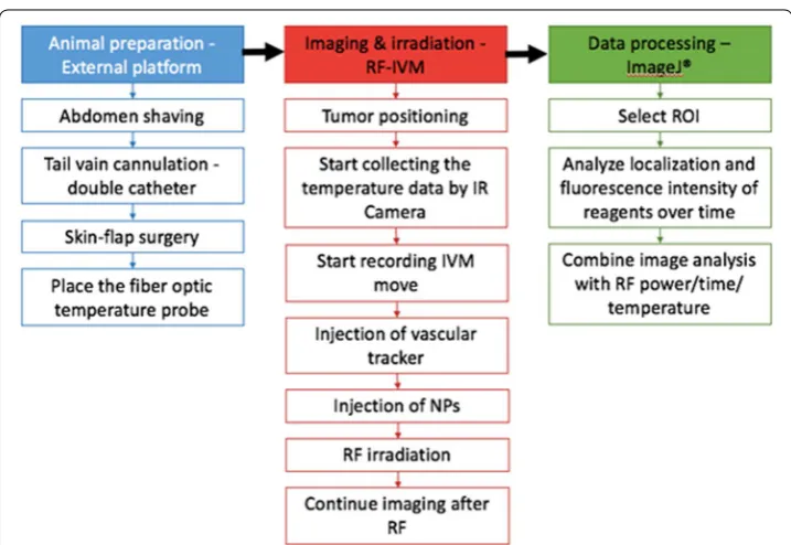
Fig. 1 Schematic summarizing all relevant procedures including animal preparation and IVM imaging during RF field exposure, followed by data processing and analysis
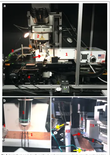
Fig. 2 (Panel a RF setup including RF amplifier (1), confocal microscope objective lens (2), anesthesia tubing 3) coming from behind the microscope stage and XYZ servo‑controller (4) for the mouse stage (indicated by the red arrow) above the microscope sta
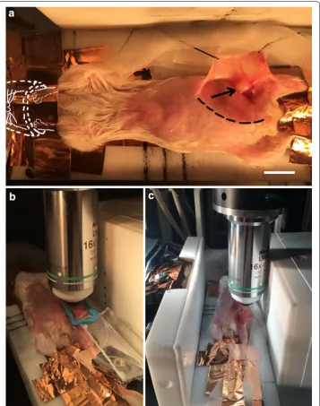
Fig. 3 Integrated Balb/c mouse IVM‑RF setup. field. The region of the incision (but not the tumor) has been blurred out for instructional purposes, so that extremities, including ears, paws, feet, and tail) and secures the nose to the anesthesia tube
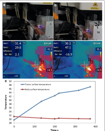
Fig. 4 Temperature measurement during the course of IVM imaging under RF field exposure
+6

References

Related documents