ORIGINAL ARTICLE
IJPHY
ABSTRACT
Background: This study might have been directed to some degree because of clashing results in the past studies re-garding the impacts for different SS protocols on muscle strength and possibility for injury. The objective of the study was to investigate the acute effects of different static stretching (SS) durations (20, 30, and 60s) on isokinetic concentric quadriceps (Q) and hamstrings (H) peak torque (PT), eccentric H PT and conventional and functional H:Q ratios un-der different stretching conditions and angular velocities (60°and180°/s) in active women.
Methods: Isokinetic tests were performed on 108 active women. A HUMAC system was used to measure unilateral concentric Q and H PT, and eccentric H PT at 60 and 180º/s at baseline and after a bout of H-only, Q-only, and com-bined H and Q muscles SS. The data were statistically treated using five separate three-way (time x conditions x veloc-ity) ANOVA.
Results: There were no significant differences among groups at baseline (P > 0.05). Significant reductions of all outcome measures have been shown to occur after 30 and 60s of SS (P < 0.05). The highest reductions of concentric Q and H PT, eccentric H PT and H:Q ratios were observed after 60s of SS. With no significant effects with the 20s SS (P > 0.05).
Conclusion: Short-lasting stretching can be done before exercises that require strength. However, since 30s or 60s stretching protocols adversely affect the muscle strength, performance and lower H:Q ratios they are not recommended prior to activities demanding the production of high forces.
Keywords: Static stretch, Isokinetic tests, Muscle strength, Hamstrings-Quadriceps ratios
Received 24th August 2016, revised 23rd September 2016, accepted 02nd October 2016
www.ijphy.org
10.15621/ijphy/2016/v3i5/117448
CORRESPONDING AUTHOR
Int J Physiother. Vol 3(5), 609-618, October (2016) ISSN: 2348 - 8336
ACUTE EFFECTS OF DIFFERENT STATIC STRETCHING
PROTOCOLS ON PEAK TORQUE, CONVENTIONAL AND
FUNCTIONAL HAMSTRINGS-TO-QUADRICEPS RATIOS
IN ACTIVE WOMEN
¹Ghada M. ALQaslah PT, M.Sc *2,3Afaf AM Shaheen PT, Ph.D
*2Afaf AM Shaheen PT, Ph.D
Assistant professor,
Department of Basic Sciences, Faculty of Physical Therapy, Cairo University, Cairo, Egypt. ¹Physiotherapist, Physical Therapy Department,
Jubail General Hospital, Ministry of Health, Jubail, Saudi Arabia.
³Rehabilitation Health Sciences Department, College of Applied Medical Science,
King Saud University, Riyadh, Saudi Arabia.
INTRODUCTION
Static stretching (SS) is a method that is frequently inte-grated into a large number of warm-up routines[1,2].It is commonly utilized to improve flexibility to achieve optimal performance, and possibly decreasing the danger of mus-culoskeletal damage throughout strenuous exercise [3]. Re-cent literatures indicated that a session of stretching might temporarily diminish performance when the stretching is performed preceding activities requiring force and power generation [4,5]. In fact, few studies have concluded that SS had no apparent benefits for injury risk reduction [6,7]. While, few authors recommended that pre-event SS might increase the hazard of injury [8,9,10,11].On other hand, muscle strength is a standout amongst those key factors for fruitful sports performance and is an important pointer of the adequacy from claiming damage restoration clinched alongside players [12].
Hamstrings-to-quadriceps peak torque ratio (H:Q ratio) may be a standout amongst those the vast majority essen-tial examination to monitor the performance of athletes and the rehabilitation progress of injured players. This pro-portion of strength of agonist to antagonist knee muscles has been used to investigate function and stability of knee joint as well as balance between H and Q during velocity dependent movements [9,12, 13,14].
Customarily, the H:Q ratio is studied by dividing max-imal H concentric PT by the maxmax-imal Q concentric PT, and this conventional proportion demonstrates a strength comparison between the opposing muscles [15]. However, throughout mankind’s movement the H frequently work eccentrically to resist, control, and furthermore contradict the strong contraction of Q throughout knee extension that occurs during running or kicking. These eccentric muscle movements produce large amounts for strain inside the H muscles. So, it has been proposed that a functional H:Q ratio(defined as maximal H eccentric PT divided by maxi-mal Q concentric PT) is appropriate to determine damage hazard [8].The H:Q ratios have received a lot of attention regarding their use to quantify muscular imbalance as well as rehabilitation and physical conditioning [16].
Specifically, H:Q ratio have been used to evaluate the pos-sibility for H and knee-related injuries [9, 13, 14,17,18].It has been recommended that woman with relatively lower H:Q ratios may be predisposed to a higher risk of lower extremity damage [18].
Previous studies explained that acute SS might reduce the conventional H:Q [8, 15] and eccentric PT [8,19-21]. On the other hand, the impacts of SS on functional H:Q ra-tio had not been evaluated except by few studies [8,21,22]. Furthermore, In light of survey of the accessible literature, there is even now difference around a number of authors regarding the impacts of separate stretching routines on muscle strength and performance. Studies utilized different variables for stretching interference and procedures, such as stretching condition, number of repetitions, stretching time and angular velocities. These impacts have implications for
sportsmen implicated in activities that demand explosive strength and power production, and have led some authors to advice against the practice of SS before such activities, and these contradicting views cause confusion among the coaches, athletes, and the common fitness enthusiasts. This study was performed in part due to conflicting findings in the previous literature regarding the effects of different du-ration of the SS under different stretching conditions and angular velocities on muscle strength, performance and potential of injury. As stretching time is one of the most important variable affecting muscle strength and perfor-mance, we hypothesized that the longer stretching would cause a greater decrease in muscle strength, performance (concentric and concentric PT) and lower H:Q ratios and consequently increase the perceived risk of injury as eval-uated by H:Q ratios. Also we can hypothesized that there was an interaction between stretching time, condition and velocity. Thus, the objective of this research was to investi-gate the acute effects of different SS durations (20,30, and 60s) on concentric Q and H PT, eccentric H PT and the conventional and functional H:Q ratios during isokinetic muscle actions under different stretching conditions and angular velocities (60°and180°/s) in healthy recreational active women.
MATERIALS AND METHODS
Participants
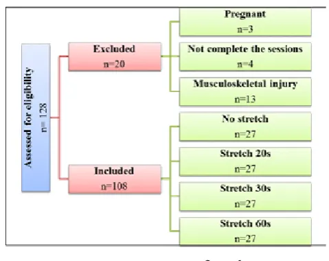
A sample of 128 healthy recreationally active women ini-tially participated in the study. They were recruited from the female section- King Saud University (KSU). Active women defined as the women engage in some form of mod-erate intensity physical activity for 30 minutes or more for at least three times / week [23,24]. All of participants were sound because there were no records of any past hip, knee, or ankle-related injuries, lower-extremity contracture, an operation performed on their back or lower extremity within the past two years, neurological findings, and take hormone or muscle-affecting drugs. Thirteen participants were excluded due to preexisting musculoskeletal limita-tions or injuries, also, four participants did not complete the sessions and three participants were pregnant.
to participate in the study. The study was approved by the ethical committee, Collage of Applied Medical Sciences, KSU. It was granted by the Research Center of the Female Scientific and Medical Colleges, Deanship of Scientific Re-search, KSU. Data were collected from May until the end of November 2014.
Figure 1: Participants flow chart
Isokinetic testing procedure
A randomized controlled quaziexperimental design was used. Before the experimental sessions the participants completed the familiarization session. This session includ-ed anthropometric measurements [(body weight, height and body mass index (BMI)] followed by warming-up on a stationary cycle ergometer. The participants practiced sub-maximal and sub-maximal isokinetic muscle action of Q and H muscles at 60 and 180°/s until they were convenient with the protocol. Furthermore, the stretching exercises were conducted during the familiarization session to ensure that each participant could tolerate the stretches. The eligible participants visited the laboratory on three experimental sessions (H stretching condition, Q stretching condition and combined H and Q stretching condition) separated by at least 48 hours [25].
At baseline the participants performed isokinetic tests to measure the maximal concentric Q and H PT, eccentric H PT and H:Q ratios at different angular velocities (60 and180°/s) (slow to fast velocity) [26]to ensure no differ-ences identified between groups. The test evaluated the dominant leg, which was the right leg for approximately 89% (n=96) of participants while, only 11% preferred their left leg (n=12). The leg that the participants used to kick a ball was determined as the dominant leg [26].
For the experimental sessions, each participant complet-ed a five-minute warm- up on a stationary cycle ergom-eter with the resistance set to 50 watt and a pedaling ca-dence of 60 to 70 rpm prior to the initial isokinetic testing [27]. Participants were positioned on the HUMAC system (HUMAC 2009 NORM, Computer Sports Medicine, Inc. Stoughton, MA, and the USA) using a standard protocol for a test of Knee Extension /Flexion in a seated position in accord with the manufacture’s protocol. They were in a sitting position with pads securing the dominant leg and a restraining strap over the pelvis and trunk. The isokinetic
dynamometer was calibrated prior to data collection. The weight of the limb was calculated using the software to as-sure that the gravity was accounted for during the meaas-sure- measure-ment testing in order to reduce the risk of inaccurate data. Also, knee movement was set from 5° to 95° of knee flex-ion to prevent Hyperextensflex-ion/hyper flexflex-ion knee injury from occurring [6]. The slower angular velocity (60°/s) was completed before the high speed (180°/s) as this facilitates learning during measurements at high angular velocities of the knee. The angular velocities were chosen based on the recommendation of Nelson et al [28] that the impacts of SS were velocity specific.
The input axis of the dynamometer was aligned with the axis of the knee; while the contra-lateral leg was braced against the limb stabilization bar. Each test consisted of three maximal repetitions performed for each velocity. A one-minute rest was allowed between testing at each veloc-ity to prevent the buildup of fatigue. Throughout the tests, Boisterous verbal consolation might have been given by the examiner so that each participant was guided to kick out and pull back as hard and fast as possible throughout the entire range of motion [8]. The information was imple-mented within the HUMAC-NORM software (software on a NORM, 6000) to assist in calculating PT. PT was estimat-ed as the highest torque value of the repetition that yield-ed the highest PT value [29]. Conventional H:Q ratio was calculated by dividing each participant’s highest concentric H PT by the highest concentric Q PT[30].Functional H:Q ratio was estimated by dividing the highest eccentric H PT by the highest concentric Q PT [14].
Static Stretching protocol
Following the pre-stretching isokinetic tests for all groups, each participant of the three experimental groups (II, III and IV) underwent four SS exercises designed to stretch the Q-only, H-only and combined Q and H muscles of the dominant leg only. The SS routine composed of one unas-sisted and three asunas-sisted exercises using a protocol from previous studies [29, 30].
ing, respectively for 60s SS. Immediately after the stretch-ing exercises, the average time that elapsed from the end of the stretching to the start of the post-stretching isokinetic test was 5.4±1.1 minutes.Promptly after the stretching ex-ercises, the normal time that slipped by from the end of the stretching to the begin of the post-stretching isokinetic test was 5.4±1.1 minutes.
H muscle stretching condition
The SS protocol for H muscle has been portrayed in point of interest by Costa et al.[15].Each participant performed one unassisted stretching exercise followed by three assist-ed stretching exercises. The unassistassist-ed stretching exercise might be a standing toe contact. With the dominant leg completely extended and the left thigh externally rotated and supporting the body weight, the participant flexed the middle so that both hands draw closer the dominant foot with no guide from the examiner. The first assisted SS exer-cise was finished in a modified - hurdler position.
The participant sat on a mat with the dominant leg com-pletely extended, and the non-dominant thigh flexed and laterally rotated and flexed thus, the non-dominant foot was squeezed against the medial side of the dominant knee. The participant was motivated to reach with both hands to-ward the dominant toes by flexing the middle, which was assisted by the examiner pushing against the participant’s back. To play out the second assisted SS exercise, the par-ticipant laid recumbent on a mat with her dominant thigh flexed at the hip and dominant leg completely extended. While securing the non-dominant leg, the investigator passively flexed the dominant thigh by pushing against the back leg and heel toward the head.
The last assisted SS exercise started with the participant lying recumbent on a mat with the non-dominant thigh flexed and dominant leg extended thus, the dominant thigh and leg were straight and opposite to the floor. The investigator passively dorsiflexed the foot by pushing down on the toes and supporting the heel.
Q muscle stretching condition
The protocol intended to stretch the Q muscles was de-picted in point of interest by Cramer et al., [29]. Every participant played out an unassisted stretching exercise took after by three assisted stretching exercises. For the un-assisted stretching exercise, the participant stood upright with one hand against a wall for balance. The participant then flexes the dominant leg at the knee joint for 90°.The ankle of the flexed leg was held by the ipsilateral hand, and the foot was raised, so that the heel of the dominant foot drew closer the rump. The initially assisted stretching ex-ercise was performed with the participant lying inclined on a cushioned table with her legs completely extended. The dominant leg was flexed at the knee joint and grad-ually pushed down thus, the participant’s heel drew clos-er the rump. In the event that the heel could contact the rump, the knee was gently lifted off the supporting surface, causing a slight hyperextension at the hip joint, to finish the stretch. To play out the second assisted SS exercise, the
participant remained with her back to a table and rested the dorsal surface of her dominant foot on the table by flex-ing the leg at the knee joint. From this position, the dom-inant leg extensors were stretched by gently pushing back on both the knee of the flexed leg and the related shoulder. The last assisted SS exercise started with the participant ly-ing recumbent along the edge of the cushioned table with the dominant leg hanging off of the table. The dominant leg was flexed at the knee and the thigh was slightly hyper ex-tended at the hip by delicately pushing down on the knee.
H and Q stretching condition
The same H and Q stretching exercises described before
Data Analysis
The Statistical Package for Social Science (SPSS) form 22 was utilized to examine the data. Mean, standard devia-tion and percentage of differences were calculated. A one-way analysis of variance ANOVA was used to test the dis-tinctions among the groups regarding their isokinetic PT production of Q and H muscles as well as H: Q ratios at baseline before each stretching condition. Five separate three-way ANOVA time [control-vs post-20s stretching vs post-30s stretching vs post-60s stretching] x condition [H-only stretching vs Q-only stretching vs combined H and Q stretching vs control] x velocity [60° vs 180°/s] was used to analyze the H and Q concentric PT, H eccentric PT, and the conventional and functional H:Q ratios. In case of significant effect or interaction Post hoc test was performed to examine the difference between and within groups. The Significance level was set at 5%.
RESULTS
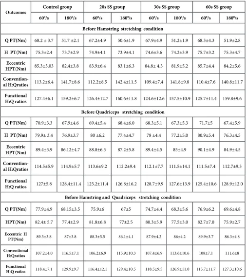
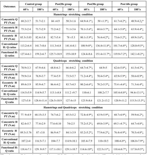
Table 1 depicts the demographic attributes of the partic-ipants at baseline. The results showed non-significant differences between groups regarding demographic char-acteristics (age, height, weight and BMI) (P>0.05). As presented in Table 2, there were no significant differences between and within groups at baseline for all the outcome measures before the three stretching conditions (P>0.05). Table 3shows significant reductions in all outcome mea-sures after 30 and 60s of SS at different angular velocities compared to control group. With no significant effects with the 20s SS (P > 0.05).
Table 1: Demographic characteristics of all participants at baseline
Groups N (Yrs.)Age Height (cm) Weight (Kg) BMI
Control 27 ±1.220.7 159.2 ±2.2 53.3 ±1.6 21.2±.83
20s stretch 27 ±2.121.3 159.3 ±4.5 54.6 ±3.4 21.4±.97
30s stretch 27 21 ±1.5 158.8 ±3.7 52.6 ±3.5 20.9±.81
60s stretch 27 ±1.821.1 159 ±3.2 52.7 ±2.6 21.3±2.6
Concentric Q PT
There was a significant main effect for velocity, time and condition (P<0.05). The three-way ANOVA indicated no three-way interaction (time x condition x velocity) (P=0. 825). Also, no two-way interaction for time x ve-locity (P>0.05), but significant two-way interactions were observed for condition x velocity and time x condition (P<0.05). Moreover, Post-hoc comparisons showed a sig-nificant difference between the conditions (P<0.05) in fa-vor to H stretching condition. In between-groups, one-way
ANOVA revealed significant reductions after 30 and 60s of SS under H stretching condition, Q stretching condition and combined H and Q stretching condition at both angu-lar velocities (Table 3). In particuangu-lar, the highest reduction of concentric Q PT was recorded after 60s of SS under H stretching condition at 60 and 180º/s (Mean =61.7±4.2 Nm and 48.9±4.2 Nm, respectively). The percentages of re-duction compared to control group were 9.5% and 5.4%, respectively.
Table 2: Mean (±SD) values of concentric Q and H PT, eccentric H PT and the conventional and functional H: Q ratios at 60 and 1800/s for all groups at baseline before different stretching conditions
Outcomes Control group 20s SS group 30s SS group 60s SS group
600/s 1800/s 600/s 1800/s 600/s 1800/s 600/s 1800/s Before Hamstring stretching condition
Q PT(Nm) 68.2 ± 3.7 51.7 ±2.1 67.2±4.9 50.6±1.9 67.9±4.9 51.2±1.9 68.3±4.3 51.9±2.8
H PT(Nm) 75.3±2.4 73.7±2.9 74.9±4.1 73.9±4.1 74.6±3.6 74.2±3.9 75.7±3.2 75.3±4.7 Eccentric
HPT(Nm) 85.3±3.03 82.4±3.8 83.9±6.4 83.1±6.3 84.8± 4.3 81.9±5.2 85.7±4.4 84.2±5.6
Convention-al H:Qratios 113.2±6.4 141.7±8.6 112.2±8.5 142.4±11.5 109.4±7.4 141.8±9.8 110.4±7.6 140.8±11.7 Functional
H:Q ratios 127.4±6.1 159.2±6.7 126.4±12.7 160.6±11.8 124.6±12.6 157.5±10.9 125.7±11.4 159.8±9.6 Before Quadriceps stretching condition
Q PT(Nm) 70.9±3.3 67.9±4.6 69.4±5.4 68.4±6.0 68.3±5.1 67.3±5.3 71.7±5 67.4±5.9
H PT(Nm) 79.9± 3.4 76.9±3.7 80 ±6.2 77.4±4.7 78 ±4.4 77.2±5.0 80.9±5.4 76.3±4.5
Eccentric
HPT(Nm) 89.4±3.9 86.12±4.7 88.8±6.3 87.2±5.8 89.4±4.5 85±4.9 90.1±4.9 84.9±4.5
Convention-al H:Qratios 114.5±5.9 114.9±5.7 113.6±9.2 112.2±9.4 112.1±7.7 111.5±14.1 111.5±7.4 112.7±9.3
Functional
H:Q ratios 127±5.8 128.4±11.4 125.2±11.4 126.8±16.2 128.7±9.9 127.6±13.9 125.4±10.6 128.9±12.0 Before Hamstring and Quadriceps stretching condition
Q PT(Nm) 77.9±4.9 68.15±3.5 75.9±6 67±5 74.7±4.4 68.3±5.6 76.9±6.2 69.6±4.8
HPT(Nm) 82.4± 5.7 77.4±2.9 81.8±6.8 77±2.5 80.3±5.9 77.5±3.0 82.7±7.0 75.9±2.7
Eccentric H
PT(Nm) 89.3±3.8 87±3.8 88.3±5.5 86.1±4.1 87.9±4.2 86±4.2 89.9±3.7 86.3±4.8
Conventional
H:Qratios 107.2±4.0 116.5±7.1 106.2±6.9 115.9±10.3 107.4±6.9 113.6±10.6 108±7.1 111.6±8
Functional
Concentric H PT
There was a significant main effect for velocity, time and condition (P<0.05). According to the three- way ANOVA, the concentric H PT for the H muscle showed three-way interaction (time x condition x velocity) (P= 0.037). Fur-thermore, two-way interactions for condition x velocity and time x condition were reported (P<0.05). On the other hand, there was no two-way interaction for time x veloci-ty (P> 0.05). In addition, Post-hoc comparisons showed a significant difference between the conditions (P<0.05) in favor to H stretching at 600/s and Q stretching condition at 1800/s. The results of ANOVA displayed a significant reduction of concentric H PT (P<0.05) following 30 and
60s SS under the three stretching conditions at different angular velocities (Table 3). The highest reduction was ob-served after 60s SS under H stretching condition at 60º/s (Mean=64.1±5.8 Nm) and Q stretching condition at 180º/s (Mean=58.6±8.9N.m) with 14.8% and 23.7% differences from control group respectively.
For eccentric H PT, there was a significant main effect for velocity, time and condition (P<0.05).The three-way ANO-VA for eccentric H PT revealed no three-way interaction (time x condition x velocity) (P=0.514), and no two-way interactions for condition x velocity and time x velocity (P>0.05).While, two-way interaction for condition x time (P< 0.05) was recorded. In addition, Post-hoc comparisons
Table 3: Mean (±SD) values of concentric Q and H PT, eccentric H PT and the conventional and functional H: Q ratios after SS at 60 and 1800/s for all groups at different conditions
Outcomes Control group Post20s group Post30s group Post 60s group 600/s 1800/s 600/s 1800/s 600/s 1800/s 600/s 1800/s
Hamstrings stretching condition Concentric Q
PT (N.m) 68.2±3.7 51.7±2.1 66 ±4.9 50.3±1.6 64.9±4.1*↓ 50 ±1.5*↓ 61.7±4.2*↓ 48.9±4.2↓* Concentric H
PT (N.m) 75.3±2.4 73.7±2.9 73.2±4.2 71.1±3.6 70.1±3.6*↓ 68.6±3.7*↓ 64.1±5.8*↓ 63.9±4.4*↓ Eccentric H
PT (N.m) 85.3±3.03 82.4±3.8 82.7±5.4 78 ±5.3 80.1±3.9*↓ 76.4±4.2*↓ 73.6±5.2*↓ 69.9±3.6*↓ Conventional
H: Q ratios 113.2±6.4 141.7±8.6 111.3±6.8 141.4±8.1 108.9±8*↓ 136.4±11.8*↓ 101.7±6.8*↓ 128.8±9.5*↓ Functional H:
Q ratios 127.4±6.1 159.2±6.7 125.7±10.9 155±10.9 124.4±8.6 151.4±11.7*↓ 119.9±7.2*↓ 142.1±8.2*↓ Quadriceps stretching condition
Concentric Q
PT (N.m) 70.9±3.3 67.9±4.6 68.8±6.3 66.4±6.2 64.7±4.7*↓ 64.9±5 62.6±5.8*↓ 61.5±4.3*↓ Concentric H
PT (N.m) 79.9±3.4 76.9±3.7 77.4±5.9 73.5±5.7 71.2±4.4*↓ 70.4±5.4*↓ 65.9±5.9*↓ 58.6±8.9*↓ Eccentric H
PT (N.m) 89.4±3.9 85.9±4.7 86.4±4.2 83.7±4.5 80.2±6.6*↓ 78.2±5.5*↓ 75.4 ±4.6*↓ 71.3±6.4*↓ Conventional
H: Q ratio 114.5±5.9 114.9±5.7 113.1±8.8 111.2 ±9.7 110±6.1 108.2±7.7 105.4±8.8*↓ 94.4±13.1*↓ Functional H:
Q ratio 127±5.8 128.4±11.4 126.5±10.9 127.4±15 123.9±8.6 121.2±13.2 120.9±11.2 115.5±13.3*↓ Hamstrings and Quadriceps stretching condition
Concentric Q
PT (N.m) 77. 9±4.9 68.15±3.5 74.7±6.2 65.5±5.2 72.8±4.9*↓ 63.9±5.9*↓ 68.7±4.9*↓ 59.9±6.2*↓ Concentric H
PT (N.m) 82.4±5.7 77.4±2.9 77.6±8.10 74±2.5 75.2±5.2*↓ 69.8±3.9*↓ 69.3 ±4.7*↓ 64.7±4.8*↓ Eccentric H
PT (N.m) 89.3±3.76 87 ±3.8 86.9±4.7 84.1±3.9 83.2±5.2*↓ 77.9±6.2*↓ 76.4±4.9*↓ 70.5±4.8*↓ Conventional
H: Q ratio 107.2±4 116.5±7.1 104±7.7 114.9±10.1 103.4±7.9 110±10.5 100±6.8*↓ 106.8±7.9*↓ Functional H:
Q ratio 118.4±7.1 129. 9±9.7 117.1±10.1 129.1±10.7 114.6±10*↓ 122.5±11*↓ 110.4±8.7*↓ 117.8±9.5*↓
recorded significant difference between the three stretch-ing conditions (P<0.05) in favor to H stretchstretch-ing condition after 30 and 60 s of SS at both angular velocities. Eccentric H PT was found to decrease significantly after 30 and 60s of SS (Table 3). Additionally, the greatest reduction of PT compared with control group was detected for the 60s of SS under H stretching condition at 60 and 180º/s (Mean =73.6±5.2N.m and 69.9±3.6N.m, respectively). The per-centages of PT reduction were 13.7% and 15.2%, respec-tively.
H:Q ratios
For the conventional H:Q ratio, there was a significant main effect of velocity, time and condition (P<0.05). The three-way ANOVA indicated no three-way interaction (time x condition x velocity) (P=0.249), no two-way inter-actions for condition x time (P>0.05). But, there were two-way interactions for time x velocity and condition x veloc-ity (P<0.05). In addition, Post-hoc comparisons recorded significant difference between the conditions (P<0.05) in favor to combined H and Q stretching condition at 60 º/s and Q stretching condition at 180º/s after 60s of SS. Under H stretching condition, the results of ANOVA indicated a significant reduction of conventional H:Q ratio after 30s of SS at both angular velocities. On the other hand, signif-icant decrements were presented after 60s of SS under all three stretching conditions at different angular velocities (Table 3). The highest reduction was recorded after 60s of SS under combined H and Q stretching condition at 60º/s (Mean=100±6.8) and Q stretching condition at 180º/s (Mean=94.4±13.1) with 6.7% and 17.8%, differences from control group respectively.
Regarding the functional H:Q ratio, there was a signifi-cant main effect for velocity, time and condition (P<0.05). The three-way ANOVA indicated no three-way interac-tion (time x condiinterac-tion x velocity), no two-way interacinterac-tion for condition x time (P>0.05). But, there were two-way interactions for time x velocity and condition x velocity (P<0.05).In addition, Post-hoc comparisons revealed a sig-nificant difference between the conditions (P<0.05) in fa-vor to combined H and Q stretching condition at 60 º/s and Q stretching condition at 180º/s. ANOVA indicated signif-icant reductions after 30s of SS under H stretching condi-tion at 180º/s and combined H and Q stretching condicondi-tion at both angular velocities. Also, significant decrements of functional H:Q ratio was presented after 60s of SS under H stretching condition and combined H and Q stretch-ing condition at different angular velocities and under Q stretching condition at 180º/s (Table 3). In particular, the 60s SS revealed the highest reduction under combined H and Q stretching condition at 60º/s (M=110.4±8.7) and Q stretching condition at 180º/s (M=115.5±13.3). The per-centages of reduction from control group were 6.8% and 10%, respectively.
DISCUSSION
The primary focus of the current study was to examine the acute effects of different SS duration (20, 30 and 60s) on
isokinetic concentric Q and H PT, eccentric H PT and H:Q ratios at different stretching conditions and angular veloc-ities in active women. In this manner, the current study sought to fill an important gap in the current studies on the impacts of different SS routines on muscle quality, perfor-mance and potential for overuse injuries of the knee. The most important findings were the reductions in isokinetic concentric Q and H PT, eccentric H PT as well as, H:Q ratios under the three stretching conditions after 30 and 60s of SS at 60 and 180º/s. While the examined parameters were unchanged after 20s of SS.
Regarding the concentric and eccentric PT of Q and H muscles, the results showed that SS of H-only, Q-only and combined H and Q for 30 and 60s significantly decreas-es isokinetic concentric Q PT at both angular velocitidecreas-es. However, the greatest significant reduction of Q PT was observed after 60s vs 30s of SS occurring under H stretch-ing condition at 60º/s (10% vs. 4.8%) and at 180º/s (5.4% vs. 3.3%) compared to control group. Similarly, significant reduction of concentric H PT was recorded under the three stretching conditions for 30 and 60s at both angular ve-locities. In particular, the 60s SS yielded more pronounced decreases in isokinetic concentric H PT than the 30s SS, occurring under H stretching condition at 60º/s where the percentages of reduction compared to control group were 14.8% vs. 6.9% and under Q stretching condition at 180º/s with 23.7% vs.8.4%. Generally, these results were constant with past studies that conveyed reductions in isokinetic concentric PT after a session of 30s [19, 29,30,32-35] and 60s of SS [36].
In this study the reduction of concentric Q PT under Q stretching condition was 8.74% after 30s of SS at60º/s. This percentage is higher than that reported by Costa et al. They reported 6.15% decrease in Q PT after 30s of SS under the same stretching condition and velocity [9]. Furthermore, the percentage of reduction of concentric Q PT after 30s of Q stretching reported in the previous studies was (3- 4.4 %) which is lower than that reported in current research [22, 23, 35]. Then again, Q muscle SS for 60s decreased concentric Q PT by (11.7% and 9.4%) at 60 and 180º/s, respectively. The findings are in line with past study com-pared the acute impacts of four different SS durations (10, 20, 30, and 60s) on isokinetic concentric Q PT [36]. The authors recorded Q PT reduction with only 30 and 60s of SS. Concentric Q PT was decreased by (5.5% and 11.6%, respectively) at 60º/s, and (5.8% and 10%, respectively) at 180º/s under Q stretching condition.
concen-tric Q and H PT at both low and high velocities, when the participants performed 30s SS protocol [11, 20,37].
With regard to eccentric H PT, a significant reduction was noted at both angular velocities when the participants performed a SS protocol for 30 and 60s under the three stretching conditions. Whereas, the highest eccentric H PT reduction was observed after the 60s vs. 30s of SS, under H stretching condition (13.7 % vs.6.1%) at 60º/s and (15.2% vs.7.2%) at 180º/s. This result is consistent with Costa et al. who reported eccentric H PT reduction after 30s of H stretching by 15% and 18.3% at 60 and 180º/s, respective-ly[8].Besides, this finding was not quite the same as dif-ferent studies that have reported no significant changes in eccentric H PT after 30s of the SS [21,26,27].
Isokinetic PT in this study showed similar trends to that re-corded in past studies [30,37]. Isokinetic concentric Q and H PT and eccentric HPT decreased as duration increased. Along these lines, there might be an immediate relation-ship between the stretch duration and stretching-induced declines in muscle strength and performance. Also, it was noted that the reduction of PT was affected by the stretch-ing condition, and inversely related to angular velocity. Since eccentric muscle activities produce a generally high amount of intrinsic force and if the H is considerably weak-er than the Q, this muscular imbalance may expand the danger of damage. Also, H injuries are normal in sports including running and hop [10]. In like mannerCosta et al., revealed that the potential risk of damage happened in healthy recreational active women is related to reduction of eccentric H PT [8].
Therefore, it might be vital to restrain any activity that could conceivably diminish H concentric and/or eccentric strength, especially if the stretching can be completed at some other time during the day than before strength test-ing or athletic performances. It is postulated that tempo-ral reduction in strength (PT) taking after SS have been referred to techniques such as changes in the mechanical elements of skeletal muscle contraction [15,29,32] and/or neural factors related to muscle activation [29, 33].
The findings of H:Q ratios showed that the conventional and functional H:Q ratios were differentially influenced by the duration of stretching, stretching condition as well as the movement velocity. The conventional H:Q ratio sig-nificantly decreases after stretching of H for 30 and 60s at both angular velocities. The highest reduction of conven-tional H:Q ratio was noted after 60s of SS under combined H and Q stretching condition at 60º/s (6.7%) and under Q stretching condition at 180º/s (17.8%). Regarding func-tional H:Q ratio, significant reductions were observed un-der H stretching condition and combined H and Q stretch-ing condition after 30 and 60s of SS at different angular velocities. Moreover, the 60s SS yielded more pronounced decreases in functional H:Q ratio than the 30s SS, occur-ring under Q stretching condition (10%) at 180º/s and under combined H and Q stretching condition (6.8% vs. 3.2%) at 60º/s.
In the present study, the H:Q ratios decreased as angular ve-locity increased compared to control group, which is near-ly constant with a past study that showed reduction in the functional H:Q ratio after 30s of SS under combined H and Q stretching by 7% at 180º/s [30]. Similarly, 7% and 9.1% reductions of conventional H:Q ratio under H stretching at 60 and 180°/s were noted by Costa et al., 2013 in active women [8].However, the findings are in contrast with the previous studies, which showed no significant reduction of conventional [22,30]and functional H:Q ratio[21, 22]at both low and high velocities, when the participants per-formed 30s SS protocol.
Devan et al., 2004, costa et al., 2009 and Holcomb et al.,2007 concluded that H muscle is about 50-80% as strong as the Q muscle [18,30,38]. It has been proposed in this manner, that the disproportionate H:Q strength ratio might be conversely identified with the danger of lower ex-tremity injuries [18, 30].That is, as the H:Q ratio decreas-es, the danger of lower extremity injuries may increase. In addition, some studies have suggested that the functional H:Q ratio might be more representative of the functional variations between H and Q strength than the convention-al H:Q ratio[10]. Nevertheless, both H:Q ratios have been utilized as a precaution method to screen for potential H and knee-related injuries [13,17]. The general suggestion is that the H:Q ratio should be 0.6 or more noteworthy for injury prevention[18], and strengthening exercises can ad-just low H:Q ratios [38].
The discrepancy in the results of the current study and the previous studies might be because of the distinction s in the training condition of the subjects. In this study, the participants were healthy, college-aged, recreationally ac-tive women, where the participants in most of the previous studies were competitive sports [20,21,39]. Therefore, the intense impacts of SS may be identified with the training condition s and our supposition is that this issue may should be determined and re-evaluated later in competi-tors from various sports disciplines and distinctive train-ing conditions. Furthermore, perhaps the discrepancy may be due to the difference in stretching protocol involved (stretching duration, condition, intensity, angular velocity and rest period duration).
According to authors knowledge, the present study was the first to investigate the acute effects of different stretch-ing routines includstretch-ing different duration, stretchstretch-ing both Q and H muscles as well as the effect of different angular velocity on concentric Q and H PT, eccentric H PT and the conventional and functional H:Q ratios in active Saudi women. However, we must acknowledge some of the lim-itations of the present study. Firstly, given that gender play important role in muscle performance, we only collected the information from healthy recreational active women at KSU-Riyadh city. Secondly, only dominant leg tests were measured which were not able to compare between legs. Finally, the study investigated the effects of SS on concen-tric and eccenconcen-tric isokinetic muscle strength, but it is im-portant to known how SS affects electromyography activity during Q and H contractions in both concentric and ec-centric modes.
Conclusion and Practical application
In summary, isokinetic strength production might be ad-versely influenced simply after 30 and 60s of SS. It appears to affect muscle strength at slow and fast velocities, and thus may affect all types of athletes.
In this way, it is prudent to dodge acute SS exercises just before any action requesting maximal force and power production, as this may be inconvenient to a fruitful per-formance.
On other hand, SS of a shorter span (20s) may not degrade maximal performance. Furthermore, SS may unfavorably influence the conventional as well as, the functional H:Q ratios thusly alert must be taken if stretching is directed before H:Q ratio evaluation, particularly when H:Q ratios are utilized as an index for choosing when come back to play is proper during injury rehabilitation. The findings of the present study along with that of past studies have con-jointly proposed that strength and conditioning coaches, athletic trainers, and other allied welling experts should consider the duration of SS, stretching condition as well as the velocity of movement as an approach to prevent re-duction in muscle strength, performance and risk of knee injury during physical activities.
CONFLICT OF INTEREST
The authors report no conflict of interest. The findings of the study are exhibited clearly, honestly, without fabrica-tion, falsificafabrica-tion, and without inappropriate data manip-ulation.
ACKNOWLEDGEMENTS
The study was granted from the Research Center of the Fe-male Scientific and Medical Colleges, Deanship of Scientif-ic Research, KSU.
REFERENCES
[1] Cross KM, Worrell TW. Effects of a static stretching program on the incidence of lower extremity musculo-tendinous strains. J Athl Train.1999; 34(1):11-14.
[2] Yapicioglu B, Colakoglu M, Colakoglu Z, Gulluog-lu H, Bademkiran F, Ozkaya O. Effects of a dynamic
warm-up, static stretching or static stretching with tendon vibration on vertical jump performance and EMG responses. J Hum Kinet. 2013;39:49-57.
[3] McHugh MP, Cosgrave CH. To stretch or not to stretch: the role of stretching in injury prevention and perfor-mance. Scand J Med Sci Sports. 2010;20:169-181.
[4] Behm DG, Chaouachi A. A review of the acute effects of static and dynamic stretching on performance. Eur J ApplPhysiol .2011;111:2633-2651.
[5] Simic L, Sarabon N, Markovic G . Does pre-exer-cise static stretching inhibit maximal muscular per-formance? A meta-analytical review. ScandJMedSci Sports. 2011; 23:131-148.
[6] Andrade Mdos S, De Lira CA, KoffesFde C, Mascarin NC, Benedito-Silva AA, Da Silva AC. Isokinetic ham-strings-to-quadriceps peak torque ratio: the influence of sport modality, gender, and angular velocity. J Sports Sci. 2012;30:547-553.
[7] Pope RP, Herbert RD, Kirwan JD, Graham BJ. A randomized trial of preexercise stretching for pre-vention of lower-limb injury. Med Sci Sports Ex-erc.2000;32:271-277.
[8] Costa PB, Ryan ED, Herda TJ, et al. Acute effects of static stretching on peak torque and the ham-strings-to-quadriceps conventional and functional ra-tios. Scand J Med Sci Sports.2013;23:38-45.
[9] Houweling TA, Head A, Hamzeh M A. Validity of isoki-netic testing for previous hamstring injury detection in soccer players. IsokineticsExerc Sci. 2009;17:213-220.
[10] Sugiura Y, Saito T, Sakuraba K, Sakuma K, Suzuki E.
Strength deficits identified with concentric action of the hip extensors and eccentric action of the ham-strings predispose to hamstring injury in elite sprint-ers. J Orthop Sports Phys Ther.2008;38:457-46.
[11] Thacker SB, Gilchrist J, Stroup DF, KimseyCD Jr. The
impact of stretching on sports injury risk: a system-atic review of the literature. Med Sci Sports Exerc. 2004;36:371-378.
[12] Cheung RT, Smith AW, Wong DP. H:Q ratios and
bi-lateral leg strength in college field and court sports players. J Hum Kinet. 2012;33:63-71.
[13] Myer GD, Ford KR, Barber Foss KD, Liu C, Nick
TG, Hewett TE. The relationship of hamstrings and quadriceps strength to anterior cruciate ligament in-jury in female athletes. Clin J Sport Med. 2009;19:3-8.
[14] Yeung SS, Suen AM, Yeung EW. A prospective cohort
study of hamstring injuries in competitive sprinters: preseason muscle imbalance as a possible risk factor. Br J Sports Med. 2009;43:589-594.
[15] Costa PB, Ryan ED, Herda TJ, Defreitas JM, Beck
TW, Cramer JT. Effects of static stretching on the hamstrings to-quadriceps ratio and electromyo-graphic amplitude in men. J Sports Med Phys Fitness. 2009a; 49:401-409.
[16] Kong PW, Burns SF. Bilateral difference in hamstrings
[17] Croisier JL, Ganteaume S, Binet J, Genty M, Ferret JM. Strength imbalances and prevention of hamstring injury in professional soccer players. A prospective study. Am J Sports Med. 2008;36:1469-1475.
[18] Devan MR, Pescatello LS, Faghri P, Anderson J. A
prospective study of overuse knee injuries among fe-male athletes with muscle imbalances and structural abnormalities. J Athl Train. 2004;39:263-267.
[19] Brandenburg JP. Duration of stretch does not
influ-ence the degree of force loss following static stretch-ing. J Sports Med Phys Fitness.2006;46: 526-534.
[20] Sekir U, Arabaci R, Akova B, Kadagan SM. Acute
ef-fects of static and dynamic stretching on leg flexor and extensor isokinetic strength in elite women athletes. Scand J Med Sci Sports.2010;20:268-281.
[21] Sekir U, Arabaci R, Akova B. Acute effects of static
stretching on peak and end-range hamstring-to-quad-riceps functional ratios. World J Orthop. 2015;6:719-726.
[22] Ayala F, De Ste Croix M, Sainz De Baranda P, Santonja F. Acute effects of static and dynamic stretching on hamstring eccentric isokinetic strength and unilateral hamstring to quadriceps strength ratios. J Sports Sci. 2013; 31:831-839.
[23] Al-Hazzaa HM. Health-enhancing physical activity
among Saudi adults using the International Phys-ical Activity Questionnaire (IPAQ). Public Health Nutr.2007;10: 59-64.
[24] Al-Nozha MM, Al-Hazzaa HM, Arafah MR, et
al. Prevalence of physical activity and inactivity among Saudis aged 30-70 years. A population-based cross-sectional study. Saudi Med J. 2007;28:559-568.
[25] De Weijer VC, Gorniak GC, Shamus E. The effect
of static stretch and warm-up exercise on hamstring length over the course of 24 hours. J Orthop Sports PhysTher. 2003;33:727-733.
[26] Cramer JT, Housh TJ, Coburn JW, Beck TW, Johnson
GO. Acute effects of static stretching on maximal ec-centric torque production in women. J Strength Cond Res.2006;20:354-358.
[27] Winke MR, Jones NB, Berger CG, Yates JW.
Moder-ate static stretching and torque production of the knee flexors. J Strength Cond Res.2010;24: 706-710.
[28] Nelson AG, Kokkonen J, Arnall DA, Li L. Acute stretch-ing increases postural stability in non balance trained individuals. J Strength Cond Res.2012;26:3095-3100.
[29] Cramer JT, Housh TJ, Johnson GO, Miller JM, Coburn
Citation
ALQaslah, G., & Shaheen, A. (2016). ACUTE EFFECTS OF DIFFERENT STATIC STRETCHING PROTOCOLS ON PEAK TORQUE, CONVENTIONAL AND FUNCTIONAL HAMSTRINGS-TO-QUADRICEPS RATIOS IN AC-TIVE WOMEN. International Journal of Physiotherapy, 3(5), 609-618.
JW, Beck TW. Acute effects of static stretching on peak torque in women. J Strength Cond Res.2004;18:236-241.
[30] Costa PB, Ryan ED, Herda TJ, DeFreitas JM, Beck
TW, Cramer JT. Effects of stretching on peak torque and the H:Q ratio. Int J Sports Med. 2009b;30:60-65.
[31] Rossi L, Pereira R, Simão R, Brandalize M, Gomes A.
Influence of static stretching duration on quadriceps force development and electromyographic activity. Human movement. 2010;11:137-143.
[32] Cramer JT, Housh TJ, Johnson GO, Weir JP, Beck
TW, Coburn JW. An acute bout of static stretching does not affect maximal eccentric isokinetic peak torque, the joint angle at peak torque, mean power, electromyography, or mechanomyography. J Orthop Sports PhysTher. 2007b;37:130-139.
[33] Cramer JT, Housh TJ, Weir JP, Johnson GO, Coburn
JW, Beck TW. The acute effects of static stretching on peak torque, mean power output, electromyography, and mechanomyography. Eur J ApplPhysiol. 2005;93: 530-539.
[34] Marek SM, Cramer JT, Fincher AL, et al. Acute effects
of static and proprioceptive neuromuscular facilita-tion stretching on muscle strength and power output. J Athl Train.2005;40:94-103.
[35] Papadopoulos G, Siatras T, Kellis S. The effect of stat-ic and dynamstat-ic stretching exercises on the maximal isokinetic strength of the knee extensors and flexors. IsokineticsExerc Sci. 2005;13:285-291.
[36] Siatras TA, Mittas VP, Mameletzi DN,
Vamvak-oudisEA.The duration of the inhibitory effects with static stretching on quadriceps peak torque produc-tion. J Strength Cond Res.2008;22:40-46.
[37] Alangari AS, Al-Hazzaa HM. Normal isometric and
isokinetic peak torques of hamstring and quadriceps muscles in young adult Saudi males. Neurosciences (Riyadh) .2004;9:165-170.
[38] Holcomb WR, Rubley MD, Lee HJ, Guadagnoli MA.
Effect of hamstring-emphasized resistance training on hamstring: quadriceps strength ratios. J Strength Cond Res. 2007;21:41-47.
[39] Egan AD, Cramer JT, Massey LL, Marek SM. Acute