Page 71 www.ijiras.com | Email: contact@ijiras.com
Comparative Evaluation Of Alteration In Retention Force Values
Of Different Attachment Systems For Implant Overdenture Over
Various Time Intervals: An In Vitro Study
Dr. Savy Arora
M.D.S. Student, Department of Prosthodontics, Maharishi Markandeshwar College of Dental Sciences and
Hospital, M.M. University, Mullana
Dr. Sanjeev Mittal
Professor, Department Of Prosthodontics,
Maharishi Markandeshwar College of Dental Sciences and Hospital, M.M. University, Mullana
I. INTRODUCTION
Epidemiologic and demographic studies performed lately have anticipated an expansion in the quantity of maturing edentulous patients in most countries. The prosthetic therapy of the edentulous patient, since long, has been a noteworthy challenge for dentists. The compulsion of suffering misery from uncomfortable dentures was countered by the advent of dental implants in the field of dentistry in the mid 1980s. Specifically, the issues of stability and retention of lower
prostheses have been improved by creating a fixed prosthesis or fabrication of an overdenture (OVD) to implants when the number is constrained in light of anatomical or social complexities. With the inclusion of implants in overdentures, there is a decrease in displacement of prosthesis due to lateral forces, leading to better stability and a repeatable centric occlusion. It also improves the masticatory function, quality of life, speech and even nutrition.
In the course of recent years, mandibular two-implant supported overdentures (opposing conventional maxillary Abstract:
Aim: To compare the retention force value alterations of four different types of implant overdenture attachments over various time intervals.
Materials And Methods: 28 cuboidal blocks were fabricated using autopolymerising acrylic resin. Four of these were used as master blocks, one for each group. Master blocks for Group A and B contained an implant analog with ball abutment, for Group C contained a single piece implant with ball abutment and for Group D contained an implant analog with Locator abutment. Six blocks for each group were used as prosthetic blocks, which included the overdenture attachment to be studied. Prosthetic blocks for Group A contained nylon cap - clear attachments, Group B contained nylon cap - pink attachments, Group C contained O-ring attachments and Group D contained Locator - clear attachments. The retention force was tested at four time intervals - baseline, after 1 month (after 90 cycles of insertion-removal), after 6 months (540 cycles) and after 1 year of simulated clinical use (1080 cycles), using Universal testing machine. These values were compared and statistical analysis was performed on the data obtained.
Results: Locator attachments were found to be the most retentive among all the stud attachments. The attachments showed significant retention loss over a period of 1 year except for nylon cap- pink. Maximum retention loss occurred for O-ring attachments (76.6%). Nylon cap - pink was found to require least force for removal but showed more consistency over a period of 1 year of use.
Conclusion: It was concluded that all overdenture attachments lose retention over time. However, the Locator attachment showed maximum retention values after 1080 cycles of insertion-removal.
Page 72 www.ijiras.com | Email: contact@ijiras.com dentures) have become the standard of care for edentulous
patients. A definitive objective would be the smallest intervention that offers an enhancement in the support, stability and retention of complete dentures. Henceforth, with the alternative of less intrusive implant surgery in the anterior region of the mandible, with decreased implant components and prosthodontic expenditure, the idea of the mandibular single-implant retained overdentures (opposing the conventional complete maxillary prostheses) is a reality for elderly edentulous patients.
Currently, numerous attachment systems are available for utilisation with implant - tissue - supported overdentures. Attachments can be classified on basis of their variability in flexibility, geometrical shape and cross section, casting precision and process of manufacture.Frequently used implant overdenture attachments comprise bar - clip attachments, stud attachments and magnetic attachments. The utility of stud attachments has been classically restricted to implants with divergence of less than 10 degrees. They are relatively economical, less technique sensitive, easy to use and easy to repair.Stud attachments could be nylon cap attachments, O-rings, ERA (extracoronal resilient attachments), Sterngold attachments or Locator attachment systems.
O-rings are doughnut shaped attachments which offer several advantages, including easy insertion/ removal by the patient, better hygiene, ease of maintenance, low cost and exclusion of the superstructure bar. The Locator attachment system, which was introduced in 2000, has dual retention (inner and outer), is self-aligning and has the least profile height of all the attachment systems available.
However, stud attachments tend to wear over time of clinical use and thus, lose retention. Wear occurs primarily during insertion and removal of the prosthesis and during functional as well as parafunctional activities. Thus, an alteration in the retention force of the attachment systems is expected with time, which leads to more maintenance visits and reduced patient satisfaction. A controversy exists regarding the comparison of retention loss over time with these attachment systems. Therefore, this in vitro study was conducted to compare the retention values of locator attachment, ball/O-ring and ball/nylon-cap, over a specified simulated period of time of use.
II. MATERIALS AND METHOD
A cuboidal wooden block of dimensions 40*25*8mm was constructed. A silicone mould was obtained using polyvinyl siloxane impression material and twenty eight acrylic cuboidal blocks of the same dimension were thus obtained in autopolymerising acrylic resin. These were divided into four groups; each group containing seven blocks - one block being the master block and six blocks used as prosthetic blocks which would incorporate the attachments. The samples were grouped according to the attachment systems to be used as:
Group A: Ball/Nylon Cap Attachment Group B: Ball/Nylon Cap Attachment Group C : Ball/O-ring Attachment Group D: Locator Attachment
A. PREPARATION OF MASTER BLOCKS
Using an acrylic trimming bur, one recess was prepared exactly in the centre of the master block. The block was placed on the surveying table such that its upper and lower surfaces were parallel to the horizontal plate of the surveyor. For group A, B and D, the implant analogues were attached to the impression transfer and autopolymerising acrylic resin was used to secure the impression transfer to the analysing rod of the surveyor. For group C, the single piece implant was vertically attached to the analysing rod. The implant/analogue was placed exactly vertically into the prepared recess such that the implant abutment junction would correspond to the level of the horizontal upper surface. Autopolymerising acrylic resin was used to fill the space between the master block and the implant/analogue to rigidly fix it, simulating osseointegration. The abutments were tightened using torque ratchet to 25 Ncm.
The cylindrical metal posts were incorporated using autopolymerising resin into the recesses prepared 2mm away from each end such that 4mm of the height was exposed out of the acrylic block. This shall allow exact vertical insertion and removal of the prosthetic blocks from the master blocks in a single pathway.
B. CONNECTING THE ATTACHMENTS TO THE
PROSTHETIC BLOCKS
Three recesses were prepared in the prosthetic block such that the prosthetic block would passively seat on the master blocks. A small circular piece of glove was placed on the abutment to prevent the flow of acrylic resin into the areas with undercuts, during pick up procedure. The attachment system was assembled and placed on the implant abutment. The attachments were incorporated into the centre of these blocks using autopolymerising acrylic resin, using direct pick up technique. After polymerisation, excess acrylic around the attachments was cleaned with a small round bur. (Fig 1)
Page 73 www.ijiras.com | Email: contact@ijiras.com Hooks were prepared using 19 gauge wire and secured on
the upper surface in the centre of the blocks, using autopolymerising acrylic resin. The prosthetic blocks were pulled away from the master block during retention force measurement by these hooks. The samples were stored in artificial saliva at room temperature to simulate oral conditions.
C. RETENTION FORCE TEST
The master and prosthetic blocks were positioned on the machine table to ensure that all abutments and inserts were fully and accurately engaged. Engagement and disengagement of the attachments were carried out at right angles to the horizontal level of the blocks. Assuming that a patient removes and inserts his prosthesis thrice daily for hygiene purpose (three meals a day), retention force values were measured at baseline, after 90 cycles of insertion - removal (after 1 month of simulated clinical usage), after 540 cycles (after 6 months) and after 1080 cycles (after 1 year). A time interval of 10 seconds was given between each removal insertion cycle to allow elastic recovery of the attachments system.
The Universal Testing Machine was used to measure the force which is required to separate the prosthetic block from the master block. The samples were kept moist with artificial saliva as it acts as a lubricant to simulate potential in-vivo conditions. The maximum vertical dislodging force required to separate the two blocks was recorded (in Newtons) at a crosshead speed of 50mm/min, using a load cell of 20 kN. This speed approximates the actual speed of movement of an overdenture away from its retentive elements in the mouth under a vertical dislodging force. Only vertical uniaxial insertion and removal movements were performed during testing. (Fig 2)
Figure 2: Retention force Measurement Using Universal Testing Machine
D. STATISTICAL ANALYSIS
The data was compared the data using one-way ANOVA, keeping the significance at p ≤ 0.05, followed by POST HOC test with Tukey HSD analysis
III. RESULTS
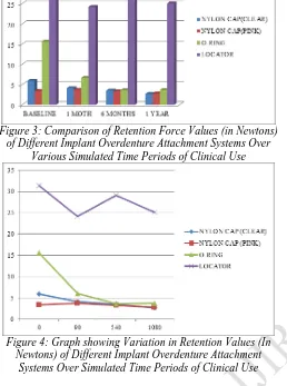
The retention force measurements at baseline showed highest retention for Locator attachment system followed by O-ring, nylon cap - clear and nylon cap - pink attachments.
The nylon cap-pink and O-ring attachment systems showed significant decrease in their retention over 1 year of simulated clinical use (Table 1). Most of the attachments showed significant retention loss over the first month of use and the Locator attachment showed insignificant retention loss over time (Figure 3 and 4).
GROUPS AT BASELINE
90 CYCLES
540 CYCLES
1080 CYCLES
GROUP A (NYLON CAP
CLEAR)
SAMPLE 1 6.8 4 3.6 0.8
SAMPLE 2 5.8 4.8 4.2 3.2
SAMPLE 3 6.8 4.4 3.2 4.2
SAMPLE 4 6.2 3.8 2 2.2
SAMPLE 5 5.4 4.6 4.2 2.6
SAMPLE 6 4.8 3.2 3.8 3.2
MEAN 5.967 4.133 3.5 2.7
PERCENTAGE
DECREASE 30.7% 41.3% 54.7%
GROUP B (NYLON CAP
PINK)
SAMPLE 1 3 3.4 3.8 2.4
SAMPLE 2 2.8 2.8 2.6 2.8
SAMPLE 3 3.6 3.6 2.8 3.6
SAMPLE 4 4.2 3.6 4 3.8
SAMPLE 5 3.8 5 3.6 2.6
SAMPLE 6 3.2 3.8 3.2 1.6
MEAN 3.433 3.7 3.33 2.8
PERCENTAGE
DECREASE -7.7% 3% 18.4%
GROUP C (O-RING)
SAMPLE 1 18.4 7.2 4.8 3.2
SAMPLE 2 15.2 5.4 3.4 4.4
SAMPLE 3 18.6 7.4 2.8 4.8
SAMPLE 4 12.4 3.4 3 3.6
SAMPLE 5 13.2 6.8 4.6 2.8
SAMPLE 6 16.2 6.2 3.2 3.2
MEAN 15.667 6.667 3.633 3.667
PERCENTAGE
DECREASE 57.4% 76.8% 76.6%
GROUP D (LOCATOR)
SAMPLE 1 32.2 20.2 32 22.4
SAMPLE 2 25.2 23.4 30.4 26
SAMPLE 3 35.2 17.2 22.8 20.8
SAMPLE 4 28.8 28.6 32.2 30.2
SAMPLE 5 31.2 23.6 26.4 28.2
SAMPLE 6 35.8 32.2 30.8 22.8
MEAN 31.4 24.2 29.1 25.067
PERCENTAGE
DECREASE 22.9% 7.3% 20.2%
Page 74 www.ijiras.com | Email: contact@ijiras.com Figure 3: Comparison of Retention Force Values (in Newtons)
of Different Implant Overdenture Attachment Systems Over Various Simulated Time Periods of Clinical Use
Figure 4: Graph showing Variation in Retention Values (In Newtons) of Different Implant Overdenture Attachment
Systems Over Simulated Time Periods of Clinical Use
IV. DISCUSSION
For nylon cap - clear attachment, the retention decreases significantly over the first month of simulated overdenture use and then becomes more or less constant while in case of nylon cap - pink, the retention force value alterations are insignificant throughout the period of the study. A loss of retention for nylon cap attachments was also observed by Cohen et al and Tabatabaian et al, which can be attributed to distortions of plastic component due to wear on repeated insertion and removal. Lehmann stated in his study that "forces from 5 to 7 N would be enough for a set of attachment to retain an overdenture during function". Pink nylon cap was the only attachment to show insufficient initial retention value, which can be because it is slightly oversized in comparison to the clear nylon cap. Such a loose attachment may be used in patients with manual dexterity problems or at initial delivery of immediately loaded prosthesis to ensure easy adaptability of the patient and it would decrease chances of screw loosening during the first few months. This can be replaced later by a higher retentive attachment.
For O-ring matrices, the retention decreases significantly over first 90 cycles (15.667N to 6.667N), then shows an insignificant reduction to a value of 3.667N at 6 month interval (540 cycles) and then remains constant. A similar
pattern was observed by Branchi et al, where 50% retention loss occurred over first 500 cycles and then value reduced very gradually uptil 5500 cycles. The retentive force of O-ring attachments is an outcome of the undercut structure of the patrix, elasticity of its rubber matrix and the frictional resistance between the matrix and patrix. The frictional resistance produces a contact force between the contacting surfaces, leading to deformation of the rubber component. When subject to wear, metal housing and plastic inserts generate scratches parallel to the direction of wear. However, rubber generates a rigid pattern perpendicular to the wear direction. The wear and tear caused may elicit a slight increase in diameter of the matrix, leading to loss of retention. Previous studies also found a significant reduction in retention force values over time for O-ring attachment. After 540 cycles, retention force tested was below the theoretical limit of 5N required to achieve an acceptable retention of a removable prosthesis, as also seen by Branchi et al.
For the Locator attachments, a significant decrease was observed from baseline to 1 month of use, then remained relatively constant. An overall 20.2% retention loss was observed till 1 year of simulated use. Previous studies have also reported a decrease in retention force values of locator attachments. This decrease is attributed to the wear and deformation of the nylon insert. Scanning electron microscopy in study conducted by Rutkunas et al revealed that the plastic core remained relatively stable while the inner surface of the outer ring, which is in contact with the metal undercuts of the locator abutment, showed significant wear. The initial retention value of Locator attachment system (31.4 N) was seen to be similar with findings by Kobayashi et al (33.5N).
The retention values increased slightly from 1 month to six months of usage and then decreased till 1 year of use. An increase in retention values followed by a decrease for Locators was also observed in previous studies.There is an increase in hardness and surface roughness due to change in surface charge on repetitive insertion-removal cycles. This leads to fine mechanical friction, consequently causing increase in the retention force values. This may also occur due to thermal expansion and water absorption of nylon inserts.
Page 75 www.ijiras.com | Email: contact@ijiras.com absorbed during insertion may be divided into elastic
(recoverable) and plastic (permanent) components. If the deformation is elastic, no loss of retention is expected. If permanent deformation occurs, incomplete recovery occurs leading to rapid loss of retention." According to Craig, "a material that is momentarily submitted to stress below its yield strength returns to its original form without any internal or structural change. However, if this stress is repetitive as in a fatigue process, the material can suffer definitive deformations."
The initial and final retention values were in the order - Locator (31.4N to 25.1N) > O-ring (15.667N to 3.67N) > Nylon cap - clear (5.97N to 2.8N) > Nylon cap - pink (3.43N to 2.7N). Maximum retention loss was observed for O-ring(76.6%) and minimum for nylon cap (pink) and Locator attachment system (20.2%) after 1 year of simulated clinical use.
V. LIMITATIONS
To simplify the experimental study, a homogenous rectangular model made of acrylic with an attachment linked to the implant was used which may have caused limitations in this study. Thus, overdenture samples fabricated on edentulous models or in vivo studies can provide more realistic results. In this study, only vertical dislodgement forces were simulated. Clinically, a combination of vertical, horizontal and oblique and rotational forces act on the prosthesis during masticatory function and parafunctional habits.
VI. CONCLUSION
Within the limitations of this study, the following conclusions can be drawn:
The retention for all attachments seemed to decrease with time.
Retention loss was maximum for O-ring. The least retention loss was seen for nylon cap (pink) and Locator attachment system.
Initial and final retention of Locator attachment system is maximum among all groups, followed by O-ring > nylon cap (clear) > nylon cap (pink).
Retention loss was maximum within the first month of simulated clinical use for nylon cap (clear), O-ring and Locator attachments.
Retention force value for nylon cap (pink) was found to be insufficient for removable prostheses, even at baseline. The retention force for O-ring and nylon cap (clear) decreased to below 4N after six months of clinical use simulation.
REFERENCES
[1] Douglass CW, Shih A, Ostry L. Will there be a need for complete dentures in the United States in 2020?. J Prosthet Dent. 2002;87:5–8.
[2] Mojon P, Thomason JM, Walls AW. The impact of falling rates of edentulism. Int J Prosthodont. 2004; 17:434–40.
[3] Türk PE, Geckili O, Türk Y, Günay V, Bilgin T. In Vitro Comparison of the Retentive Properties of Ball and Locator Attachments for Implant Overdentures. Int J Oral Maxillofac Implants. 2014;29:1106–13.
[4] Kordatzis K, Wright PS, Meijer HJ. Posterior mandibular residual ridge resorption in patients with conventional dentures and implant overdentures. Int J Oral Maxillofac Implants. 2003;18:447–52.
[5] Jemt T, Stålblad PA. The effect of chewing movements on changing mandibular complete dentures to osseointegrated overdentures. J Prosthet Dent. 1986;55:357–61.
[6] Busetti J, De Carli JP, Rodrigues Neto DJ, Pereira JR. Overdentures and masticatory efficiency: literature review. Dental Press Implantol. 2013;7(4):34-7.
[7] Feine J, Carlsson G, Awad M, Chehade A, Duncan W, Gizani S et al. The McGill consensus statement on overdentures as first choice of care for edentulous patients. Gerodontology. 2002;19(1):3–4.
[8] Attard N, Zarb G. Long-term treatment outcomes in edentulous patients with implant overdentures: the Toronto study. Int J Prosthodont. 2004;17(4):425–33. [9] Naert I, Alsaadi G, van Steenberghe D, Quirynen M. A
10-year randomized clinical trial on the influence of splinted and unsplinted oral implants retaining mandibular overdentures: peri-implant outcome. Int J Oral Maxillofac Implants. 2004;19(5):695–702.
[10]Engquist B, Bergendal T, Kallus T, Linden U. A retrospective multicenter evaluation of osseointegrated implants supporting overdentures. Int J Oral Maxillofac Implants. 1988;3(2):129–34.
[11]Jemt T, Chai J, Harnett J, Heath MR, Hutton JE, Johns RB et al. A 5-year prospective multicenter follow-up report on overdentures supported by osseointegrated implants. Int J Oral Maxillofac Implants. 1996;11(3):291– 8.
[12]Mraiwa N, Jacob R, van Steenberghe D, Quirynen M. Clinical assessment and surgical implications of anatomic challenges in the anterior mandible. Clin Imp Dent Res. 2003;5(4):219–25.
[13]Geertman ME, Boerrigter EM, Van Waas MA, van Oort RP. Clinical aspects of a multicenter clinical trial of implant-retained mandibular overdentures in patients with severely resorbed mandibles. J Prosthet Dent. 1996;75(2):194–204.
[14]Shafie H. Clinical and Laboratory Manual of Implant Overdentures. 1st ed. St Louis;The CV Blackwell Co.:2007. p.32-33.
[15]Banton B, Henry MD. Overdenture retention and stabilization with ball-and-socket attachments: Principles and technique. J Dent Technol. 1997;14(7):14–20. [16]Alsabeeha NH, Payne AG, Swain MV. Attachment
systems for mandibular two-implant overdentures: A review of in vitro investigations on retention and wear features. Int J Prosthodont. 2009;22(5):429-40.
Page 76 www.ijiras.com | Email: contact@ijiras.com locator attachment system with different implant
angulations. Int J Oral Maxillofac Implants. 2015;30(3):556–63.
[18]Kleis WK, Kämmerer PW, Hartmann S, Al-Nawas B, Wagner W. A Comparison of three different attachment systems for mandibular two-implant overdentures: One-year report. Clin Implant Dent Relat Res. 2010;12(3):209–18.
[19]Cune M, van Kampen F, van der Bilt A, Bosman F. Patient satisfaction and preference with magnet, bar-clip, and ball-socket retained mandibular implant overdentures: A cross-over clinical trial. Int J Prosthodont. 2005; 18(2):99–105.
[20]Shastry T, Anupama NM, Shetty S, Nalinakshamma M. An in vitro comparative study to evaluate the retention of different attachment systems used in implant-retained overdentures. J Indian Prosthodont Soc. 2016;16(2):159-66.
[21]Besimo CH, Guarneri A. In vitro retention force changes of prefabricated attachments for overdentures. J Oral Rehabil. 2003;30(7):671-8.
[22]Atsahazrm P, Ansari H, Khorsand M, Fatemi M, Sadeghpour SM, Azarmeh S. The influence of inclined implants and attachments on the retention and longevity of implant-retained overdentures: An in vitro study. J Dent Shiraz Univ Med Scien. 2012;13(3):90-6.
[23]Petropolous VC, Smith W, Kousvelari E. Comparison of retention and release periods for implant overdenture attachments. Int J Oral Maxillofac Implants. 1997; 12(2):176-85.
[24]Sarnat AE. The efficiency of cobalt samarium (Co5Sm) magnets as retentive units for overdentures. J Dent. 1983; 11(4):324-33.
[25]Cohen BI, Pagnillo M, Condos S, Deutsch AS. Comparative study of two precision overdenture attachment designs. J Prosthet Dent. 1996;76(2):145-52. [26]Tabatabaian F, Alaie F, Seyedan K. Comparison of three
attachments in implant-tissue supported overdentures: An in vitro study. J Dent (Tehran). 2010;7(3):113-8.
[27]Lehmann KM, Arnim FV. Studies on the retention forces of snap-on attachments. Quintessence Dent Technol. 1978;7:45–48.
[28]Branchi R, Vangi D, Virga A, Guertin G, Fazi G. Resistance to wear of four matrices with ball attachments for implant overdentures: A fatigue study. J Prosthodont. 2010;19(8):614–9.
[29]Schallamach A. A theory of dynamic rubber friction. Wear 1963;6:375-82.
[30]Ludwig K, Hartfil H, Kern M. Analysis of the wear and tear of ball attachments. Quintessence J Dent Technol. 2006;4:46–55.
[31]Cheng T, Sun G, Huo J, He X, Wang Y, Ren YF. Patient satisfaction and masticatory efficiency of single implant-retained mandibular overdentures using the stud and magnetic attachments. J Dent. 2012;40(11):1018-23.
[32]Rodrigues RC, Faria AC Macedo AP, Sartori IA, de Mattos Mda G, Ribeiro RF. An in vitro study of non-axial forces upon the retention of an O-ring attachment. Clin Oral Impl Res. 2009;20(12):1314–9.
[33]Rabbani S, Juszczyk AS, Clark R, Radford DR. Investigation of retentive force reduction and wear of the locator attachment system with different implant angulations. Int J Oral Maxillofac Implants. 2015; 30(3):556–63.
[34]Abi Nader S, De Souza RF, Fortin D, De Koninck L, Fromentin O, Albuquerque Junior RF. Effect of simulated masticatory loading on the retention of stud attachments for implant overdentures. J Oral Rehabil. 2011;38(3):157– 64.
[35]Carvalho ER, Figueirala MH, Fonsecaa P, Vazb MA, Brancoa FM. In vitro study of the insertion and disinsertion effect on retention of two attachment systems of an overdenture on two implants. Rev Odonto Cienc. 2014;29(1):1-5.
[36]Uludag B, Polat S, Sahin V, Comut AA. Effects of implant angulations and attachment configurations on the retentive forces of locator attachment–retained overdentures. Int J Oral Maxillofac Implants. 2014; 29(5):1053–7.
[37]Kim SM, Choi JW, Jeon YC, Jeong CM, Yun MJ, Lee SH et al. Comparison of changes in retentive force of three stud attachments for implant overdentures. J Adv Prosthodont. 2015;7(4):303-11.
[38]Reda KM, El-Torky IR, EL-Gendy MN. In vitro retention force measurement for three different attachment systems for implant-retained overdenture. J Indian Prosthodont Soc. 2016;16(4):380-5.
[39]Rutkunas Y, Mizutani H, Takahashi H, Iwasaki N. Wear simulation effects on overdenture stud attachments. Dent Mater J. 2011;30(6):845–53.
[40]Fromentin O, Lassauzay C, Abi Nader S, Feine J, de Albuquerque Junior RF. Testing the retention of attachments for implant overdentures – validation of an original force measurement system. J Oral Rehabil. 2010;37(1):54–62.
[41]Kobayashi M, Srinivasan M, Ammann P, Perriard J, Ohkubo C, Müller F et al. Effects of in vitro cyclic dislodging on retentive force and removal torque of three overdenture attachment systems. Clin Oral Impl Res. 2014;25(4):426–34.
[42]Alsabeeha N, Atieh M, Swain MV, Payne AG. Attachment systems for mandibular single-implant overdentures: An in vitro retention force investigation on different designs. Int J Prosthodont. 2010;23(2):160–6. [43]Petropolous VC, Smith W, Kousvelari E. Comparison of
retention and release periods for implant overdenture attachments. Int J Oral Maxillofac Implants. 1997; 12(2):176-85.