International Journal of Current
Medical and Pharmaceutical
Research
Available Online athttp://www.journalcmpr.com
RESEARCH ARTICLE
HEPATOPROTECTIVE EFFECT OF CAFFEINE AGAINST ISONIAZID-INDUCED
HEPATIC DAMAGE IN ALBINO RATS
Vikash Kumar Chaudhari*
1, Amit Singh
2, Dinesh Chauhan
3, Firoj Alam
4, Praveen Verma
5, Vijay
yadav
6and Pradeep Singh
71,2,3,4,5
Department of Pharmaceutical Sciences, Kunwar Haribansh Singh College of Pharmacy,
Jaunpur-222001, Uttar Pradesh, India
7
Department of Pharmacy, Institute of Technology and Management, Gorakhpur-273209
,
Uttar Pradesh, India
6
Department of Pharmacy, Dr. B. R. Ambedkar University
,
Agra-282004
,
Uttar Pradesh, India
ARTICLE INFO ABSTRACT
Objective: The study was designed to investigate the hepatoprotective activity of caffeine against
isoniazid-induced hepatotoxicity in rats.
Materials & Method: Hepatotoxicity was induced in albino rats by administering isoniazid (250
mg/kg, p.o.) once daily for 14 days. Simultaneously, caffeine (100, 200, 300 mg/kg p.o) was administered 1 h prior to the administration of isoniazid (250 mg/kg, p.o.) once daily for 14 days. Silymarin (50 mg/kg, p.o) was used as a reference drug.
Results: Elevated levels of aspartate aminotransferase (AST), alanine aminotransferase (ALT) and
total bilirubin following isoniazid administration were significantly lowered due to pretreatment with caffeine.
Conclusion: The results of this study indicated that the hepatoprotective effect of caffeine might be
attributed to its safe and effective treatment of isoniazid hepatotoxicity.
Copyright © 2015 Vikash Kumar Chaudhari et al. This is an open access article distributed under the Creative Commons Attribution License, which permits unrestricted use, distribution, and reproduction in any medium, provided the original work is properly cited.
INTRODUCTION
The liver is a vital organ present in vertebrates and some other animals. It lies below the diaphragm in the abdominal-pelvic region of the abdomen. It produces bile, an alkaline compound which aids in digestion via the emulsification of lipids. The direction of bile flow is opposite to the direction of the blood flow through the sinusoids[1]. This organ plays a major role in
metabolism and has a number of functions in the body, including glycogen storage, decomposition of red blood cells, plasma protein synthesis, hormone production, detoxification, and production of biochemical’s which is necessary for body [2].
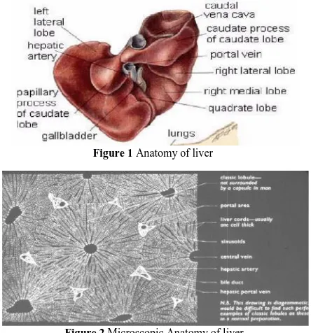
Anatomy of liver
The liver is the largest organ of the human body (Fig. 1), weights approximately 1500 g. It is located in the upper right corner of the abdomen. The organ is closely associated with the small intestine, processing the nutrient-enriched venous blood that leaves the digestive tract [3]. The liver is divided into 4 lobes: right, left, caudate and quadrate. The right and
left lobes are the largest, while the caudate and quadrate are smaller and located posteriorly. Two ligaments are visible anteriorly. The falciform ligament present superiorly, which separates the right and left lobes. The round ligament sitituated inferior to the falciform ligament, which protrudes from the liver slightly. Adjacent to the caudate lobe is the sulcus for the inferior vena cava. Just inferior to the caudate lobe is the portal hepatic, where the hepatic artery and hepatic portal vein enter the liver. The portal vein carries nutrient laden blood from the digestive system. Inferior to the portal hepatic is the bile duct which leads back to the gallbladder[4].
Microscopic Anatomy of liver
The basic functional unit of the liver is the liver lobule (Fig. 2). A single lobule is hexagonal in shape and its size similar to the sesame seed. It is made up of liver cells arranged in one-cell-thick plate like layers that radiate from the central vein to the edge of the lobule [5]. The primary structures in a lobule include plates of hepatocytes (liver cell) form the bulk of the lobule, portal triads at each corner of hexagon, central vein, liver sinusoids that run from the central vein to the portal
Key words:
Caffeine, hepatotoxicity, isoniazid, silymarin
triads, hepatic macrophages (Kupffer cells), bile canaliculi
(“little canals”)-formed between walls of adjacent hepatocytes, space of disse-a small space between the sinusoids and the hepatocytes[6].
Liver Cell Types
The liver has 5 cell types: Hepatocytes, kupffer cells, sinusoidal endothelial cells, bile duct epithelial cells, and ito cells. Mostof the liver’s synthetic and metabolic capabilities
stem from the work of hepatocytes. Kupffer cells are macrophages that reside in the sinusoids. These cells help to clear out old red blood cells and bacteria. These cell help in breakdown of heme (the iron-containing pigment in haemoglobin) into bilirubin, which is one of the chief pigments of bile[1]. Sinusoidal endothelial cells have large pores that allow most proteins to pass freely through the sinusoidal endothelium into the space of Disse, where they can make direct contact with hepatocytes. Bile duct epithelial cells line the interlobular bile ducts within the portal triads. Ito cells are found in the space of disse. They are important because when the liver is injured, the Ito cells transform into cells that produce collagen, which leads to liver fibrosis. If this occurs on a large scale, it can lead to cirrhosis of the liver[1].
Liver disease
Liver disease is condition that causes liver inflammation or tissue damage and affects liver function. Liver disease is categorized both by the cause and the effect on the liver. Causes may include infection, injury, exposure to drugs or toxic compounds, an autoimmune process, or a genetic defect that leads to the deposition and build-up of damaging substances such as iron or copper. Effects may include inflammation, scarring, obstructions, clotting abnormalities, and liver failure. There are various liver disease e.g. hepatitis
[7,8,9,10,11,12], acute liver failure[13,14], liver cancer[15,16], cirrhosis [17][18][19], budd-chiari syndrome[20], genetic[21,22], autoimmune
liver disease [23]. Alcoholic liver disease [24, 25, 26]obstruction
[27].
MATERIALS AND METHODS
Caffeine:
Caffeine was provided by truba institute of pharmacy bhopal
and purchased from K.M. traders, bhopal. The lethal dose of caffeine is too high to be of daily concern; the lethal dose is 170 mg/kg, or 12.5-14.6 g for an average adult male.
Isoniazide
Solonex is Isoniazide tablets. It was manufacture by macleod’s
pharmaceutical ltd and purchased from das medical store, karod (Bhopal). Hepatotoxicity was induced by daily dose of Isoniazide (250mg/kg p.o.) for 14 days.
Silymarin
Silymarin provided by Truba institute of pharmacy Bhopal. Silymarine manufactured by sigma aldrich and parches from K.M. treders bhopal (M.P.)
Animal care and handling
Westar albino rats of either sex weighing 150-250 g will use to observe for the hepatotoxicity and hepatoprotective activity of isoniazide and caffeine. The animals provide by animal house of Truba institute of Pharmacy, Bhopal. The animal were acclimatized to the standard laboratory condition in well cross ventilated animal house at temperature 25±2˚C relative humidity 44-56% and light and dark cycle of 10 and 14 hour respectively for one week before and during the experiment. The animal was fed with standard diet and water ad libitum. The experiment was approved by CPCSEA and the institutional ethics committee.
Grouping and dosing
Albino Westar rats of either sex (150-250g) will used. All the animals will divide into the six groups each group consists of 6 animals and they will receive the treatment as follows.
Group I: Control will receive propylene glycol for 14 days Group II: Test will receive INH (250mg/kg p.o.) for 14 days Group III: Standard will receive Standard drug (Silymarine 100 mg/kg, ip) + INH (250mg/kg p.o.) for 14 days
Group IV: Receive caffeine (100 mg/kg orally) + INH (250mg/kg p.o.) for 14.days
Group V: Receive caffeine (200 mg/kg orally) + INH (250mg/kg p.o.) for 14 days.
Group VI: Receive caffeine (300 mg/kg orally) + INH (250mg/kg p.o.) for 14 days.
Procedure for induce hepatotoxicity
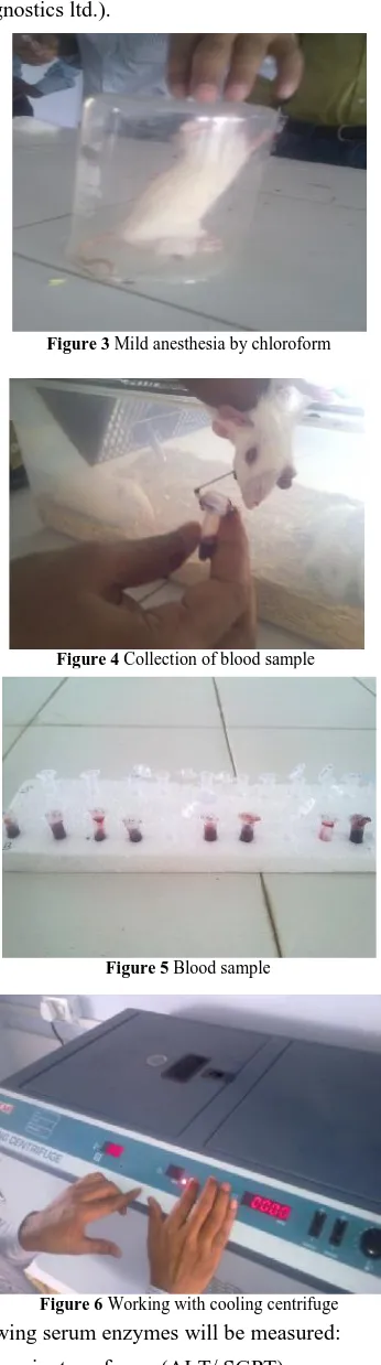
250 mg/kg Isoniazide administer orally for 14 day. The test drug will administered orally by suspending in water solution. Twenty-four hours after last dose of INH, bloods will obtain from all groups of rats by puncturing retro-orbital plexus. The blood samples will allow coagulating for 45 min at room temperature. Serum will separate by centrifugation at 3000 rpm at room temperature for 20 min and used for the biochemical estimation given in fig. 3, 4, 5, 6.
Biochemical estimation
The blood samples were collected without any anticoagulant and were allowed to clot for 10 min. at room temperature. The blood was centrifuge at 3000 rpm for 10 min up to 4˚C. The
Figure 1 Anatomy of liver
Figure 2 Microscopic Anatomy of liver
triads, hepatic macrophages (Kupffer cells), bile canaliculi
(“little canals”)-formed between walls of adjacent hepatocytes, space of disse-a small space between the sinusoids and the hepatocytes[6].
Liver Cell Types
The liver has 5 cell types: Hepatocytes, kupffer cells, sinusoidal endothelial cells, bile duct epithelial cells, and ito cells. Mostof the liver’s synthetic and metabolic capabilities
stem from the work of hepatocytes. Kupffer cells are macrophages that reside in the sinusoids. These cells help to clear out old red blood cells and bacteria. These cell help in breakdown of heme (the iron-containing pigment in haemoglobin) into bilirubin, which is one of the chief pigments of bile[1]. Sinusoidal endothelial cells have large pores that allow most proteins to pass freely through the sinusoidal endothelium into the space of Disse, where they can make direct contact with hepatocytes. Bile duct epithelial cells line the interlobular bile ducts within the portal triads. Ito cells are found in the space of disse. They are important because when the liver is injured, the Ito cells transform into cells that produce collagen, which leads to liver fibrosis. If this occurs on a large scale, it can lead to cirrhosis of the liver[1].
Liver disease
Liver disease is condition that causes liver inflammation or tissue damage and affects liver function. Liver disease is categorized both by the cause and the effect on the liver. Causes may include infection, injury, exposure to drugs or toxic compounds, an autoimmune process, or a genetic defect that leads to the deposition and build-up of damaging substances such as iron or copper. Effects may include inflammation, scarring, obstructions, clotting abnormalities, and liver failure. There are various liver disease e.g. hepatitis
[7,8,9,10,11,12], acute liver failure[13,14], liver cancer[15,16], cirrhosis [17][18][19], budd-chiari syndrome[20], genetic [21,22], autoimmune
liver disease [23]. Alcoholic liver disease [24, 25, 26]obstruction
[27].
MATERIALS AND METHODS
Caffeine:
Caffeine was provided by truba institute of pharmacy bhopal
and purchased from K.M. traders, bhopal. The lethal dose of caffeine is too high to be of daily concern; the lethal dose is 170 mg/kg, or 12.5-14.6 g for an average adult male.
Isoniazide
Solonex is Isoniazide tablets. It was manufacture by macleod’s
pharmaceutical ltd and purchased from das medical store, karod (Bhopal). Hepatotoxicity was induced by daily dose of Isoniazide (250mg/kg p.o.) for 14 days.
Silymarin
Silymarin provided by Truba institute of pharmacy Bhopal. Silymarine manufactured by sigma aldrich and parches from K.M. treders bhopal (M.P.)
Animal care and handling
Westar albino rats of either sex weighing 150-250 g will use to observe for the hepatotoxicity and hepatoprotective activity of isoniazide and caffeine. The animals provide by animal house of Truba institute of Pharmacy, Bhopal. The animal were acclimatized to the standard laboratory condition in well cross ventilated animal house at temperature 25±2˚C relative humidity 44-56% and light and dark cycle of 10 and 14 hour respectively for one week before and during the experiment. The animal was fed with standard diet and water ad libitum. The experiment was approved by CPCSEA and the institutional ethics committee.
Grouping and dosing
Albino Westar rats of either sex (150-250g) will used. All the animals will divide into the six groups each group consists of 6 animals and they will receive the treatment as follows.
Group I: Control will receive propylene glycol for 14 days Group II: Test will receive INH (250mg/kg p.o.) for 14 days Group III: Standard will receive Standard drug (Silymarine 100 mg/kg, ip) + INH (250mg/kg p.o.) for 14 days
Group IV: Receive caffeine (100 mg/kg orally) + INH (250mg/kg p.o.) for 14.days
Group V: Receive caffeine (200 mg/kg orally) + INH (250mg/kg p.o.) for 14 days.
Group VI: Receive caffeine (300 mg/kg orally) + INH (250mg/kg p.o.) for 14 days.
Procedure for induce hepatotoxicity
250 mg/kg Isoniazide administer orally for 14 day. The test drug will administered orally by suspending in water solution. Twenty-four hours after last dose of INH, bloods will obtain from all groups of rats by puncturing retro-orbital plexus. The blood samples will allow coagulating for 45 min at room temperature. Serum will separate by centrifugation at 3000 rpm at room temperature for 20 min and used for the biochemical estimation given in fig. 3, 4, 5, 6.
Biochemical estimation
The blood samples were collected without any anticoagulant and were allowed to clot for 10 min. at room temperature. The blood was centrifuge at 3000 rpm for 10 min up to 4˚C. The
Figure 1 Anatomy of liver
Hepatoprotective effect of caffeine against isoniazid-induced Hepatic damage in albino rats
41
obtained serum was stored at 4˚C for estimation of SGOT, SGPT and total bilirubin. This estimation was done according to the standard procedure given along with the perches kit (span diagnostics ltd.).
The following serum enzymes will be measured: Alanine aminotransferase (ALT/ SGPT) Aspartate aminotransferase (AST/SGOT) Total bilirubin (TB)
Estimation of serum SGOT
Principle
Aspartate aminotransferase (AST) catalyzes the transfer of the
amino group from L-aspartate to α-Ketoglutarate to yield oxalacetate and L-glutamate. The oxalacetate undergoes reduction with simultaneous oxidation of NADH to NAD in the malate dehydrogenase (MDH) catalyzed indicator reaction. The resulting rate of decrease in absorbance at 340nm is directly proportional to the AST activity. Lactate dehydrogenase (LDH) is added to prevent interference from endogenous pyruvate which is normally present in serum.
AST
L-Aspartate + α-Ketoglutarate Oxalacetate + L-Glutamate MDH
Oxalacetate + NADH + H+ L-Malate + NAD+ +H2O
Working reagent preparation:
Reagent 1, 2 and 4 are ready to use
Solution 1: dilute 1ml of reagent 3 to 10 ml with purified water.
Reagent storage and stability:
Reagent 1, 2 and 4 are stable at 2-8˚C and reagent 3 is stable at
15-30˚C till the expiry date mentioned.
Solution 1 is quite stable at room temperature (15-30˚C)
Procedure
Estimation of serum SGOT given in table 1.
Formula
Absorbance of test–
Absorbance of control
SGOT = × concentration of Absorbance of standard - standard
Absorbance of blank
Estimation of SGPT
Principle
ALT catalyzes the transfer of the amino group from L-alanine
to α- ketoglutarate resulting in the formation of pyruvate and L-glutamate. Lactate dehydrogenase catalyzes the reduction of pyruvate and the simultaneous oxidation of NADH to NAD. The resulting rate of decrease in absorbance is directly proportional to ALT activity.
ALT
L-Alanine + α-Ketoglutarate Pyruvate + L-Glutamate LDH
Pyruvate + NADH + H+ L-Lactate + NAD+ +H2O
Table 1 Estimation of serum SGOT
Pipette in to
the marked Blank Standard Test Control
Volume in ml
Reagent 1 0.25 0.25 0.25 0.25
Serum --- --- 0.05 ---Standard --- 0.05 ---
---Mix well and incubate at 37˚C for 60 minutes Reagents 2 0.25 0.25 0.25 0.25 Deionised water 0.05 --- ---
---Serum --- --- --- 0.05 Mix well and allow to stand at room temperature (15- 30˚C) for
20 minutes
Solution 1 0.25 0.25 0.25 0.25
Figure 3 Mild anesthesia by chloroform
Figure 4 Collection of blood sample
Figure 5 Blood sample
Working reagent preparation:
Reagent 1, 2 and 4 are ready to use
Solution 1: dilute1ml of reagent 3 to 10 ml with purified water. Reagent storage and stability:
Reagent 1, 2 and 4 are stable at 2-8˚C and reagent 3 is stable at
15-30˚C till the expiry date mentioned. Solution 1 is quite
stable at room temperature (15-30˚C)
Procedure
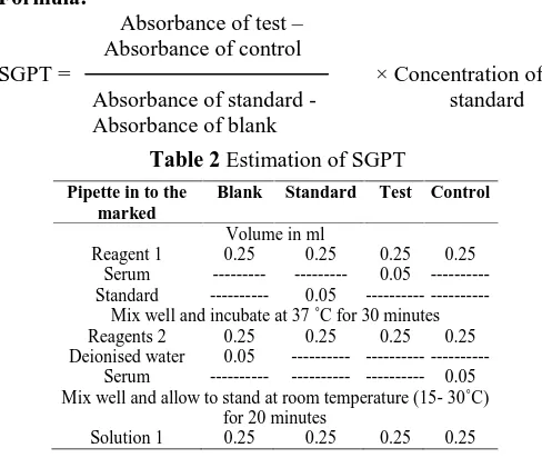
Estimation of SGPT given in table 2.
Formula:
Absorbance of test–
Absorbance of control
SGPT = × Concentration of Absorbance of standard - standard Absorbance of blank
Estimation of serum Bilirubin:
Principle
Bilirubin reacts with diazotized sulphanilic acid in acidic medium to form a pink colored azobilirubin with absorbance directly proportional to Bilirubin concentration.
Reagents:
Reagents 1: Diazo-A Reagents 2: Diazo-B Reagents 3: Diazo Blank Reagents 4: Methanol
Reagents 5: Artificial Slandered (10 mg % Bilirubin)
Preparation of working solution:
Diazo Reagents: just before use mix 1.0 ml of reagent 1with 0.03 ml of reagent 2
Procedure
Estimation of serum Bilirubin given in table 3.
Read optical density agents distilled water at 540 nm wavelength.
Formula
Optical density of T1–
Optical density of T2
Total A = × 10
Optical density of standard
RESULTS AND DISCUSSION
The prime objective of study was to investigate the hepatoprotective activity and effective dose of caffeine used to reduce the liver injury. The hepatoprotective activity screened by carbon tetrachloride, paracetamol and Isoniazide induced hepatotoxicity in male wistar albino rat. In this protocol hepatoxicity induce by oral administratation of Isoniazide in rat which leads to the hepatocellular necrosis in 14 day. The degree of liver damage was determined by biochemical parameter such as SGOT, SGPT, and total serum bilirubine and probably by histopathology. During metabolism of INH, hydrazine is produced directly (from INH) or indirectly (from acetyl hydrazine).
It is evident that hydrazine plays a role in INH-induced liver damage. It can cause moderate abnormalities in serum transaminase leading to hepatotoxicity; hence the measurement of serum transaminase is often advocated during INH administration, to assess the extent of INH-induced hepatotoxicity [28]. Administration of INH significantly elevated levels of AST, ALT, bilirubin, due to damaged structural integrity of the liver because these are cytoplasm in location and are released into circulation after cellular damage
[29]
.
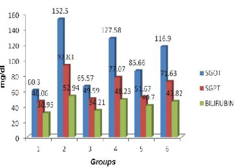
Caffeine treatment with INH prevented the INH-induced perturbations in the activities of AST, ALT, TB, in both the serum and liver tissue. It is likely that the INH-derived hepatotoxicity metabolites, namely acetyl. The results in this study were confirmed by Histopathological observation. In contrast to the control group, INH-intoxicant rat showed mild inflammatory cell infiltration and fatty changes, but rimonabant administration for 14 days attenuated this histopathological changes. Administration of INH at a dose of 250 mg/kg (p.o.) caused a significant rise (P<0.01) in level of serum marker enzymes such as AST, ALT, TB, Silymarin significantly (P<0.01) reduced these levels near to normal. A significant (P<0.01) decrease was observed in the AST, ALT, TB (Table 1) level in the animals treated with different doses of caffeine (10 mg/kg, 20 mg/kg and 30 mg/kg ) and showed dose dependant activity. At the dose of 20 mg/kg of caffeine showed comparable activity with the standard drug silymarin.
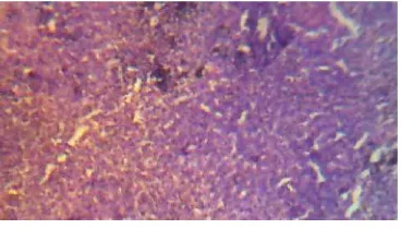
The observed Histopathological changes under medical microscope at 100X and then photomicrography of liver of experimental groups are shown in fig. 7,8,9,10,11,12. Group 1 showed the histopathology of normal control rats which showed in control rat, liver section showed normal hepatic cell with well preserved cytoplasm, prominent nucleus and nucleolus and central vein. The INZ group II treated rat liver showed there was hydropic change hepatocyte with single cell necrosis surrounded by neutrophils. Congestion of central vein and sinusoid were seen with acute inflammatory cell in filtering sinusoids mainly in central zone. The INZ +
Table 2 Estimation of SGPT
Pipette in to the marked
Blank Standard Test Control
Volume in ml
Reagent 1 0.25 0.25 0.25 0.25 Serum --- --- 0.05 ---Standard --- 0.05
---Mix well and incubate at 37˚C for30 minutes Reagents 2 0.25 0.25 0.25 0.25 Deionised water 0.05
---Serum --- --- --- 0.05 Mix well and allow to stand at room temperature (15- 30˚C)
for 20 minutes
Solution 1 0.25 0.25 0.25 0.25
Table 3 Estimation of serum Bilirubin
Reagents T1 T2
Serum/plasma, ml 0.05 0.05 Distilled Water, ml 0.45 0.45 Reagents3: Diazo Blank, ml -- 0.125
Diazo Reagents, ml 0.125 --Distilled Water, ml -- --Reagent 4: Methanol, ml 0.625 0.625
Hepatoprotective effect of caffeine against isoniazid-induced Hepatic damage in albino rats
43
Silymarine treated (group III) rat liver there was milled central venous congestion and milled fatty vacoulation.
Caffeine of dose 20 mg/kg show significant result as compared to other dose of caffeine treatment for 14 day as there liver section showed residual hepatocellular necrosis with cords of regenerating hepatocytes. INZ induce hepatotoxicity showing extensive area of confluent necrosis and also showing fatty change and hydropic degeneration.
Pre-treatment with silymarine showing complete normalization of liver architecture. Pre-treatment with caffeine showing better histology for 14 day treatment as there liver section showed partial protection of hepatocytes. Effect of caffeine on serum biochemical parameter in isoniazide induced hepatotoxicity in rat given in table 4. The observed Histopathological changes under medical microscope at 100X and then photomicrography of liver of experimental groups are shown in fig. 7,8,9,10,11,12 and result shown in graph 1.
No of animal in each group (n) = 6 INZ = Isoniazide
The data were analyzed by one way ANOVA followed by tukey multiple comparison Test.
Each values represents the mean ± SEM; n=6, *p<0.05, **p<0.01, ***p<0.001
a- Significant difference as compare to normal control group
b- Significant difference as compare to negative control group
c- Significant difference as compare to standard group d- Significant difference as compare to test I group e- Significant difference as compare to test II group f- Significant difference as compare to test III group
Histopathological study
Table 4: Effect of caffeine on serum biochemical parameter in Isoniazide induced hepatotoxicity in rat
Group Treatment Dose SGOT(U/L)
mean ± SEM
SGPT(U/L) mean ± SEM
Total bilirubine (mg/dl) mean ± SEM
I Vehicle 0.2 ml of
propylene glycol
60.30 ± 1.22 46.06 ± 1.49 30.95 ± 1.68
II INZ 250 mg/kg 152.50 ± 4.41a*** 92.83 ± 8.68a*** 52.94 ± 1.38a***
III Silymarin+ INZ 100 mg/kg + 250 mg/kg
65.57 ± 3.34 b***
49.59 ± 1.41 b***
34.21 ±1.46 b*** IV Caffeine+ INZ 10 mg/kg +
250 mg/kg
127.58 ± 6.61 a***c***
77.07 ± 2.67 a**c**
48.23 ± 2.46 a***c*** V Caffeine+ INZ 20 mg/kg +
250 mg/kg
85.66 ± 1.94 b***d**
51.67 ± 7.39 b***d*
40.70 ± 1.76 a**b*** VI Caffeine+ INZ 30 mg/kg +
250 mg/kg
116.90 ± 16.21 a***b*c***
71.63 ± 4.21 a*
45.82 ± 1.55 a***c***
Figure 7 Normal group
Figure 8 INZ treated group
Figure 9 Silymarine + INZ treated group
Figure 11 INZ + Caffeine (20 mg/kg) treated group Figure 10 INZ + Caffeine (10 mg/kg) treated group
CONCLUSION
The study was designed to evaluate the hepatoprotective activity of caffeine and effective dose, against albino rat in experimental liver damage induced by Isoniazide. Caffeine show significant protective effect by lowering the serum level of SGOT, SGPT, and total bilirubine of the liver. This biochemical observation was supplemented by histopathological examination of liver section. The activity of various doses was comparable to that of silymarine, a known hepatoprotective. In present pharmacological evaluation, the caffeine was extensively indigested for its hepatoprotective potential against INZ induces hepatotoxicity. At the end of this study, a strong conclusion can be drowning that caffeine
posse’s significant hepatoprotective activity. Caffeine of dose
20 mg/kg show better result as compared to 10 mg/kg and 30 mg/kg.
ACKNOWLEDGMENT
I’Dinesh Kr. Chauhan’ offer my sincere thanks to Mr. Hemant
nagar, head of department pharmacology, truba institute of pharmacy bhopal (M.P.) for his valuable suggestions and encouragement throughout the work.
References
1. Eric S. Allen. Human anatomy & physiology northeastern university. 2002; 3-12.
2. Maton, Anthea, Jean Hopkins, Charles William McLaughlin, Susan Johnson, Maryanna Quon Warner, David LaHart, Jill D. Wright. Human biology and health. Englewood Cliffs, New Jersey, USA: Prentice Hall. 1993; 1-13.
3. http://www.hepatitis.org.uk/s-crina/menu.htm 4. http://en.wikipedia.org/wiki/Main_Page
5. Henry Gray, Gray's Anatomy: The Anatomical Basis of Medicine and Surgery. 1995.
6. Romil saxena, Neil D Theise, James M Crawford. Microanatomy of the human liver, exploring the hidden interfaces, by the american association for the study of liver diseases 1999; 1339-1341.
7. Little JW, Falace DA, Miller CS, Rhodus N L. Dental management of the medically compromised patient. 5th ed. St. Louis: Mosby; 1997.
8. Gillcrist JA. Hepatitis viruses A, B, C, D, E and G: implications for dental personnel. J Am Dent Assoc. 1999;130:509-20.
9. Wolters LM, Niesters HG, de Man RA. Nucleoside analogues for chronic hepatitis B. Eur J Gastroenterol Hepatol 2001; 13:1499-506.
10. DePaola LG. Managing the care of patients infected with blood borne diseases. J Am Dent Assoc 2003; 134:350-8.
11. Matthews GV, Nelson MR. The management of chronic hepatitis B infection. Int J STD AIDS. 2001;12: 353-7.
12. Wisnom C, Siegel MA. Advances in the diagnosis and management of human viral hepatitis. Dent Clin North Am 2003; 47:431-47.
13. Mark Feldman, Lawrence S. Friedman, Lawrence J. Brandt. Sleisenger & Fordtran's gastrointestinal and liver disease pathophysiology, diagnosis, management. 9th ed. St. Louis, Mo.: MD Consult. 2009; 978-1-4160-6189-2.
14. Lee WM, Hynan LS, Rossaro L. Intravenous N-acetylcysteine improves transplant-free survival in early stage non-acetaminophen acute liver failure. Gastroenterology. 2009; 137: 856–64.
15. Kruskal JB, Goldberg SN. Emerging therapies for hepatocellular carcinoma: opportunities for radiologists. J Vasc Interv Radiol. 2002; 13:253-8. 16. Bergsland EK, Venook AP. Hepatocellular
carcinoma. Curr Opin Oncol. 2000; 12:357-61. 17. Zavaglia C, Airoldi A, Pinzello G. Antiviral therapy
of HBV and HCV-induced liver cirrhosis. J Clin Gastroenterol. 2000; 30:234-41.
18. Sokol RJ. Liver cell injury and fibrosis. J Pediatr Gastroenterol Nutr. 2002; 35:7-10.
19. Jones DE. Auto antigens in primary biliary cirrhosis. J Clin Pathol. 2000; 53:813-21.
20. http://labtestsonline.org/search-help.
21. William F. Balistreri, MD. Genetic Liver Disease, Medscape Today. 2003.
22. Gochee PA, Powell LW. What’s new in
hemochromatosis. Curr Opin Hematol. 2001; 8:98-104.
23. Hiromi Ishibashi, MD, Atsumasa Komori, MD, Shinji Shimoda, MD, M Eric Gershwin, MD. Guidelines for therapy of autoimmune liver disease. 2007, 214-217. 24. http://www.betterhealth.vic.gov.au/
25. Robert S. O’Shea, Srinivasan Dasarathy, Arthur J
McCullough. Alcoholic liver disease, Aasld practice guidelines. 2009; 309-319.
26. Gramenzi F, Caputo, M Biselli, F Kuria, E loggi, P Andreone, M Bernardi. Review article: alcoholic liver disease pathophysiological aspects and risk factors. Alimentary pharmacology and therapeutics. 2006; 1155-1157.
27. http://slabtestsonline.org/search-help
28. Mitchell JR, Zimmerman HJ, Ishak KG, Thorgiersson UP, Timbrell JA, Snodgrass WR: Isoniazide liver injury: clinical spectrum, pathology and probable pathogenesis. Ann Intern Med. 1976; 84:181-92. 29. Vaghasiya J, Bhalodia Y, Shailesh M, Jivani N,
Rathod S. Protective effect of polyherbal formulation on isoniazid induced hepatotoxicity in rats. Journal of Pharmacy Research. 2009; 2:4:610-614.
Figure 13 Effect of silymarine and caffeine on SGOT, SGPT, bilirubine