2014; 2(6): 345-350
Published online November 21, 2014 (http://www.sciencepublishinggroup.com/j/ajls) doi: 10.11648/j.ajls.20140206.13
ISSN: 2328-5702 (Print); ISSN: 2328-5737 (Online)
Genome-wide screen for
Escherichia coli
genes involved in
repressing cell-to-cell transfer of a nonconjugative
pSC101-derived plasmid
Yuka Shibata
1, Akiko Matsumoto
1, Mutsumi Horino
2, Akiko Hirabayashi
2, Kozue Shirota
2,
Chinatsu Kawano
2, Sumio Maeda
1, 2, *1
Graduate School of Humanities and Sciences, Nara Women's University, Nara, Japan
2
Faculty of Human Life and Environment, Nara Women's University, Nara, Japan
Email address:
[email protected] (S. Maeda)
To cite this article:
Yuka Shibata, Akiko Matsumoto, Mutsumi Horino, Akiko Hirabayashi, Kozue Shirota, Chinatsu Kawano, Sumio Maeda. Genome-Wide Screen for Escherichia coli Genes Involved in Repressing Cell-To-Cell Transfer of a Nonconjugative pSC101-Derived Plasmid. American Journal of Life Sciences. Vol. 2, No. 6, 2014, pp. 345-350. doi: 10.11648/j.ajls.20140206.13
Abstract:
Acquiring new genetic traits by lateral gene transfer is a bacterial strategy for environmental adaptations. We previously showed that Escherichia coli laterally transmits nonconjugative plasmids in cocultures that contain strains with or without the plasmid. Using a pMB1-derived plasmid and the Keio collection, a comprehensive library of E. coli knockout mutants for nonessential genes, we recently screened for genes responsible for promoting or repressing cell-to-cell plasmid transfer in recipient cells. In this study, we used a pSC101-derived plasmid, instead of a pMB1-derived plasmid, to screen for repressing genes and identified 29 “transfer-up” mutants. Among these, four mutants are common to those previously screened using a pMB1-derived plasmid. Although the roles of the 29 gene products in plasmid transfer mechanism remain uncertain, it is interesting that 28 of the 29 screened genes map to two limited regions on the E. coli chromosome: 18 genes at 34.25–35.31 min and 10 genes at 12.62–13.35 min. Because these two regions commonly contain termination (Ter) sites for DNA replication (TerC: 34.64 min and TerH: 12.91 min), it is possible that chromosomal mutations around specific Ter sites may affect plasmid acquisition in the recipient cells.Keywords:
Lateral Gene Transfer, Keio Collection, pSC101-Derived Plasmid, Ter Site, Escherichia coli1. Introduction
Bacteria adapt to varying environmental conditions through the application of lateral gene transfer between bacterial cells, ultimately resulting in bacterial evolution [1–3]. However, within human environments this results in the undesirable spread of pathogenic, antibiotic-resistant or artificially engineered genes [1, 4–8]. Bacteria use the following three mechanisms for lateral gene transfer: conjugation, transduction, and transformation [1]. For DNA transfer from donor to recipient cells, conjugation and transduction involve specific structures such as conjugative pili and phage capsids, respectively. However, transformation primarily involves recipient cells that express genetic competence for the uptake of free extracellular DNA [9, 10]. Transformation competence can be induced both naturally and artificially, although not all bacterial species develop natural competence [2, 9, 10].
Under natural conditions, Escherichia coli is not assumed to be transformable; however, it can develop high genetic competence under artificial conditions such as exposure to high Ca2+ concentrations [11, 12]. Recent reports showed that
E. coli could express modest genetic competence under certain conditions that may arise within its environment [13–20]. Relevant to these findings, we demonstrated that spontaneous lateral transfer of non-conjugative plasmids occurred in an E. coli mixed cell culture [21, 22]. On the basis of subsequent analyses, it was hypothesised that this cell-to-cell plasmid transfer resulted from a kind of transformation of plasmid DNA released from co-cultured cells [23]. However, the detailed molecular mechanisms underlying this process, including the genes involved, remain unknown.
responsible for regulating cell-to-cell plasmid transfer in recipient cells. The Keio collection is a comprehensive library of E. coli knockout mutants for 3,985 nonessential genes, which constitute 90% of all genes in the E. coli K-12 genome. This collection was used in several genome-wide screens for genes involved in various cellular functions [27–29]. We devised a 96-well microplate assay system for cell-to-cell plasmid transfer and screened eight “transfer-down” mutants [24] and 55 “transfer-up” mutants [25] (hereafter referred to as “down” mutants and “up” mutants, respectively) using the Keio strains as plasmid recipients.
In this study, we screened for “up” mutants using the pSC101-derived nonconjugative plasmid pGBM1 [23, 30], instead of the pMB1-derived plasmid pHSG399-F6, which was used in our previous study [25]. pHSG399-F6 exhibited a high transfer frequency, whereas pGBM1 exhibited a low transfer frequency [23, 31]; therefore, we considered pGBM1 more suitable for screening “up” mutants. In this study, we present our screening results and analyses of 29 new “up” mutants.
2. Materials and Methods
2.1. E. coli Strains, Plasmids and Materials
CAG18439 [32] [MG1655 derivative; F−, λ−, lacZ118(Oc), lacI3042::Tn10(tetr), rph-1], Keio strains [26] [F−, rrnB, ∆lacZ4787, HsdR514, ∆(araBAD)567, ∆(rhaBAD)568, rph-1,
∆(single gene)::kanr] and pGBM1 [30] were obtained from the National BioResource Project (NIG, Japan): E. coli
(http://www.shigen.nig.ac.jp/ecoli/strain/top/top.jsp).
Tetracycline (tet), streptomycin (str), polyethylene glycol (PEG; molecular mass = 8000) and Luria–Bertani powder (LB, Lennox) were purchased from Sigma. Tryptic soy broth (TSB) was purchased from Becton, Dickinson. Distilled water (DNase- and RNase-free, molecular biology grade) and kanamycin (kan) purchased were from Invitrogen. Microplates (96-well) and pin replicators were purchased from Nippon Genetics. Nylon 66-membrane filters (pore size: 0.45 µm, Biodyne A) were purchased from Pall. Agar powder (guaranteed reagent grade) and other general reagents were purchased from Wako.
2.2. Screen for “up” Mutants Involved in Cell-to-Cell Plasmid Transfer
We screened for “up” mutants using the Keio strains as recipient cells using 96-well microplates, according to a previously described protocol [25] with some modifications. Screening details are provided below.
To screen for “up” mutants in 96-well microplates, transformants were selected twice in liquid media containing two antibiotics. Plasmid-donor cells (CAG18439-harboring pGBM1) were pre-cultured in 10 ml of LB broth (tet: 75 µg/ml; str: 100 µg/ml) at 37°C for 22 h. Cultured donor cells were recovered by centrifugation and re-suspended in 7 ml of LB broth. Recipient cells (each Keio strain) were pre-cultured at 37°C for 22 h in 200 µl of LB broth (kan: 75 µg/ml) in
microplate wells, recovered by centrifugation and re-suspended in 50 µl of the above donor cell suspension. Each mixture (5 µl) of Keio and donor cells was inoculated onto TSB agar (1.5%) prepared in microplate wells and cultured in quadruplicate at 25°C (duplicate) and 37°C (duplicate) for 16 h.
The cultured cells in wells were re-suspended in 100 µl of LB broth. Small amounts (approximately 0.2 µl) of this suspensions were transferred with a 96-pin replicator to 100 µl of the first selection LB broth, which contained str (75 µg/ml for plasmids) and kan (75 µg/ml for Keio strains), and subsequently cultured at 37°C for 16 h. The same manipulations were repeated for the second selection. The turbidities (optical density 600 nm) of the resulting second selection cultures were determined using a microplate reader (Multiskan JX, Thermo Fisher Scientific). Wells that showed apparent cell growth were counted.
These quadruplicate screens showed in four results per Keio strain. Under these conditions, most Keio mutants resulted in 0/4 positive wells. The mutants that produced 2/4–4/4 positive wells were regarded as “up” mutants.
For a plating assay for plasmid transfer in “up” mutants, mixed cultures in each microplate well were plated onto LB agar plates that contained two antibiotics (str: 75 µg/ml and kan: 75 µg/ml) to confirm the production of double-resistant cells. To estimate plasmid transfer frequency in each “up” mutant, a plating assay was also used with a colony biofilm on TSB agar (1.5%) prepared in a polystyrene plate (ø 90 mm), as previously described [25].
2.3. Plasmid Isolation from the Produced Transformants
Double-resistant colonies that appeared in a plating assay were cultured in fresh LB broth that contained str and kan overnight at 37°C and were subsequently used for plasmid preparation. Plasmids were isolated, digested with EcoRI and analyzed by 0.8% agarose-gel electrophoresis using conventional methods [33].
2.4. Data Analyses for Screened Genes
Information for the screened genes was obtained from the
following databases: PEC
(http://www.shigen.nig.ac.jp/ecoli/pec/index.jsp), Uniprot
(http://www.uniprot.org/), EchoBASE
(http://www.york.ac.uk/res/thomas/index.cfm), GenoBase (http://ecoli.naist.jp) and EcoCyc (http://ecocyc.org/).
3. Results and Discussion
3.1. Screen for “up” Mutants and Confirmation of Transformants
agar medium in microplate wells. The transformants were subsequently selected twice by culturing portions of these cocultured cells in liquid media that contained two antibiotics (kan and str). If Keio strains had acquired plasmids, double-resistant cells appeared and selectively grew in these wells. The numbers of positive wells for four experiments for each Keio strain were counted.
Under these conditions, most samples resulted in 0/4 positive wells. However, 133 strains produced 1/4 or more positive wells and 36 strains produced 2/4–4/4 positive wells. We analyzed these 36 strains to confirm the occurrence of double-resistant cells using a plating assay (transformant selection on agar plates). Ultimately, we obtained 29 positive strains.
To ensure that no pseudo-positive mutants may result from unexpected str resistance arising in the original Keio mutants, we examined the str sensitivities of the original 29 plasmid-free Keio strains. None of the 29 original strains exhibited str resistance. In addition, the presence of plasmids in the screened cells was determined by plasmid isolation from new colonies that appeared in a plating assay. This confirmed that all 29 strains had full-length plasmids (Fig. 1). Because these 29 strains also exhibited obvious kan resistance, which is characteristic of the Keio strains, we concluded that the double-resistant cells that appeared after the cocultures were indeed transformants of the Keio strains that had
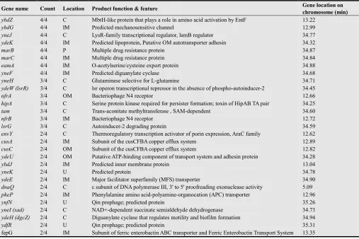
acquired plasmids from cocultured donor cells. Therefore, we selected these 29 mutants as “up” mutants (Table 1). The plasmid transfer frequencies of these “up” mutants were in the range of approximately 10−9–10−8 using a plating assay, whereas those of most other Keio strains were below the limit of detection (approximately < 10−10).
Figure 1. Agarose gel electrophoresis results for EcoRI-digested plasmids isolated from the double-resistant colonies obtained by a plating assay used for plasmid-transfer experiments. The arrows indicate the bands for pGBM1 (4028 bp). Lane M: size markers (λ Hind III); lanes n1–n3: negative controls [DNA prepared from CAG18439 (n1) and original Keio strains (n2: ybdG, n3: yneJ)]; lanes p1–p2: positive controls (pGBM1); and lanes 1–29: plasmids prepared from new double-resistant colonies of Keio strains (1: ybdZ, 2: ybdG, 3: yneJ, 4: ydeK, 5: marB, 6: marC, 7: eamA, 8: yneF, 9: yneH, 10: ydeW, 11: nfrA, 12: hipA, 13: tam, 14: nfrB, 15: lsrG, 16: envY, 17: cusA, 18: cusC, 19: ydeU, 20: ybdJ, 21: yneK, 22: ydeE, 23: dnaQ, 24: pheP, 25: ynfN, 26: yneI, 27: ydeH, 28: ydfR and 29: fepG).
Table 1. Repressive genes that were screened in this study and their features
Gene name Count Location Product function & feature Gene location on chromosome (min) ybdZ 4/4 C MbtH-like protein that plays a role in amino acid activation by EntF 13.22
ybdG 4/4 IM Predicted mechanosensitive channel 12.99 yneJ 4/4 C LysR-family transcriptional regulator, lamB regulator 34.77 ydeK 4/4 IM Predicted lipoprotein, Putative OM autotransporter adhesin 34.32
marB 4/4 P Multiple drug resistance protein 34.87
marC 4/4 IM Multiple drug resistance protein 34.84
eamA 4/4 IM O-acetylserine/cysteine export protein 34.88
yneF 4/4 IM Predicted diguanylate cyclase 34.68
yneH 3/4 C Glutaminase selective for L-glutamine 34.71
ydeW (lsrR) 3/4 C lsr operon transcriptional repressor in the absence of phospho-autoinducer-2 34.45
nfrA 3/4 OM Bacteriophage N4 receptor 12.66
hipA 3/4 C Serine protein kinase required for persister formation; toxin of HipAB TA pair 34.25
tam 3/4 C Trans-aconitate methyltransferase , SAM-dependent 34.60
nfrB 3/4 IM Bacteriophage N4 receptor 12.72
lsrG 3/4 C Autoinducer-2 degrading protein 34.59
envY 2/4 C Thermoregulatory transcription activator of porin expression, AraC family 12.62
cusA 2/4 IM Subunit of the cusCFBA copper efflux system 12.89
cusC 2/4 OM Subunit of the cusCFBA copper efflux system 12.82
ydeU 2/4 OM Putative ATP-binding component of transport system and adhesin protein 34.28
ybdJ 2/4 IM Predicted inner membrane protein 13.04
yneK 2/4 U Predicted protein 34.78
ydeE 2/4 IM Major facilitator superfamily (MFS) transporter 34.90
dnaQ 2/4 C ε subunit of DNA polymerase III, 3' to 5' proofreading exonuclease activity 5.09
pheP 2/4 IM Phenylalanine amino acid-polyamine-organocation (APC) transporter 12.96
ynfN 2/4 U Qin prophage; predicted protein 35.26
yneI (sad) 2/4 C NAD+-dependent succinate semialdehyde dehydrogenase 34.73
ydeH (dgcZ) 2/4 C Diguanylate cyclase that regulates motility and biofilm formation 34.94
ydfR 2/4 U Qin prophage; predicted protein 35.31
fepG 2/4 IM Subunit of ferric enterobactin ABC transporter and Ferric Enterobactin Transport System 13.35
3.2. Analyses of Data of the Screened Genes
Table 1 summarizes the known characteristics of the 29 screened genes. Although we could not find direct linkages between the functions of the screened genes and plasmid transfer, we analyzed the following.
Half of these genes (15 of 29) were associated with cell-surface proteins, such as those in the inner membrane, outer membrane, and periplasmic space, which is consistent with the transmembrane phenomenon. Four genes were estimated to be involved in intra- and intercellular signaling with cyclic-di-GMP (ydeH and yneF) [34, 35] and autoinducer-2 (ydeW and lsrG) [36, 37], suggesting the involvement of various known signal transduction pathways. Another four genes (ybdG, yneJ, marC and envY) were the same as those that were screened in our previous study [25] using a pMB1-derived plasmid; however, the remaining screened genes (25 of 29) were different. No obvious functional connections were observed between the genes screened in this study and the eight genes for the “down” mutants previously screened [24] using a pMB1-derived plasmid. These results suggests that the above four common genes (ybdG, yneJ, marC and envY) may share plasmid-transfer mechanisms with different plasmids, although there may be some dissimilarities in these mechanisms. None of the 29 genes in Table 1 have been reported to be involved in DNA transfer or DNA uptake in E. coli or other bacteria, which suggests the uniqueness of this phenomenon, as also previously suggested [23–25, 31].
Moreover, by carefully surveying our results, interestingly, 28 of the 29 screened genes mapped to two limited regions on the E. coli chromosome; 18 genes at 34.25–35.31 min and 10 genes at 12.62–13.35 min (Table 1). The well positions of the 29 screened strains in the storage and culture microplates were distributed into separate plates and wells; therefore the concentration in gene loci is not an artifact resulting from the accidental contamination of one or a few specific plates. As additional supporting evidence, among the genes for the 97 strains that showed 1/4 positive wells in our initial screening, another 16 genes also mapped to these two chromosomal regions. Moreover, among the 55 “up” mutants screened in our previous study [25], 10 genes [6 genes (ydeW, dcp, ydbK,
ydfT, ybcS and fimZ) in addition to the above 4 common genes (ybdG, yneJ, marC and envY)] belong to these two regions.
We observed that these two chromosomal regions commonly contained termination (Ter) sites for DNA replication (TerC: 34.64 min and TerH: 12.91 min) [38–40, http://www.ecogene.org/old/topic.php?topic_id=228]. TerC is considered the predominant Ter site utilized in vivo. TerH is a weak but functional site, and TerI (13.46 min) is also in close proximity to TerH. Therefore, chromosomal mutations around specific Ter sites may induce structural or physiological disturbance in replication of chromosomal DNA and affect plasmid acquisition in the recipient cells. The product of dnaQ, the other gene screened in this study, is the ε subunit of DNA polymerase III [41] (Table 1). This appears to be consistent with the above idea that some form of DNA synthesis disorder
may affect plasmid acquisition.
It is currently uncertain which is more important: individual functions of screened genes, DNA synthesis disorder or both. There may be rather complex mechanisms that vary according to the types of plasmid replicons. However, the results of this study include several interesting directions for investigating the molecular mechanisms of cell-to-cell plasmid transfer. Investigations that are more detailed will be necessary to unravel the overall mechanism of cell-to-cell plasmid transfer.
4. Conclusion
In this study, we have screened for genes responsible for repressing cell-to-cell plasmid transfer in E. coli using a pSC101-derived plasmid and the Keio collection. We identified 29 “transfer-up” mutants. Among these, four mutants are common to those previously screened using a pMB1-derived plasmid. It is interesting that 28 of the 29 screened genes map to two limited regions on the E. coli
chromosome: 18 genes at 34.25–35.31 min and 10 genes at 12.62–13.35 min. Because these two regions commonly contain termination (Ter) sites for DNA replication (TerC: 34.64 min and TerH: 12.91 min), it is possible that chromosomal mutations around specific Ter sites may affect plasmid acquisition in the recipient cells.
Acknowledgements
This work was supported by JSPS KAKENHI Grant Number 25292051. We are grateful for the Keio strains provided by the National BioResource Project (NIG, Japan) and Prof. H. Mori (NAIST, Japan). We would like to thank Enago (www.enago.jp) for the English language review.
References
[1] F. Bushman, Lateral DNA Transfer, Cold Spring Harbor Laboratory Press, Cold Spring Harbor, New York, USA, 2002.
[2] M. G. Lorenz and W. Wackernagel, “Bacterial gene transfer by natural transformation in the environment,” Microbiol Rev, vol. 58, pp. 563–602, 1994.
[3] C. M. Thomas and K. M. Nielsen, “Mechanisms of, and barriers to, horizontal gene transfer between bacteria,” Nat Rev Microbiol, vol. 9, pp. 711–721, 2005.
[4] P. S. Duggan, P. A. Chambers, J. Heritage, and J. M. Forbes, “Survival of free DNA encoding antibiotic resistance from transgenic maize and the transformation activity of DNA in ovine saliva, ovine rumen fluid and silage effluent,” FEMS Microbiol Lett, vol. 191, pp. 71–77, 2000.
[5] P. Keese, “Risks from GMOs due to horizontal gene transfer,” Environ Biosafety Res, vol. 7, pp. 123–149, 2008.
[7] B. G. Kelly, A. Vespermann, and D. J. Bolton, “Horizontal gene transfer of virulence determinants in selected bacterial
foodborne pathogens,” Food Chem Toxicol
doi:10.1016/j.fct.2008.02.007, 2008.
[8] A. Pontiroli, A. Rizzi, P. Simonet, D. Daffonchio, T. M. Vogel, and J. M. Mnier, “Visual evidence of horizontal gene transfer between plants and bacteria in the phytosphere of transplastomic Tobacco,” Appl Environ Microbiol, vol. 75, pp. 3314–3322, 2009.
[9] I. Chen and D. Dubnau, “DNA uptake during bacterial trans formation,” Nat Rev Microbiol, vol. 3, pp. 241–249, 2004
[10] D. Dubnau, “DNA uptake in bacteria,” Annu Rev Microbiol, vol. 53, pp. 217–244, 1999.
[11] D. Hanahan, “Studies on transformation of Escherichia coli
with plasmids,” J Mol Biol, vol. 166, pp. 557–580, 1983.
[12] M. Mandel and A. Higa, “Calcium-dependent bacteriophage DNA infection.” J Mol Biol, vol. 53, pp. 159–162, 1970.
[13] F. Bauer, C. Hertel, and W. P. Hammes, “Transformation of
Escherichia coli in foodstuffs,” Syst Appl Microbiol, vol. 22, pp. 161–168, 1999.
[14] B. Baur, K. Hanselmann, W. Schlimme, and B. Jenni, “Genetic transformation in freshwater: Escherichia coli is able to develop natural competence,” Appl Environ Microbiol, vol. 62, pp. 3673–3678, 1996.
[15] Y. Ishimoto, S. Kato, and S. Maeda, “Freeze-thaw-induced lateral transfer of non-conjugative plasmids by in situ transformation in Escherichia coli in natural waters and food extracts,” World J Microbiol Biotechnol, vol. 24, pp. 2731–2735, 2008.
[16] S. Maeda, N. Kakihara, and Y. Koishi, “Competency development of Escherichia coli in foodstuffs,” Microbes Environ, vol. 18, 100–103, 2003.
[17] S. Maeda, A. Sawamura, and A. Matsuda, “Transformation of colonial Escherichia coli on solid media,” FEMS Microbiol Lett, vol. 236, pp. 61–64, 2004.
[18] D. Sun, Y. Zhang, Y. Mei, H. Jiang, Z. Xie, H. Liu, X. Chen, and P. Shen, “Escherichia coli is naturally transformable in a novel transformation system,” FEMS Microbiol Lett, vol. 265, pp. 249–255, 2006.
[19] S. D. Tsen, S. S. Fang, M. J. Chen, J. Y. Chien, C. C. Lee, and D. H. Tsen, “Natural plasmid transformation in Escherichia coli,” J Biomed Sci, vol. 9, pp. 246–252, 2002.
[20] M. Woegerbauer, B. Jenni, F. Thalhammer, W. Graninger, and M. Burgmann, “Natural genetic transformation of clinical isolates of Escherichia coli in urine and water,” Appl Environ Micobiol, vol. 68, pp. 440–443, 2002.
[21] S. Maeda, M. Ito, T. Ando, Y. Ishimoto, Y. Fujisawa, H. Takahashi, A. Matsuda, A. Sawamura, and S. Kato, “Horizontal transfer of nonconjugative plasmids in a colony biofilm of Escherichia coli,” FEMS Microbiol Lett, vol. 255, pp. 115–120, 2006.
[22] T. Ando, S. Itakura, K. Uchii, R. Sobue, and S. Maeda, “Horizontal transfer of non-conjugative plasmid in colony biofilm of Escherichia coli on food-based media,” World J Microbiol Biotechnol, vol. 25, pp. 1865–1869, 2009.
[23] R. Etchuuya, M. Ito, S. Kitano, F. Shigi, R. Sobue, and S. Maeda, “Cell-to-cell transformation in Escherichia coli: a novel type of natural transformation involving cell-derived DNA and a putative promoting pheromone, PLoS ONE, vol. 6, e16355, 2011.
[24] N. Kurono, A. Matsuda, R. Etchuya, R. Sobue, Y. Sasaki, M. Ito, T. Ando, and S. Maeda, “Genome-wide screening of
Escherichia coli genes involved in execution and promotion of cell-to-cell transfer of non-conjugative plasmids: rodZ
(yfgA) is essential for plasmid acceptance in recipient cells,” Biochem Biophys Res Commun, vol. 421, pp. 119–123, 2012.
[25] A. Matsuda, N. Kurono, C. Kawano, K. Shirota, A. Hirabayashi, M. Horino, R. Etchuya, R. Sobue, Y. Sasaki, S. Miyaue, A. Sekoguchi, C. Sugiura, Y. Shibata, M. Ito, T. Ando, and S. Maeda, “Genome-wide screen for Escherichia coli
genes involved in repressing cell-to-cell transfer of non-conjugative plasmids,” Biochem Biophys Res Commun, vol. 482, pp. 445–450, 2012.
[26] T. Baba, T. Ara, H. Hasegawa, Y. Takai, Y. Okumura, M. Baba, K. Datsenko, M. Tomita, B. L. Wanner, and H. Mori, “Construction of Escherichia coli K-12 in-frame, single-gene knockout mutants: the Keio collection,” Mol Syst Biol, vol. 2, 2006.0008, 2006.
[27] T. Inoue, R. Shingaki, S. Hirose, K. Waki, H. Mori, and K. Fukui, “Genome-wide screening of genes required for swarming motility in Escherichia coli K-12,” J Bacteriol, vol. 189, pp. 950–957, 2007.
[28] V. Sanchez-Torres, T. Maeda, and T. K. Wood, “Global regulator H-NS and lipoprotein NlpI influence production of extracellular DNA in Escherichia coli,” Biochem Biophys Res Commun, vol. 401, pp. 197–202, 2010.
[29] O. Sharma, K. A. Datsenko, S. C. Ess, M. V. Zhalnina, B. L. Wanner, and W. A. Cramer, “Genome-wide screens: novel mechanisms in colicin import and cytotoxicity,” Mol Microbiol, vol. 73, pp. 571–585, 2009.
[30] D. Manen, M. Pougeon, P. Damay, and J. J. Geiselmann, “A sensitive reporter gene system using bacterial luciferase based on a series of plasmid cloning vectors compatible with derivatives of pBR322,” Gene, vol. 186, pp. 197–200, 1997.
[31] R. Sobue, N. Kurono, R. Etchuya, and S. Maeda, “Identification of a novel DNA element that promotes cell-to-cell transformation in Escherichia coli,” FEBS Lett, vol. 585, pp. 2223–2228, 2011.
[32] M. Singer, T. A. Baker, G. Schnitzler, S. M. Deischel, M. Goel, W. Dove, K. J. Jaacks, A. D. Grossman, J. W. Erickson, and C. A. Gross, “A collection of strains containing genetically linked alternating antibiotic resistance elements for genetic mapping of Escherichia coli,” Microbiol Rev, vol. 53, pp. 6408–6411, 1989.
[33] J. Sambrook, E. F. Fritsch, and T. Maniatis, Molecular Cloning: a Laboratory Manual, 2nd ed. Cold Spring Harbor Laboratory Press, Cold Spring Harbor, New York, USA, 1989.
[34] T. L. Povolotsky and R. Hengge, “Life-style' control networks in Escherichia coli: signaling by the second messenger c-di-GMP,” J Biotechnol, vol. 160, pp. 10–16, 2012.
[36] W. R. Galloway, J. T. Hodgkinson, S. D. Bowden, M. Welch, and D. R. Spring, “Quorum sensing in Gram-negative bacteria: small-molecule modulation of AHL and AI-2 quorum sensing pathways,” Chem Rev, vol. 111, pp. 28–67, 2011.
[37] C. S. Pereira, J. A. Thompson, and K. B. Xavier, “AI-2-mediated signalling in bacteria,” FEMS Microbiol Rev, vol. 37, 156–181, 2013.
[38] E. Esnault, M. Valens, O. Espe´ li, and F. Boccard, “Chromosome structuring limits genome plasticity in
Escherichia coli,” PLoS Genet, vol. 3, e226, 2007.
[39] C. Neylon, A. V. Kralicek, T. M. Hill, and N. E. Dixon,
“Replication termination in Escherichia coli: structure and antihelicase activity of the Tus-Ter complex,” Microbiol Mol Biol Rev, vol. 69, pp. 501–526, 2005.
[40] A. Thiel, M. Valens, I. Vallet-Gely, O. Espeli, and F. Boccard, “Long-range chromosome organization in E. coli: a site-specific system isolates the Ter macrodomain,” PLoS Genet, vol 8, e1002672, 2012.
[41] R. S. Scheurmann, S. Tam, P. M. Burgers, C. Lu, and H. Echols, “Identification of the E-subunit of Escherichia coli