Effect Of Fluoride Release Of Different Restorative
Materials On Remineraliation Of Dentin
Alaa M. Moharam, Sameh M. Nabih, Nady M. Ibrahim
Restorative Dentist, Cairo University Assistant chairman of Restorative Department, Police Hospital Nasr City, Cairo, Egypt, + 01064452291
Professor & Head of Operative Dentistry Department, Faculty of Dental Medicine, Al Azhar University Cairo, Egypt, + 01005267707
Assistant Professor of Operative Dentistry Department, Faculty of Dental Medicine, Al Azhar University Cairo, Egypt, + 01227802241
Abstract: Background: Dental caries is one of the most prevalent chronic diseases affecting humanity, its progression or control depends on the balance between pathological and protective factors, and the best strategy for caries management is focused on methods of improving the remineralizing process. Three different fluoride restorative materials were used and compared to study the effect of fluoride release on remimeralization of dentin. Objectives: To evaluate the fluoride release of three different fluoride releasing restorative materials, Fuji IX GC, Fuji II LC and Beautifil injectable and their effects on remineralization of dentin before and after recharging. Materials and Methods:
The experiement required measurement of cumulative fluoride release, through a microprocessor based portable fluoride meter by subjecting three groups of fluoride release restorative materials (Fuji IX GC, Fuji II LC and Beautifil injectable) to pH cycling three times daily for twenty minutes between the artificial saliva for neutralization at pH 7 and citric acid at PH4. The cumulative fluoride release measured at one week, four weeks and twelve weeks. The remineraliation of dentin is assessed through dentin restorative materials samples exposed to pH cycling and measured by measurement of fluoride uptake into dentinal tubules at one week and twelve weeks before and after recharging. Through using quantitative environmental scanning electron microscope (QESEM). Results: Regarding cumulative fluoride release assessment Fuji IX GC is the highest statistically significant than other tested fluoride releasing restorative material at the three aging periods tested at this study and at the two aging media exposed the three tested materials ,and regarding fluoride uptake assessment beautifil injectable highly significant difference than other two tested restorative materials at both aging periods and both aging media tested at this study. Also, the results revealed that there was no statistically significant difference between fluoride uptake values after recharging. Conclusion: Under the circumstances of this study conventional glass ionomer restorative material is the martial of choice regarding fluoride release represented by Fuji IX GC, the glass ionomer modified resin composite (Giomer represented by newly introduced beatifil injectable) has the advantage of fluoride uptake and remineralization.no statistical significance before and after recharging of glass ionomer rstoratives.
Keywords: Giomr,Fluoride release, Fluoride uptake, Remineralization.
1. Introduction
Dental caries is one of the most prevalent chronic disease affecting humanity. Its progression or control depends on the balance between pathological and protective factors, and the best strategy for caries management is focused on methods
of improving the remineralizing process(1). Tooth
remineralization is a naturally occurring process in the oral cavity(2). It is defined as a process in which calcium and phosphate ions are sourced to promote ion deposition into crystal voids in demineralised enamel. Remineralization remains imperative towards the management of non-cavitated carious lesions and prevention of disease progression within the oral cavity. The process also has the ability to contribute towards restoring strength and function within tooth structure(3). Tooth demineralization chemical process by which minerals (mainly calcium) are removed from any of the hard tissues - enamel, dentine, and cementum(4). The process of demineralization begins at the crystal surface found inside the hard tooth tissue and may progress into cavitation unless arrested or overridden by remineralization. The effect of demineralization can be reversed if there is sufficient time to allow remineralization to occur to counteract the acids in the oral cavity(5). Together, demineralization and remineralization contribute towards a
(6)
necessary, the use of materials with minimal aggression to
the tooth structure and cariostatic, adhesive, and
bit wandering to the amount that was really released(12). In spite of the fact that fluoride releasing products directly contact the cavity wall, but there is little evidence regarding their diffusion and their effect on surrounding tooth structure(13). The ability of a restorative to act as a fluoride reservoir is mainly dependent on the type and permeability of filling materials, on the frequency of fluoride exposure and on the kind and concentration of fluoridating agent(14). Various methods of aging (deionized water, distilled water and artificial saliva), periods of aging (days or months) and methods of fluoride recharge are contributing factors(15). From the previous review, it was speculated that it would be of importance to evaluate the effect of fluoride release of different restorative materials on remineralization uptake of dentin. through measurement of Measurement of fluoride release of each restorative material used in this study,
Measurement and comparing fluoride uptake
remineralization of dentin before and after recharging.
2. Materials and Methods:
Three Restorative materials used in this study; Conventional glass ionomer system, Fuji IXGC capsule with its. GC conditioner. Resin modified glass ionomer, Fuji IILC capsules. Glass ionomer modified resin composite, giomer, ,Beautiful injectable restorative material with its FL-Bond II bonding agent. Three Solutions used for pH cycling, Citric acid at pH 4, artificial saliva at pH 7. Two standard fluoride calibration solutions HI70702 and HI70703; were used for checking the electrode potential and calibrating the fluoride meter. TISAB (total ionic strength adjustment buffer); it was used for measuring fluoride ions as it provides a constant background ionic strength.
1. Assessment of fluoride release
1.1. Grouping of specimens:
A total of 30 specimens were prepared in this study, they were divided into three equal groups according to the type of the restorative material used Fuji IX, Fuji II LC and Beautifil injectable, specimens of each group were divided into two subgroups (five specimens each) according to the immersion media; either artificial saliva or citric acid,the specimens of the subgroup artificial saliva were always kept immersed in the artificial saliva at pH 7 the specimens of the subgroup citric acid were exposed to pH cycling between the artificial saliva at pH 7 and the citric acid at pH 4. For each group the cumulative fluoride release was assessed at one week, four weeks and twelve weeks.
1.2. Specimen preparation:
A specially designed Teflon mold was used to prepare the specimens discs. The mold was 2mm thick and has a circular hole in its middle with an internal diameter of 3mm. The Teflon mold was placed on a glass slab over a transparent polyester strip, and then the material was packed and the excess was removed using sharp # 12 bard barker scalpel blade. Another polyester strip and another glass slab were placed on the top of the mold; to ensure surface smoothness a constant 200g load was applied. After the removal of the upper glass slab, the material was light cured with LED visible light curing unit (500mW/cm2 intensity) at zero distance according to the manufacturer's instructions except
for the chemically cured conventional glass ionomer (Fuji IXGC) material which was left to set at room temperature.
1.3. Storage of specimens:
Each specimen was placed in a glass tube containing 5ml artificial saliva which was changed weekly and stored at room temperature. All tubes were properly covered with rubber stoppers to prevent evaporation. Specimens of the subgroup; M1 the immersion media was artificial saliva at pH 7, throughout the whole study period. While specimens of subgroups M2 citric acid were exposed to pH cycling regimen; between citric acid at pH 4 and artificial saliva at pH 7. The pH cycling regimen consisted of three cycles in acidic medium; in which the specimens were immersed in 5ml of the acid for 20 minutes three times daily with the intervening period in the artificial saliva. This pH cycling protocol was done at room temperature and repeated daily for 12 weeks.
1.4. Fluoride release assessment:
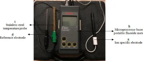
The artificial saliva was analyzed for fluoride with the use of microprocessor based portable fluoride meter * figure (1). Through the use of the ion specific electrode (a) connected to a microprocessor based portable fluoride meter (b). Every week prior to fluoride release measurements from the different materials, the fluoride meter was calibrated. The calibration was in the middle of the range at 100mg/L (ppm) using 100 ppm fluoride solution (HI 70703) and the other point was selected for low fluoride measurements at 10 mg/L (ppm) using 10 ppm fluoride solution (HI 70702). In order to obtain an accurate result, the meter was calibrated at a temperature not more than 20±3C. A separate stainless steel temperature probe (c) was supplied and connected to the meter. It was used to measure and adjust the temperature of the artificial saliva. The reading was shown on the lower part of liquid crystal display. A separate reference electrode (d) was also supplied and connected to the portable fluoride meter. It was immersed with the fluoride electrode and the
temperature probe in the sample solution during
measurement of fluoride concentration in order to guarantee an accurate and repeatable measurement every time. The 5 ml storage medium of each specimen was added to 0.5 ml of readymade total ionic strength adjustment buffer solution TISAB II (HI 4010-05) in ratio 10:1 according to the manufacturer's instructions. The three connected electrodes were properly immersed in the solution in order to assure accurate readings of fluoride released in the artificial saliva. The results were displayed on the upper part of liquid crystal display (LCD) directly in fluoride concentration and expressed in ppm (mg/L).
2. Assessment of fluoride uptake of dentin, remineralization assessment
2.1. tooth selection:
Non-carious and non-restored freshly extracted human posterior teeth were used in this study. After extraction, the teeth were thoroughly washed under running water to remove soft debris. Defected or cracked tooth was excluded and replaced by another one. The teeth used for this study were stored in deionized distilled water at room temperature till the start of the study.
2.2. Grouping of specimen:
A total of 48 specimens were prepared in this study, they were divided into three equal groups according to the type of restorative material used Fuji IX, Fuji IILC and Beautifil injectable 18 specimens each. Each group were divided into two subgroups nine specimens each according to the immersion media, either artificial saliva or citric acid. Each subgroup was classified into three classes according to storage period three specimens each either one week, four weeks, and twelve weeks which represent specimens that was measured after fluoride recharging. Following each storage period the teeth were sectioned and the amount of fluoride uptake was assessed at the axial wall of the cavity at distance 50 u from dentin/restoration interface.
2.3. Specimen preparation:
Each tooth was prepared for standardized class V cavity preparation (2 mm diameter and 1 mm depth). Application of the restorative material into the prepared cavities were done according to the manufacture instruction. GC Fuji conditioner was applied to the cavities using microbruch for 10 seconds at the sample which restored with Fuji IX GC according to manufacturing and then rinsed with copious amount of water.
2.4. Restoration of prepared:
Before bonding procedures the prepared cavities were cleaned using air/water spray then blot dried with a cotton pellet. Application of the dental restorative materials tested in each group was manipulated according to the manufacturer's instruction. Then after the application of the three tested restorative materials, two coats acid resistant nail varnish was applied one mm away from the cavity margin on all the teeth surfaces.
2.5. Storage of the teeth:
Each tooth was placed in a glass tube containing 5 ml artificial saliva which was changed weekly and stored at room temperature. Teeth of the subgroup artificial saliva, the immersion media was artificial saliva at pH 7, throughout the whole study, while of subgroup citric acid were exposed to pH cycling between citric acid pH 4 and artificial saliva at pH 7 at artificial saliva. This pH cycling were done 20 minutes daily, 3 times daily. This pH cycling protocol was done at room temperature and repeated daily for 12 weeks. Fresh solutions citric acid according to the subgroup were used and was changed three times daily, the pH of the new solutions were measured using pH meter to avoid any change in the pH. Teeth of the two classes periods of one week and four weeks collected after the completion of the different storage periods for the fluoride uptake measurement. While
in third class which represented the recharging specimens at twelve weeks; teeth were exposed for twelve weeks of the same regimens used with the other two classes. After the twelve weeks period, teeth were exposed to a topically applied fluoride regimen using Clinpro White varnish (3M ESPE, St Paul MN, USA). Then the varnish was applied evenly to the teeth for 15 minutes each as recommended by the manufacturer. After water flushing, each specimen was stored again in its glass test tubes containing 5 ml artificial saliva at room temperature. Specimens were then exposed again to the same regimen for an additional of four weeks.
2.6. Specimens preparation for fluoride uptake and recharging assessment:
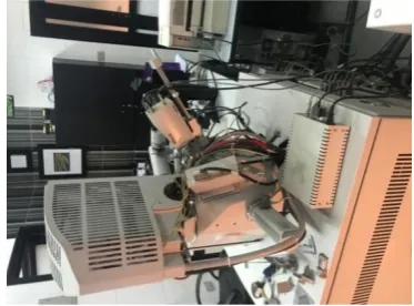
The specimens were then placed on stainless steel stubs and examined by Quanta Environmental Scanning Electron Microscope (QESEM) Figure (2), to determine the percentage of fluoride uptake by dentin before and after recharging.
Figure (2): Quanta Environmental Scanning Electron Microscope (QESEM).
2.7. Fluoride uptake analysis:
The fluoride uptake analysis was carried out using the QESEM. Scanning quantification were performed along the restoration/tooth interface to measure the amount of fluoride uptake by dentin axially at depth of 50 m from the interface (represented by weight percentage). Spot analysis was done at three points occlusally, cervically (100 m away from the axial line angles) and in the middle of the axial wall at 200X magnification and at voltage 15.00 KV. The weight percentage of fluoride was quantified with other elements (O2, Na, Al, Si, P and Ca). This quantification results provided normalization to 100% for all the selected elements. The fluoride with other elements was presented in a spectrum curve with identification peaks and in a quantified result box. Three reading of fluoride Wt% were averaged to obtain one reading from each specimen.
Statistical analysis:
performed using Kruskal-Wallis test followed by Mann-Whitney U test, for the cumulative fluoride release data. While for fluoride uptake, detailed comparisons between materials, storage media and storage periods were performed using one-way ANOVA test followed by Tukey's test. In addition, paired t-test was also used to compare between fluoride uptake after 12 weeks and after recharging.
3. Results:
For the fluoride release results:
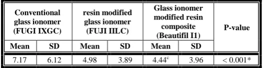
The results showed, the means and standard deviation (SD) of the cumulative fluoride release values in ppm demonstrating the effect of the different tested materials; on the mean fluoride release irrespective of other variables. It was revealed that conventional glass ionomer Fuji IX GC showed the statistically significant highest mean cumulative fluoride release followed by resin modified glass ionomer Fuji IILC, while glass ionomer modified resin composite Beautifil II) showed the statistically significant lowest mean cumulative fluoride release at P < 0.001. The results also showed that after storage in artificial saliva for one week, four weeks and twelve weeks; conventional glass ionomer Fuji IXGC, showed the statistically significant highest mean cumulative fluoride release values. There was no statistically significant difference between resin modified glass ionomer Fuji IILC and glass ionomer modified resin composite Beautifil II; both showed the statistically significant lowest mean cumulative fluoride release values at four weeks and twelve weeks Also, the results showed that after pH cycling in citric acid for one week, four weeks and twelve weeks; conventional glass ionomer ,Fuji IXGC ,showed the statistically significant highest mean cumulative fluoride release values There was no statistically significant difference between resin modified glass ionomer (Fuji 2lc) and glass ionomer modified resin composite (Beautifil II); both showed the statistically significant lowest mean cumulative fluoride release values (0.71±0.05ppm and 0.86±0.27ppm) respectively at one week, (3.26±1.07ppm
and 2.83±0.90ppm) respectively at 4 weeks and
(11.39±4.28ppm and 9.48±3.27ppm) respectively at 12 weeks.
Table (1): The mean cumulative fluoride release ± standard deviation (SD) values in ppm and results of comparison
between materials irrespective of other variables in the study.
Conventional glass ionomer
(FUGIIXGC)
resin modified glass ionomer (FUJI IILC)
Glass ionomer modified resin composite (Beautifil I1)
P-value
Mean SD Mean SD Mean SD
7.17 6.12 4.98 3.89 4.44c 3.96 < 0.001* *: Significant at P 0.05, Different letters are statistically
significantly different
Figure (3): Bar chart representing the effect of the three tested materials on the mean cumulative fluoride release values in ppm irrespective of other variables in the study.
For the cumulative fluoride uptake remineralization results:
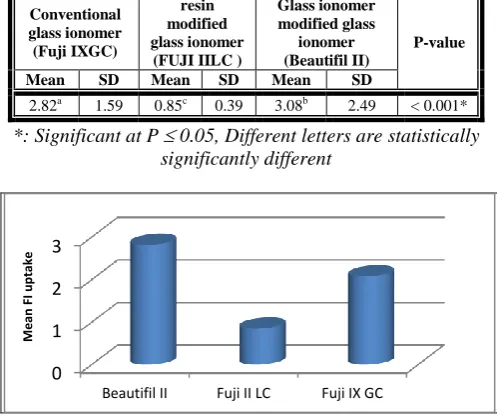
The data showed the means and standard deviation (SD) of the fluoride uptake values in Wt% demonstrating the effect of the different tested material on the mean fluoride uptake irrespective of other variables in the study. Descriptive statistics and the test of significance were performed using Tukey's post-hoc test for pair-wise comparison, the mean fluoride uptake values were also plotted in bar chart. The mean of fluoride uptake values of conventional glass ionomer FujiIX GC, resin modified glass ionomer Fuji IILC and glass ionomer modified resin composite (Beautifil II) were 2.08±1.59Wt%, 0.85±0.39Wt% and 2.28±0.49Wt% respectively. It was revealed that glass ionomr modified rein composite, showed the statistically significant highest mean fluoride uptake followed by conventional glass ionomer Fuji IXGC, While resin modified glass ionomer Fuji IILC showed the statistically significant lowest mean fluoride uptake at P < 0.001. The results also showed that storage in artificial saliva at one week; glass ionomer modified resin composite (Beautifil II) showed the statistically significant highest mean fluoride uptake value 1.61±0.07Wt%, but there
was no statistically significant difference between
conventional glass ionomer (Fuji 9) and resin modified glass ionomer Fuji IILC; both showed the statistically significant lowest mean fluoride uptake value (0.76±0.17Wt% and 0.42±0.16Wt%) respectively. At 12 weeks; there was no statistically significant difference between conventional glass ionomer (Ketac Fil) and glass ionomer modified resin composite (Beautifil II); both showed the statistically
significant highest mean fluoride uptake value
(2.28±0.35Wt% and 1.98±0.70Wt%) respectively, but resin modified glass ionomer Fuji IIlc showed statistically
significant lowest mean fluoride uptake value
0.66±0.33Wt%. The results showed that pH cycling in citric acid at one week and 12 weeks glass ionomer modified resin composite Beautifil II,showed the statistically significant highest mean fluoride uptake value 1.66±0.16Wt%, but there
was no statistically significant difference between
conventional glass ionomer Fuji IXGC and resin modified glass ionomer (Fuji IILC); both showed the statistically
significant lowest mean fluoride uptake values
(0.84±0.12Wt% and 0.59±0.26Wt%) respectively. 0
2 4 6 8
Fuji II LC Fuji IX Beautifil II
M
e
an
FI
r
e
le
Table (2): The mean fluoride uptake ± standard deviation (SD) values in Wt% and results of comparison between
materials irrespective of other variables in the study.
Conventional glass ionomer (Fuji IXGC)
resin modified glass ionomer
(FUJI IILC )
Glass ionomer modified glass
ionomer (Beautifil II)
P-value
Mean SD Mean SD Mean SD
2.82a 1.59 0.85c 0.39 3.08b 2.49 < 0.001* *: Significant at P 0.05, Different letters are statistically
significantly different
Figure (4): Bar chart representing the effect of the three tested restorative materials the mean fluoride uptake values
irrespective of other variables in the study.
For the fluoride recharging results:
The results showed that there was no statistically significant difference between fluoride uptake values after 12 weeks and after fluoride recharging in the two tested storage media (artificial saliva, and citric acid), with conventional glass ionomer Fuji IXGC, which was; 3.58±0.31Wt% and 4.09±0.97Wt% respectively for artificial saliva, and 5.05±0.56Wt% and 5.42±0.41Wt% respectively for citric acid. While with, glass ionomer modified resin composite Beautifil II it was; 2.29±0.52Wt% and 3.40±0.26Wt% respectively for artificial saliva, and 2.59±0.59Wt% and 4.34±0.99Wt% respectively for citric acid. For resin modified glass ionomer Fuji IIlc showed that; there was no statistically significant difference between fluoride uptake values after 12 weeks and after fluoride recharging with
artificial saliva which was 0.94±0.07Wt% and
1.56±0.55Wt% respectively for artificial saliva and 1.51±0.0.26 and 2.13±0.49 respectively for citric acid.
Table (3): The mean, standard deviation (SD) values in Wt% and results of comparison between fluoride recharging.
Material Storage
media
12 weeks Recharging
P-value Mea
n SD
Mea
n SD
Conventional glass ionomer
(Fuji IXGC)
Artificial
saliva 3.58 0.31 4.09 0.97 0.054
Citric
acid 5.05 0.56 5.42 0.41 0.101
resin modified glass ionomer (Fuji IILC)
Artificial
saliva 0.94 0.07 1.56 0.55 0.216
Citric
acid 1.51 0.26 2.13 0.49
0.049 *
Glass ionomer modified resin composite (Beautifil II)
Artificial
saliva 2.29 0.52 3.40 0.26 0.110
Citric
acid 2.59 0.59 4.34 0.99 0.145
Figure (5): Bar chart representing comparison between the mean fluoride uptake values at 12 weeks and after fluoride
recharging.
4. Discussion:
Numerous studies on the amount and rate of fluoride release of different glass ionomer formulations and its modifications have been published, but an insight into the mechanisms of the fluoride release and uptake is still lacking, various studies have shown that fluoride releasing restorative materials are very effective in inhibiting caries and making tooth substrates much more reactive for remineralization(16). Several investigation have been performed on fluoride ion release from restorative material, as this property is related to their cariosatic effect(17). The fluoride ion release of from dental restorative is governed by intrinsic and extrinsic factors(18). The intrinsic factors depend on composition, powder liquid ratio, specimens geometry, temperature surface treatment, permeability and finishing However the extrinisic factors may include type of storage medium and time period of studied assessment, and include analytical methods. Since fluoride release is considered essential property in glass ionomer restorative materials, this study was carried out to elaborate the cumulative fluoride release from newly introduced product of Giomer product, claimed its ability to release fluoride, remeneralize the enamel and dentin and so increase hardness and catalyze rminralization of hard tooth structures around cavity margins(19). The assessment of fluoride release was carried out using the most tradition method and most frequently in-vitro method for fluoride ion concentration detection in solution for its sensitivity & its high precision(20) Fluoride release from different fluoride releasing restorative material would be valuable property if it is up taken by tooth substrates allowing its remeneralization and increase its resistance to acidic attack by cariogenic bacteria. Fluoride uptake by human dentin was assessed by quantitative scanning electron microscope (QESEM)(21). The pH cycling protocol done between artificial saliva and the citric acid. Selection of citric acid stimulate the effect of citrus food, juices and carbonated beverages(22). The pH cycling was carried out three times daily a day, i.e., three meals time periods; Breakfast, Lunch and Dinner to stimulate the pH experienced in the oral environment during consumption of fermentable food, stuffs(23). In the current study, the results revealed that, the highest amount of cumulative fluoride release was recorded for the conventional glass ionomer Fuji IXGC, followed by resin modified glass ionomer Fuji IILC, followed by glass ionomer resin composite (Beautifil injectable), The amount of fluoride made available to the oral cavity is not related only to the content of the material, but also to the ability for 0
1 2 3
Beautifil II Fuji II LC Fuji IX GC
M
e
an
FI
u
pt
ake
0 2 4 6
fluoride to leach from the restorative material(24). Two mechanisms have been proposed by which fluoride may be release from the restorative material into an aqeous environment the first mechanism is short durable, short term reaction which involves initial burst effect and rapid leach dissolution of the glass particles attacked by acid reaction, the second mechanism is long durability and more gradual and sustained long term diffusion of ions through the bulk of the cement(25). According to the results,the two mechanisms typically found with conventional and resin modified glass ionomer as acid base reaction by the proton attack at the surface which lead to the subsequent fluoride ions liberation from the fluoroglass particles, the leached Ca, Al ions from a
polysalts with fluoride ions which are dispersed
homogenously through the matrix regions of the set cement, seems to be responsible for the short term elution process and the initial burst effect(26). While in case of glass ionomer modified resin composite (Beautifill injectable), the setting through free radical polymerization reaction without proton attack acid base reaction, so the only source of fluoride would come from the glass filler particles and so slow diffusion release(25). Technology (SPRG) i.e., surface prereacted glass particles in glass ionomer resin composite (Beautifil injectable) so the set matrix is relatively the same of conventional resin composite i.e., hydrophobic resin monomers lead to less water sorption and subsequently less fluoride release because of lack of acid-base reaction because of S-PRG prereacted already with flurosilicate glass particles(27), so in turn affects the long term fluoride release. This result is in accordance with(28), whom found that glass ionomer modified resin composite release less fluoride than conventional glass ionomer Fuji IXGC all over the time and attributed their finding to the presense of S-PRG The results also revealed that the highest amount of fluoride release, and the highest amount of fluoride uptake weight percentage of fluoride in the dentin were evident in citric acid with its pH acid value more than normal neutral artificial salvia for all fluoride releasing restorative materials used in the current study, this result was in agreement with Kantoviz et al.(29) conserned fluoride release results. This could be attributed to that citric acid is strong organic acid, as it can be used in manufactures of beverages and foods according to Wang and Yap(30), who found that every decrease in pH value as described in the current study at citric acid pH 4 lead to increase the solubility and disintegration of the material, as the low acidity have the ability to erode the glass ionomer materials, and so the increase in fluoride ion release and increase its weight percent inside the dentinal tubules Another explanation is that the relatively acidity of the citric acid which make it able to react with basic glass fillers to form ca and aluminum acid salts(31). The artificial saliva in this study was used in high pH 7 and considered controllable group according to the results of that study. Bell et al.(32) found that fluoride release decrease after immersion of the specemins in the artificial saliva, there concluding their findings due to the retarding effect of the artificial saliva as a result of the formation of surface deposits, Hattab and Amin(18) was in agreement with that finding he found that released fluoride ions might react withcalcium ions present in artificial saliva with formation CaF2, surface coating on the speciemens that could act as a diffusion barrier. Glass ionomer modified resin composite (Beautifil injectable) showed the highest amount of fluoride uptake at period of
one week followed bu conventional glass ionomer (Fuji IX GC), while resin modified glass ionomer (Fuji II LC) is the lowest percentage in fluoride uptake in dentin. While at twelve weeks, conventional glass ionomer (Fuji IX GC) showed the highest amount of fluoride uptake followed by glass ionomer resin composite (Beautifil injectable) However resin modified glass ionomer (Fuji II LC) showed the lowest percentage in fluoride uptake at the all testing periods in dentine. The results in agreement with previous researches published by Hotta et al.(33). This may be attributed to that fluoride uptake by the adjacent dental tissues is highly influenced by the quality of tooth restoration interface. The
Beutifil injectable adhesive system contain
4-acrylooxyethyltrimellitic acid (4 AET) monomer is able to interact chemically with both hydroxyapatite and collagen fibrils. The two ionized carboxylic groups of (4-AET) molecule can react with Ca ctions of the original and remnant appatites and form Ca carboxylates salts(34). This suggests that this adhesive systems allow formation of two fold bonding mechanisms micromechanical and chemical bonding via the formation of dentin impregnated dentin layer (Hyprid layer) and chemical bonding via the acidic monomer(35). Fluoride impregnation in the dentin substrate surface would be as a consequence of the perentration of the low viscosity flowable resin into the dentinal tubules, resulting in resin tag formation, which suggests a second process of fluoride diffusion(36). This result could be attributed to fluoride content in the self etch adhesive (FL bond II) which used with Beautifil injectable, in a form of pre-reacted glass ionomer fillers and poly acid modified resin filler which are subsequently in corborated inside the hyprid layer. The recharging with fluoride varnish increased the fluoride uptake by the freshly cut dentin with all restorative materials under experimental condition but without statistical significance it could be attributed to two speculations first fluoridation mainly performed to counteract the fluoride loss of material is more likely a surface reaction, thus the inward bulk diffusion is related to the diffusion coefficient of the material, thickness and time of application(37). Second it was found that the recharging agent contain other ions, these ions may be competing with fluoride ions for binding sites at the filler particle surface, thereby impairing recharging of the materials(38).
5. Conclusion
1. The formulations of glass ionomer restorations has a significant effect on fluoride release and uptake ability of the glass ionomer restorations.
2. Conventional glass ionomer have a higher ability of fluoride release than any modifications of glass ionomer cements
3. Newely introduced glass ionomer modified resin
composite giomer have potent effect in fluoride uptake remineralization due to the role of strong impregination bond inside the dentinal tubule.
4. Exposure to acidic media has a great impact on fluoride release and remineralization uptake properties.
5. Topical fluoride application did not enhance fluoride uptake by the tooth dentin
6. Disclosure and acknowledgment
7. References:
[1]. Benelli EM, Serra MC and Rodrigues AL. In situ anticariogenic potential of glass inomer cement. Caries res 2014; 27:280-284.
[2]. Fejerskov O, Nyvad B and Kidd. Dental caries: The disease and its clinical management (Third ed.). 2015.
[3]. Cochrane NJ, Cai F and Huq NL. New approaches to enhanced remineralization of tooth enamel. Journal of Dental Research 2010; 89(11): 1187-97.
[4]. Li X, Wang J and Joiner A. The remineralisation of enamel: a review of the literature. Journal of dentistry. 2014; 42: S12-20.
[5]. Garcia-Godoy F and Hicks J. Maintaining the integrity of the enamel surface. American Dental Association 2008; 139(3).
[6]. Hicks J, Garcia-Godoy F and Flaitz C. Biological factors in dental caries: role of saliva and dental plaque in the dynamic process of demineralization and remineralization (part 1). Journal of Clinical Pediatric Dentistry 2004; 28(1):47-52.
[7]. Qvist V, Laurberg L and Poulsen A. Eight year study on conventional glass inomer and amalgam restorations in primary teeth. Acta Odontol Scand 2004; 62: 37-45.
[8]. Berg JH, Donly KJ and Posnick WR. Glass inomer-silver Quintessence Int 2003; 19:639-641.
[9]. Koga H, Kameyama A and Matsukulo T. comparison of short-term in vitro fluoride release and recharge from four different types of pit-fissure sealeants 2011; 46; 465-477.
[10]. Kusgoz A, Tuzuner T and Kemer B. Microhardness
conversion degree and fluoride release of different fissure sealants. J Mech behave biomed mater 2010; 3: 594-599.
[11]. ten Cate JM and van Duinen RN.
Hypermineralization of dentinal lesions adjacent to glass-ionomer cement restorations. J Dent Res 1995; 74: 1266-1271.
[12]. Sieixas LC, Seiaxas FH and Souza WC.
Demeneralization around restorations with different restorative materials containing fluoride. Mater Res 2004; 7: 235-240.
[13]. Magalhães CS, Hara AT and Turssi CP.
Microhardness evaluation around composite
restorations using fluoride containing adhesive systems. J Appl Oral Sci 2005; 13: 259-264.
[14]. Mukai Y, Shiiy T and Kamijo K. Formation of inhibition layer with a newly developed fluoride releasing all in one adhesive. Dent Mater J 2005; 24: 172:177.
[15]. Itota T, Carrick TE and Rusby G. Determination of
fluoride release from resin based dental materials using ion selective electrode. J Dent 2004; 32; 117-122.
[16]. Arbabzudeh Z and Gibbs T. Recharge pattern of contemporary glass ionomer restoratives. Dent Res J (Isfhan) 2012; 9: 139-145.
[17]. Wiegand A, Buchalla W and Attin T. Review on fluoride-releasing restorative materials-fluoride release and uptake characteristics, antibacterial activity and influence on caries formation. Dent Mater 2007; 23: 343-362.
[18]. Hattab FN and Amin WM. Fluoride release from glass ionomer restorative materials and the effects of surface coating. Biomater 2001; 22: 1449-1458.
[19]. Horsted-Bindslev P. Fluoride release from alternative restorative materials. J Dent 1994; 22(1): S17-20.
[20]. Harhash AY. Effect of fluoride releasing adhesives and fluoride uptake by tooth substrate on nanoleakage and shear bond strength of resin composite to dentin. PhD thesis 2007; Faculty of Oral and Dental Medicine, Cairo University.
[21]. Hashimoto M, De Munck J, Ito S, Sano H, Kaga M, Oguchi H, Van Meerbeek B and Pashly DH. In vitro effect of nanoleakage expression on resin-dentin bond strengths analyzed by microtensile bond test, SEM/EDX and TEM. Biomater 2004; 25: 5565-5574.
[22]. Yu H, Wegehaupt FJ, Wiegand A, Roos M, Attin T
and Buchalla W. Erosion and abrasion of tooth-colored restorative materials and human enamel. J Dent 2009; 37: 913-922.
[23]. Abo El Naga AIM. Effect of load and pH-cycling on
nanoeakage patterns a dentin adhesive interface of different bonding systems. PhD thesis 2006; Faculty of Oral and Dental Medicine, Cairo University.
[24]. Dionysopoulos D, Koliniotou-Koumpia E,
Hevatzoglou-Antoniades M, Kotsanos N. Fluoride release ad recharge abilities of contemporary fluoride-containing restorative materials and dental adhesives. Dent Mater J 2013; 32: 296-304.
[25]. Chan WD, Yang, Wan W and Rizkala AS. Fluoride
release from dental cements and composites: a mechanistic study. Dent Mater 2006; 22: 366-373.
[26]. Mc Lean JW, Nicholson JW and Wilson AD.
Propsed nomenclature for glass ionomer dental cements and related materials .quintessence Int.1994; 5: 587-589.
[27]. Tay FR, Pashley EL, Huang C, Hashimoto M,
[28]. Itota T, Carrick TE, Rusby S, Al-Naimi OT,
Yoshiyama M and McCabe JF. Determination of fluoride ions released from resin-based dental materials using ion-selective electrode and ion chromatograph. J Dent; 2004; 32: 117-122.
[29]. Kavaloglu Cildir S and Sandalli N. Compressive strength, surface roughness, fluoride release and recharge of four new fluoride-releasing fissure sealants. Dent Mater J 2007; 26: 335-341.
[30]. Wang XY, Yap AU. Effects of environomental
calcium and phosphate on wear and strength of glass ionomers exposed to acidic conditions. J Biomed Mater Res B Appl Biomater 2008; 88: 458-464.
[31]. Nomoto R, McCabe JF. A simple acid erosion test for dental water based cements. Dent Mater. 2001; 17: 53-59.
[32]. Bell A, Creanor SL, Foye RH and Saunders WP. The effect of saliva on fluoride release by a glass-ionomer filling material. J Oral Rehabil 1999; 26: 407-412.
[33]. Hotta M, Li Y and Sekine I. Mineraization in bovine dentin adjacent to glass-ionomer restorations. J Dent 2001; 29: 211-215.
[34]. Ikemura K, Kouro Y and Endo T. Effect of 4-Acryloxyethyltrimelletic acid in a self-etching primer on bonding to ground dentin. Dent Mater J 1996; 15: 132-143.
[35]. Ibrahim SH. Bonding mechanism of giomer to
enamel and dentin with and without pre-etching. Master's thesis 2007; Faculty of Oral and Dental Medicine, Cairo University.
[36]. Hashem A, Bader NA and El-Gezawi MF.
Assessment of fluoride release and uptake before and after recharging of clinically simulated surface of conventional and modified glass ionomers. Egy Dent J 2002; 48: 117-131.
[37]. Attin T,buchalla W, Siewert C and Helling E. Fluoride release of polyacid modified resin composite in neutral and acidic buffer solutions. J. Oral Rehbil.1999; 26:388-393.
[38]. Seppa l. Fluoride release and effect on enamel softening by fluoride treated and fluoride untreated glass ionomer specimens. Caris Res. 1994; 28: 406-408.
Author 1: received the B.S. and M.S. degrees in Operative Dentistry from Cairo International University in 2003 and 2011 respectively. He is an Assistant chairman in
Operative Dentistry Department police
hospital Cairo Egypt Since 2015, he started to study and research in the field of fluoride release and