1332
Pneumonia Detection Using Convolutional
Neural Networks
Sammy V. Militante, Brandon G. Sibbaluca
Abstract— Pneumonia is an infectious and deadly illness in respiratory that is caused by bacteria, fungi, or a virus that infects the human lung air sacs with the load full of fluid or pus. Chest X-rays are the common method used to diagnose pneumonia and it needs a medical expert to evaluate the result of X-ray. The troublesome method of detecting the pneumonia cause a life loss due to improper diagnosis and treatment. With the emerging computer technology, development on an automatic system to detect pneumonia and treating the disease is now possible especially if the patient is in a distant area and medical services is limited. This study intends to incorporate deep learning methods to alleviate the problem. Convolutional Neural Network is optimized to perform the complicated task of detecting diseases like pneumonia to assist medical experts in diagnosis and possible treatment of the disease. The authors developed several models to determine the best possible model in detecting pneumonia with the most accurate results. This study has trained five different models of CNN, namely AlexNet, LeNet, GoogleNet, ResNet and VGGNet using 1024 by 1024 resolution of 26,684 dataset images. The result achieved a 97 percent accuracy rate for VGGNet and the lowest rate is 74 percent achieved by the ResNet model. The result of statistics shows that the trained model was able to detect Pneumonia through examined images of chest X-ray.
Index Terms— Pneumonia, Convolutional Neural Networks, Disease Detection
—————————— ——————————
1
INTRODUCTION
PNEUMONIA is an illness that disturbs the lung air sacs of an infected person. It is triggered by bacteria, fungi, or a virus that infects the air sacs of lungs that fill up with discharge fluids that leads to chills, fever, coughing with mucus, and breathing trouble among persons diagnosed with this disease. Children below five years of age and elderly patients with weak immune system are vulnerable to this type of diseases. Pneumonia has killed over a million children worldwide in 2018 and remains a life-threatening disease now a days if not detected or diagnose earlier [1].Radiography, CT-scan, or MRI is the common method to discover pneumonia. Medical personnel check the patient’s radiograph of their chest to determine if they are infected with pneumonia or not. In addition, the usual method for finding pneumonia is through medical history and laboratory results of the patient.
Radiograph of chest is penetrated through X-rays where the soft tissues produces a dark color and hard tissues like bones produces a bright color [2]. Patients diagnosed with pneumonia shows the chest cavity signs of fluids filling the air sacs of lungs as for the radiograph picture appears brighter. Several abnormalities may be seen on lung cavities as brighter color may represent such as cancer cells, blood vessels swelling, and abnormality of heart [2]. To validate the range and spot of an infected area of the lungs, chest x-rays is the utmost method. In these method, emergence of the disease can be imprecise and misinterpreted with another illness.Therefore, the undertaking is pleasing in the improvement of the processing in medical situations in isolated areas for pneumonia detection. The researchers were able to train and assessed CNN model’s performance and classify chest x-rays with normal and infected with disease using different classifiers. With the recent development of Computer Aided Design (CAD) tools becomes the most important field of
research in artificial intelligence and machine learning. CAD systems has proven in facilitating the medical field such as breast cancer detection, classification of disease using mammograms, lung cancer detection, etc. CAD system is an applicable instrument in use today for diagnosis and classification of diseases in medical imaging. In achieving the precise diagnosis, the medical personnel integrate the CAD to assist and verify to support their decision making. Significant features of the images are valuable in employing machine learning techniques in this system compared to the traditional handcrafted features which has limitations in extracting significant features [3] [4] [5].The progress in a more intelligent future is now productive through generations. This technological improvement today reached new step closer in human intelligence. The deep learning has gained the ability in simulating the function of the human brain. It recommends the solution to solve real-life problems. The deep learning by means of the convolutional neural networks has ability in obtaining significant characteristics in image classification tasks [6] and provides medical promising results in image analysis [7]. CNN advantages [8] is capable in assisting the identification of some features from an image and use this feature to generate probabilities in classifying specific input [9].The contribution of this study is developed an optimized deep learning models of CNN that can detect and classify pneumonia diseases efficiently [10]. The work consists of an optimized CNN models and experimental analysis of each model towards the detection and classification of pneumonia diseases.This research article consists of the following sections: introduction, convolutional neural network, methodology, experimental results and discussion, conclusion and recommendations, acknowledgments and references.
2 CONVOLUTIONAL NEURAL NETWORKS
Process of CNN is to detect and categorize images from learned features. It is very effective in a multi-layered structure when obtaining and assessing necessary features of graphical images. Figure 1 illustrates the CNN process.
————————————————
Sammy V. Militante is the Director for Statistical Center Office and concurrently the Chair of the Computer Engineering Program at the University of Antique, Philippines, PH-9088753286. E-mail: [email protected]
Fig. 1. CNN Process.
Fig. 4 Interconnected Layers 2.1 Convolution Layer
This layer conceptualizes the characteristics of each image input. It stores three-dimensional connection among pixels by means of discovering significant image characteristics with small squares of image input. Described in figure 2 below is the overall process and functions of kernel, where in the image input is converted into 5x5 matrix pixel values and the resulting filter is 3x3 matrix or the resulting feature map in this layer. Feature map contains specific details of the original image necessary to determine an input. The feature map then down sampled with the ReLU method reducing any negative values to zero and remains all others intact.
Fig. 2 Demonstration of Convolution Layer
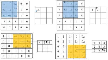
2.2 Max-Pooling Layer
This layer cut down the values further to half of its original value by choosing only max values from the kernel matrix. The sample illustration is the 4x4 matrix pixel values of an input image degraded into 2x2 filters. Its process is described in Fig.
Fig. 3 Illustration of Max Pooling Layer
2.3 Fully-Connected Layer
Multi-layer perceptron in this layer confirms that all neurons is interconnected to every neuron of every succeeding layers in the purpose of classifying input images created through the preserved features of image. Fig. 4 illustrates the outcomes of image input after it has obtained characteristics from the provided data set and extracted features.
3 M
ETHODOLOGYThe proposed methodology presents the architectural design that is divided into three stages: preprocessing, handover learning and refinement, and classification.
3.1 Dataset Preparation and Pre-Processing
1334
Fig. 5 Pneumonia Dataset were labeled properly and precisely by medical experts to
confirm correct labels for classification.
3.2 Transfer Learning and Fine-Tuning
For an effective classification, the models are set for proper setting to the needed task. Transfer learning let the model save to original parameter settings of the previously trained model to bring effective score without using intense computing power [12]. The Fine-Tuning method can extract new features from pneumonia and change the original 1000 neurons for example the VGGNet model to only four [13]. Also, FC or dense layers of 4096 down to a more significant number of 512 and 256 to prevent any waste of computing resources. Hyperparameters facilitate the deep learning model to produce significant results just as the actual training procedure takes place. Selecting the exact values can initially make a huge dissimilarity by making hyperparameter tuning a necessary process. Still, there are no precise hyperparameters for every algorithm [14]. Thus, a practical experimentation must take place.
3.3 Classification
Every single block as shown in Fig. 1 are used in architecture followed by wholly inter-connected layers and softmax activation. Feature extraction is composed of an input image, convolution, max-pooling while classification involves fully interconnected layers and output [15].
4 INVESTIGATIONAL RESULTS AND DISCUSSION
We have assessed the performance of five CNN models namely; AlexNet, GoogleNet, LeNet, ResNet, and VGGNet model. Four models achieved the highest accuracy rate ranging from 95% to 97%, GoogleNet and LeNet got the highest mark of 97% followed by VGGNet with an accuracy rate of 96% and AlexNet model has the accuracy rate of 96%. The ResNet model achieved the lowest accuracy rate with the mark of 74%. Illustrated in table 1 is the outcome of training and validation of the five identified models. The experiments were performed using an ASUS gaming laptop with Intel Core i7-8750 processor having a memory size of 16GB and a NVIDIA GeForce GTX 1050 video card. The researchers also applied the augmentation techniques in the training set of images to increase dataset by implementing the image rotation of 15 degrees, horizontal flipping, zoom of 0.2, and shifting range of 0.1 for both image width and height. Adam optimizer has been utilized in the optimization and for loss-function uses binary cross-entropy. The trained images use a batch size of 32 and it was trained for five hundred times. Fig. 5 displays the Pneumonia dataset used in the study. Table 2 shows the overall performance of five models used in this study.
Table 1. Result of Training and Validation of Five CNN models
Alex Net Model
Google Net Model
LeNet Model
ResNet Model
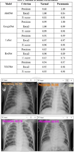
Table 2. Review of Model Performance
1336 Fig. 7 Shows the result of predicted images of normal chest x-rays.
Fig. 6 illustrates the tested images infected with pneumonia. All five models were able to predict the images with the highest rating percentage of 99.76% and the lowest recorded
was 66.42%. Fig. 7 illustrates the tested image with normal chest x-ray or not infected with pneumonia. It has predicted the images from 91.2% to 100% using five different models.
5
C
ONCLUSIONThis research explores the deep learning function in detecting pneumonia through computer vision using five convolutional neural network models. Every CNN models identified are assessed physically so that the framework in the feature extraction and fine-tuning are employed. The pneumonia infected diseases and normal chest x-ray image dataset are acquired from the Radiological Society of North America database. Our study enables to identify among the five models the best model to detect pneumonia. The AlexNet, LeNet, GoogleNet, and VGGNet are the best model based on the observations of the researchers having an accuracy rate of 95% to 97%. The ResNet model has the lowest accuracy rate of 74%. All the models have performed well on detecting pneumonia and normal chest x-rays. With this work, a distinguished process of diagnosing and detecting pneumonia can helpful in providing medical services. For future studies, adaptation of other convolutional neural network architectures like Inception-v3, shuffle Net, and Mobile Net architectures for pneumonia detection must be implemented and the optimization of hyper-parameters should also be considered to improve the accuracy of the model. This study will help medical work force in their decision making for a real-time application of the use of accurate model in detecting pneumonia and discover the potential of diagnosing pneumonia using deep learning.
ACKNOWLEDGEMENT
Aside from God Almighty, the researchers would like to express their gratitude to their families, relatives, and dear ones whose involvement made a significant impact on the accomplishment of this task.
BIBLIOGRAPHY
[1] P. Rui, K. Kang, National Ambulatory Medical Care Survey: 2018 Emergency Department Summary Tables. [2] E. Sayed, et. al, Computer-aided Diagnosis of Human
Brain Tumor though MRI: A Survey and a new algorithm, Expert System with Applications (41): 2014.
[3] D.K. Das, M. Ghosh, M. Pal, A.K. Maiti, and C. Chakraborty, Machine learning approach for automated screening of malaria parasite using light microscopic images, Micron 45, 97106, 2013.
[4] M. Poostchi, K. Silamut, R. Maude, S. Jaeger, and G. Thoma, Image analysis and machine learning for detecting malaria, Translational Research, 2018.
[5] N.E. Ross, C.J. Pritchard, D.M. Rubin, and A.G. Duse, Automated image processing method for the diagnosis and classification of malaria on thin blood smears, Medical and Biological Engineering and Computing 44, 5, 427436, 2006.
[6] A.S. Razavian, H. Azizpour, J. Sullivan, and S. Carlsson, CNN features off-the-shelf: an astounding baseline for recognition, In Proceedings of the IEEE conference on computer vision and pattern recognition workshops, 806813, 2014.
[8] Abiyev R. H., Ma’aitah M. K. S., Deep Convolutional Neural Networks for Chest Diseases Detection, Journal of Healthcare Engineering, 2018.
[9] A. Krizhevsky, I. Sutskever, & G.E. Hinton, ImageNet Classification with Deep Convolutional Neural Networks, Neural Information Processing Systems, 25. 10.1145/3065386, 2012.
[10]M. Cicero, A. Bilbily, E. Colak, T. Dowdell, B. Gra, K. Perapaladas, and J. Barfett, Training and Validating a Deep Convolutional Neutral Network for Computer-Aided Detection and Classification of Abnormalitites on Frontal Chest Radiographs, Investigative Radiology 52(5) 281-287, 2017.
[11]D. Demner-Fushman, M.D. Kohli, M.B. Rosenman, S.E. Shooshan, L. Rodriguez, S. Antani, G.R. Thoma, and C.J. McDonald, preparing a collection of radiology examinations for distribution and retrieval, J Am Med Inform Assoc, 2016 Mar;23(2):304-10. doi: 10.1093/jamia/ocv080. Epub 2015 Jul 1.
[12]Chu, V. Madhavan, O. Beijbom, J. Hoffman, and T. Darrell, Best practices for fine-tuning visual classifiers to new domains, in Proceedings European Conference on Computer Vision, pp. 435–442, Oct. 2016.
[13]Sharif Razavian, H. Azizpour, J. Sullivan, and S. Carlsson, CNN features off-the-shelf: An astounding baseline for recognition, in Proc. IEEE Conference on Computer Vision and Pattern Recognition, pp. 806–813, Sep. 2014.
[14]A. Shrestha and A. Mahmood, Review of Deep Learning Algorithms and Architectures, in IEEE Access, vol. 7, pp.
53040-53065, 2019, DOI:
10.1109/ACCESS.2019.2912200.