SHILAJIT DIBEZO-α-PYROES: MITOCHODRIA TARGETED ATIOXIDATS
Sauryya Bhattacharyya1*, Debasish Pal1, Dipankar Banerjee1, Biswajit Auddy1, Amartya K Gupta1,2, Partha Ganguly1,2, Upal K Majumder2 and Shibnath Ghosal1
1
R and D Centre, Natreon Inc. India, CL-18A, Sector II, Salt Lake City, Kolkata-700091
2
Department of Pharmaceutical Technology, Jadavpur University, Kolkata-700032 *Corresponding Author’s email id: sauryya@hotmail.com
Summary
The decrease in efficiency of mitochondria in generating energy currency (ATP) in animals and in humans is associated with aging (geriatric problems) and oxidative stress. This deficiency has a link with the systemic deficiencies of coenzyme Q10 (CoQ10) concentration
and of two of its endogenous functional associates, namely, 3-hydroxydibenzo-α-pyrone (3-OH-DBP) and 3,8- dihydroxydibenzo-α-pyrone [3,8-(OH)2-DBP]. Mitochondrial targeting of the two DBPs, isolated from shilajit (the supervitalizer of Ayurveda), and of CoQ10 could be
formidable strategies to augment antioxidant defense and energy generating elements for restoring normal functions of mitochondria. DBPs, as also their fatty-acyl and amino-acyl conjugates, occur in animal mitochondria and in blood where they act in tandem with CoQ10
in the electron transport chain. Administration of CoQ10 alone, in mitochondrial deficiency
states, therefore, could not restore normal mitochondrial functions. The concomitant targeting of DBPs and CoQ10 to mitochondria would augment energy (ATP) synthesis and protect
redox states of CoQ10 from oxidative degradation. The present findings adduce evidence of
augmentation of the concentrations of DBPs and CoQ10 in mitochondria when administered,
from exogenous sources, through intra-peritoneal/oral route. Their probable mechanism of action would involve the three redox states of DBPs (reduced form, semiquinone radical and quinone form) and similar redox states of CoQ10 as a measure to restore normal energy
synthesizing ability of mitochondria.
Introduction
Mitochondria are intracellular organelles responsible for energy metabolism. Consequently, mitochondrial dysfunction is damaging, particularly to neural and muscle tissues which have high energy demands. Mitochondrial dysfunction is central to a number of human degenerative diseases. Oxidative damage to the mitochondria is a major factor in the pathophysiology of Parkinson's disease, Friedreich's Ataxia and Wilson’s disease, mtDNA disorders, diabetes, motor neuron disease and the non-specific loss of vigor associated with aging. Oxidative damage to mitochondria also contributes to the pathophysiology of inflammation and ischaemic-reperfusion injury in stroke, heart attack and during organ transplantation and surgery.
From the mid-1970s, CoQ10 was synthetically prepared and used as a therapeutic agent in
human physiological deficiencies involving mitochondria. In the electron transport chain, according to the existing scientific tenet, CoQ10 receives electrons from NADH, forms
reduced CoQ10 (CoQH2) and passes the electron to cytochrome c, required for energy
synthesis. After transfer of the electrons, CoQH2 is again oxidized to the quinone form.
During oxidative stress, the oxidation-reduction cycle of CoQH2 ⇋ CoQ10 is impaired and
CoQ10 is randomly degraded to form polar aberrant metabolites. Oxidation of CoQH2 and
degradation of subsequently formed CoQ10 by systemic oxygen-centered free radicals
produces intractable agglomerates. Delivering CoQ10 alone, as was the common practice till
now [1], could not restore normalcy from the systemic dysfunction. Researchers are persuasively indicating presence of some “unidentified” factors involved in mitochondrial energy transduction [2]. Accordingly, there is a need for compounds, compositions and methods that limit, prevent and/or restore from damage to mitochondria by targeting energy producing molecules to the organelle. Hydroxylated dibenzo-α-pyrones (DBPs) and their acyl and aminoacyl derivatives are the key bioactive components of shilajit. Shilajit is regarded in Ayurveda as the energy currency (supervitalizer) for living animals including humans. DBPs (and equivalents) are endogenously synthesized from PUFAs (EPA/DHA) and also occur in animal mitochondria and blood [3]. In a previous study, it was reported that shilajit acted as a potent antioxidant to protect mitochondria from oxidative damage and to resurrect its ATP producing potential [4]. The components of shilajit after reaching mitochondria exerted their: (i) reductive; (ii) CoQH2 stabilizing and other attendant effects in
ATP synthesis. The present study has been designed to obtain further evidence of targeting to mitochondria of two key bioactive DBPs of shilajit, namely 3-hydroxydibenzo-α-pyrone (3-OH-DBP) and 3,8- dihydroxydibenzo-α-pyrone [3,8-(OH)2-DBP] [5]. Probable mechanism of action associated with this phenomenon has also been suggested.
Materials and Methods
Chemicals and Reagents: All chemicals and reagents were of analytical grade and obtained from Sigma-Aldrich, Merck, India or SRL, India. The solvents used for chromatographic techniques were of HPLC grade, obtained from Merck, India.
Test compounds: 3-OH-DBP and 3,8-(OH)2-DBP were isolated from shilajit, and each
Antioxidant activity of DBPs
2,2-Diphenyl-1-picrylhydrazyl (DPPH) radical scavenging assay [7]: 0.5 ml DPPH solution (3 mg in 25 ml ethanol) was mixed with 0.5 ml sample solution and the absorbance of the mixture after 20 minutes of incubation in the dark was monitored at λ 517 nm. Upon reduction, the colour of the solution fades. The concentration that causes a decrease in the absorbance of initial DPPH radicals by 50% is defined as IC50 of the sample.
Superoxide (O2.-) anion scavenging activity [7]: This assay was conducted following a
published procedure. Briefly, O2.- was generated in solutions by the system consisting of
NADH, PMS (phenazine methosulphate) and aerial O2. NBT (nitroblue tatrazolium) was
used as the probe. In presence of O2.-, yellow colored NBT transformed into blue colored
formazan which gives absorbance at λ 540 nm. Samples were mixed with the O2.- generating
solution and the decrease in absorbance was monitored after proper incubation. The concentration that causes a decrease in the intensity of initial absorbance by 50% is defined as IC50 of the sample.
ABTS radical cation decolorization assay [7]: ABTS-·, the oxidant, was generated by persulfate oxidation of 2,2′-azinobis(3-ethylbenzothiazoline)-6-sulfonic acid. This solution was diluted with phosphate buffer (pH 7.4) until the absorbance reached 0.7 to 0.8 at λ 734 nm. 1 ml of the resulting solution was mixed with the sample (0-20 µl). The absorbance was read at room temperature, 4 minutes after mixing. The concentration that causes a decrease in the absorbance of initial ABTS radicals by 50% is defined as IC50 of the sample.
Ferric reducing antioxidant potential, FRAP assay [7]: The oxidant in the FRAP assay was prepared by mixing TPTZ, acetate buffer and FeCl3.H2O and referred to as “FRAP
reagent”. The final solution has Fe(III) of 1.67 mM and TPTZ of 0.83 mM concentration, To measure reduction potential, samples were added to FRAP reagent and the absorbances were noted at λ 593 nm after 30 minutes incubation at 37°C. The concentration that causes an increase in the intensity of color with respect to control (no sample) value by 50% is defined as IC50 of the sample.
itric Oxide Radical Scavenging Activity (O assay) [7]: Nitric oxide (NO) radicals were generated from sodium nitroprusside solution in buffer saline and measured by Griess reagent. 1 ml of 10 mM sodium nitroprusside was mixed with 1 ml of sample with different concentrations in phosphate buffer (pH 7.4). The mixture was incubated at 25°C for 150 min. To1 ml of the incubated solution, 1 ml of Griess’ reagent (1% sulphanilamide in 2% o -Phosphoric acid and 0.1% NEDA in 2% o-Phosphoric acid in 1:1 v/v ratio) was added and the absorbance was read at 546 nm. The concentration that causes a decrease in the intensity of color with respect to control (no sample) value by 50% is defined as IC50 of the sample.
Assay for inhibition of erythrocyte membrane Lipid Peroxidation [8]: The degree of lipid peroxidation was evaluated by estimating the thiobarbituric acid-reactive substances (TBARS) using a standard method after minor modifications. Briefly, different concentrations of the samples were added to rat erythrocyte suspension to a final volume of 900 µl and incubated for 5 min at 37oC. Lipid peroxidation was initiated by adding 100 µl of 15 mM FeSO4 solution. After 30 min, 500 µl of this reaction mixture was placed in a tube
The mixture was heated in a water bath at 85°C for 30 min to complete the reaction. The intensity of the pink coloured complex was measured at λ 535 nm. The concentration that caused a decrease in the TBARS level with respect to control (no sample) by 50% is defined as IC50.
Animals: Male Swiss albino mice weighing 20-24 g and Sprague-Dawley rats were procured from Central Research Institute (Ayurveda), Govt. of India, Salt Lake City, Kolkata, were used. The animals were housed in an animal room with alternating light-dark cycle of 12 hr each. The animals were acclimatized for at least 7 days to the laboratory conditions before conducting experiments. Experiments were carried out between 0900 h and 1700 h. The study was conducted in accordance with Good Laboratory Practice (GLP) Regulations of WHO (WHO Document, 1998). The “Principles of laboratory animal care” (NIH Publication # 85-23, 1985) were followed in the study.
Treatment protocol – Mitochondrial targeting of the DBPs
Mice were divided into following two groups comprising 4 animals in each group.
Control: Received 0.8% (w/v) carboxymethyl cellulose (CMC) in water as vehicle intra-peritoneally.
DBPs: Received a 4:1 (w/w) mixture of 3,8-(OH)2-DBP and 3-OH-DBP in CMC
intra-peritoneally (20 mg/Kg body weight).
One hour after injection, the animals of both the groups were sacrificed and liver tissues collected.
Treatment protocol – Augmentation of total coenzyme Q by the DBPs in plasma and organs
Rats were divided into following two groups comprising 4 animals in each group. Control: Received 0.8% (w/v) carboxymethyl cellulose (CMC) in water orally. CoQ: Received CoQ10 in CMC orally (5 mg/Kg body weight).
CoQ+DBPs: Received 3,8-(OH)2-DBP in CMC orally (5 mg/Kg body weight).
The drugs were administered to the animals once per day for 5 days. Animals were subjected to hypoxic stress for 1 hour each day for 3 days from the 3rd day of the experiment. After the stipulated time-period, the content of total coenzyme Q were estimated in heart, kidney, liver (in wet weight basis) and in blood, by HPLC, after proper extraction from the tissues according to a published procedure [9].
Extraction and estimation of DBPs from liver mitochondria: Mitochondria were isolated from liver following a published procedure [10]. Briefly, liver tissues were triturated with sterilized medium comprising 0.25 M sucrose, 1 M EDTA, 10 mM Tris-HCl; pH 7.4. The homogenates were centrifuged at 900g for 20 minutes. The sediment was discarded and the trituration step was repeated again. After centrifugation, the clear supernatant was centrifuged twice at 12,000g, 20 minutes each time, to form pellets of a mitochondria-enriched fraction. The pellets were extracted with the Bligh and Dyer solvent system and the extracts were subjected to comprehensive HPTLC analyses for detection and quantitation of DBPs.
Stabilization of CoQH2in vitro by 3,8-(OH)2-DBP at different pH: 2 mg of solid CoQ10,
in an airtight vial, was dissolved in 1 ml of methanol. After thorough vortexing, 2 mg of solid NaBH4 was added, 1 mg at a time, to the yellow colored solution and kept in an ice-bath
After completion of reduction, a clear, colorless solution was obtained to which methanolic solution of 3,8-(OH)2-DBP (1 mg/ml) was added in a ratio of 1:1 (v/v) to obtain a final
concentration of the mixture – CoQH2 1 mg/ml and 3,8-(OH)2-DBP 0.5 mg/ml. This solution
(pH 8) was analyzed by HPLC for CoQH2 content at different time intervals. A control
CoQH2 solution was prepared in a similar fashion except that no 3,8-(OH)2-DBP was added.
To obtain solutions of CoQH2 with pH 7 and pH 3, aliquots of concentrated HCl were added
to the methanolic solution of CoQH2 of pH 8. The results are expressed as average of two
analyses (Tables 5-7).
Chromatographic Techniques
HPTLC Conditions: HPTLC was performed on Merck KGaA (1.05554.0007) pre-coated silica gel 60 F254 aluminium TLC plates. Solutions of marker 3-OH-DBP and 3,8-(OH)2
-DBP, along with experimental samples were applied and the plates were developed in a twin trough chamber with chloroform:methanol 9:1 (v/v) as mobile phase. Densitometric evaluation of the plates was performed at λ 240 nm by means of Camag TLC Scanner 3 in absorption mode. The scanned data were processed by means of Camag winCATS software, version 1.3.4. The plates were subsequently scanned to determine the UV reflectance spectra of each spot between 200 and 400 nm to identify the two bioactives of shilajit.
HPLC conditions for DBP analyses: HPLC analysis of DBPs were performed using Waters HPLC system comprised with PDA detector and Empower software with a NovaPak pre-packed column (RP C18, particle size 4 µm, 3.9 x 150 mm) fitted with a reverse phase guard
column and acetonitrile:water:o-phosphoric acid 32:67:1 (v/v/v) as mobile phase, with a run time of 15 minutes and flow rate 1 ml/min in an isocratic mode. Detection was done at λ 240 nm.
HPLC conditions for CoQ10 and CoQH2 analyses: HPLC analysis of CoQ10 and CoQH2
were performed using Waters HPLC system comprised with PDA detector and Empower software with a Xterra pre-packed column (RP C18, particle size 5 µm, 4.6 x 250 mm) fitted
with a reverse phase guard column and methanol:acetonitrile:ethanol 30:30:40 (v/v/v) as mobile phase, with a run time of 15 minutes and flow rate 1 ml/min in an isocratic mode. Detection was done at λ 275 nm for CoQ10 and 290 nm for CoQH2.
Results
Purity of DBPs used in the present study: Purity of the test DBPs was determined by HPLC using respective standard DBPs as marker and was found to be ≥99% (Fig. 1).
Fig. 1A – HPLC chromatogram of test Fig. 1B – HPLC chromatogram of test
A U 0.00 0.10 0.20 0.30 0.40 0.50 0.60 0.70 0.80 0.90 1.00 1.10 Minutes
1.00 2.00 3.00 4.00 5.00 6.00 7.00 8.00
4 .0 8 3 A U 0.00 0.50 1.00 1.50 2.00 2.50 3.00 Minutes
2.00 4.00 6.00 8.00 10.00 12.00 14.00 16.00
Antioxidant activity of DBPs: Antioxidant profile of the two test DBPs is furnished in Table 1. The results suggest that both the DBPs possess potent radical scavenging activity, while 3,8-(OH)2-DBP acts better than 3-OH-DBP in this aspect.
Table 1. Comparative in vitro antioxidant potency of the two test DBPs. The results are given in IC50 values of the substances.
Estimation of DBPs in mice liver mitochondria after administration of the test DBPs: 3-OH-DBP, 3,8-(OH)2-DBP and their redox products [3] were detected and quantitated in
mitochondria of control and test compounds treated mice by HPTLC (Table 2 and structures I-IV).
Table 2. HPTLC reflectance spectral data of oxido-reductive products of 3-OH-DBP and 3,8-(OH)2-DBP (formed in vivo) after treatment with the test compounds.
Product Rf Reflectance spectrum: λλλλmax (nm)
3-OH –DBP (I) 0.65 235, 277, 300, 335
3,8-(OH)2-DBP (II) 0.59 220, 235, 280, 304, 355
3,8-(OH)2-DBP-semiquinone (III) 0.52 225, 238, 280, 304, 336, 357, 370
3,8-(OH)2-DBP-quinone (IV) 0.43 234, 286
O O
OH
1
2 3
4 5 6 7 8 9
10
OH O
O OH
O O O
OH
O O
O
O
I II III IV
Assay methods 3-OH-DBP (µM) 3,8-(OH)2-DBP (µM)
DPPH radical scavenging assay - 0.54±0.03
ABTS radical scavenging assay 0.98±0.05 0.03±0.01
FRAP assay 0.41±0.03 0.08±0.01
O2.- scavenging assay 0.43±0.06 1.21±0.15
NO assay 3.27±0.13 5.09±0.22
Intra-peritoneal administration of DBPs to the animals increased the mitochondrial amounts of DBPs. The amounts of redox products of 3,8-(OH)2-DBP isolated form mice liver
mitochondria are given in Table 3. Since 3-OH-DBP is systemically converted into 3,8-(OH)2-DBP, this data include the contribution of the administered 3-OH-DBP.
Table 3. Amounts of DBP-products in mitochondria of mice liver before and after treatment (i.p.) with DBPs, analyzed by HPTLC
Compounds Control mitochondria (nmol/gm wet wt)
DBP treated mitochondria (nmol/gm wet wt)
3,8-(OH)2-DBP (II) 0.5±0.02 2.3±0.08
3,8-(OH)2-DBP-semiquinone (III) 12.7±0.15 20.2±0.22
3,8-(OH)2-DBP-quinone (IV) 26.7±0.65 24.8±0.58
Augmentation of coenzyme Q in mouse organs by 3,8-(OH)2-DBP: Co-administration
(p.o.) of CoQ10 with 3,8-(OH)2-DBP significantly improved the contents of total coenzyme Q
(CoQ9 and CoQ10) in different organs, except in plasma. The results of HPLC determinations
are incorporated in Table 4.
Table 4. Increment of total coenzyme Q after co-administration of CoQ10 and 3,8-(OH)2
-DBP in rat blood plasma and organs
Amounts of total CoQ9 and CoQ10
Treatment/
Groups Plasma (nmol/ml)
Heart (nmol/gm)
Liver (nmol/gm)
Kidney (nmol/gm) Control 0.79±0.03 18.46±0.55 15.38±0.48 11.86±0.14
CoQ10 0.81±0.02 35.69±0.86 19.41±0.38 16.51±0.09
CoQ10+DBPs 0.82±0.03 36.99±0.89 25.03±0.55 17.81±0.13
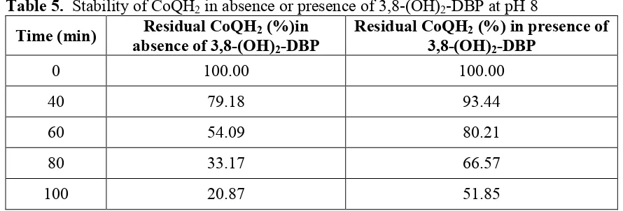
Maintenance of reduced state of CoQ10 by 3,8-(OH)2-DBP at different pH: Maintenance
of the stability of CoQH2 by 3,8-(OH)2-DBP at alkaline, neutral or acidic pH, with time, was
noted (Tables 5-7). 3-OH-DBP per se did not provide protective umbrella to the reduced state of CoQ10 to any significant extent.
Table 5. Stability of CoQH2 in absence or presence of 3,8-(OH)2-DBP at pH 8
Time (min) Residual CoQH2 (%)in absence of 3,8-(OH)2-DBP
Residual CoQH2 (%) in presence of
3,8-(OH)2-DBP
0 100.00 100.00
40 79.18 93.44
60 54.09 80.21
80 33.17 66.57
Table 6. Stability of CoQH2 in absence or presence of 3,8-(OH)2-DBP at pH 7
Time (hour)
Residual CoQH2 (%) in
absence of 3,8-(OH)2-DBP
Residual CoQH2 (%) in presence of
3,8-(OH)2-DBP
0 100.00 100.00
48 7.73 10.79
72 3.70 6.33
Table 7. Stability of CoQH2 in absence or presence of 3,8-(OH)2-DBP at pH 3
Time (hour)
Residual CoQH2 (%) in
absence of 3,8-(OH)2-DBP
Residual CoQH2 (%) in presence of
3,8-(OH)2-DBP
0 100.00 100.00
48 57.25 79.27
72 61.95 73.91
Discussion
The key bioactives of shilajit, namely, 3-OH-DBP and 3,8-(OH)2-DBP, were highly
antioxidative in nature as was observed by the conventional in vitro radical scavenging assays (Table 1). Since, shilajit was found to augment systemic ATP synthesis in debilitating physiological conditions [4], DBPs were considered to play a key role in such activity. This would require their targeting to mitochondria, when administered from exogenous sources. Administration (i.p.) of DBPs to mice showed not only the presence of DBPs and their incipient redox products (semiquinone radical and quinone) in the mitochondria (Table 2, determined by HPTLC) but also showed increment vis-à-vis control (Table 3). These observations suggest that the administered DBPs can augment their contents in the mitochondria that might play a crucial role in improved functioning of the electron transport chain.
CoQ10 is present in all tissues and membranes in highly variable amounts. By interaction with
NADH, CoQ10 is converted into CoQH2, which plays the electron-carrier role in the
mitochondrial electron transport chain. CoQH2 is also a highly efficient antioxidant in
preventing lipid, protein and DNA-oxidative damage and is continuously regenerated from CoQ10 by intracellular reduction systems. In some pathologic processes, when tissue
concentration of CoQ10 is decreased, it may be advantageous to supplement CoQ through
dietary substances [11]. However, the effect of administration of CoQ10 by dietary
supplement is difficult to interpret because loose CoQ10 might also act as a pro-oxidant [12].
Improvement in the energy generating process in animals by shilajit [4] indicates that DBPs might act in tandem with CoQ10/CoQH2 in mitochondria. In vitro experiment revealed that
3,8-(OH)2-DBP converted CoQ10 to the semiquinone radical and stabilized the latter by
spin-pairing (data not shown). The preservation of the reduced form of the coenzyme seems to be an attendant function of DBPs. This was simulated in the in vitro experiment of stabilization of CoQH2 by 3,8-(OH)2-DBP (Tables 5-7) at different pH conditions. The comprehensive
role of DBPs, in association with CoQ10, in the electron transport chain, is now being
References
1. Matthews RT, Yang L, Browne S, Baik M, Beal MF. Coenzyme Q10
administration increases brain mitochondrial concentrations and exerts neuroprotective effects. Proc Natl Acad Sci (USA). 1998; 95: 8892-8897.
2. Fato R, Bergamini C, Leoni S, Lenaz G. Mitochondrial production of reactive oxygen species: role of complex I and quinone analogs. Biofactors. 2008; 32(1-4): 31-39.
3. Ghosal S. In: Shilajit in Perspective, Narosa-Alpha Science International. 2006.
4. Bhattacharyya S, Pal D, Gupta AK, Ganguly P, Majumder UK, Ghosal S. Beneficial effect of Processed Shilajit on swimming exercise induced impaired energy status of mice. Pharmacologyonline. 2009; 1: 817-825.
5. Islam A, Ghosh R, Banerjee D, Nath P, Mazumder UK, Ghosal S. Biotransformation of 3-hydroxydibenzo-α-pyrone and aminoacyl conjugates by
Aspergillus niger isolated from native Shilajit. Electronic J Biotechnol. 2008;
11(3): 1-10.
6. Ghosal S, Lal J, Singh SK, Kumar Y, Soti, F. Chemistry of two bioactive benzopyrone metabolites from Shilajit. J Chem Res. 1989; 350-351.
7. Gupta AK, Ganguly P, Majumder UK, Ghosal S. Antioxidant and other beneficent effects of free and conjugated phytosteroids of Solanum and Withania. J Indian Chem Soc. 2008; 85: 1227-1238.
8. Moriguchi T, Takasugi N, Itakura Y. The effects of aged garlic extract on lipid peroxidation and the deformability of erythrocytes. J Nutr. 2001; 131: 1016S-1019S.
9. Rousseau G, Varin F. Determination of Ubiquinone-9 and 10 levels in rat tissues and blood by High Performance Liquid Chromatography with Ultraviolet detection. J Chromat Sci. 1998; 36(5): 247-252.
10. Liu Y, Deng F, Zhao R, Shen X, Wang C, Qu S. Microcalorimetric studies of the toxic action of La3+ in mitochondria isolated from Star-cross 288 chicken heart tissue cells. Chemosphere. 2000; 40(8): 851-854.
11. Turunen M, Appelkvist EL, Sindelar P and Dallner G. Blood concentration of coenzyme Q10 increases in rats when esterified forms are administered. J Nutr.
1999; 129: 2113-2118.