STABLE a-HELICAL PEPTIDES
Linda Mary Alexander McNamara
A thesis presented in partial fulfilment
o f the requirements for the Doctor o f Philosophy
degree o f the University o f London.
Department of Chemistry,
University College London
All rights reserved
INFORMATION TO ALL USERS
The quality of this reproduction is dependent upon the quality of the copy submitted.
In the unlikely event that the author did not send a complete manuscript and there are missing pages, these will be noted. Also, if material had to be removed,
a note will indicate the deletion.
uest.
ProQuest U642062
Published by ProQuest LLC(2015). Copyright of the Dissertation is held by the Author.
All rights reserved.
This work is protected against unauthorized copying under Title 17, United States Code. Microform Edition © ProQuest LLC.
ProQuest LLC
789 East Eisenhower Parkway P.O. Box 1346
I grew up with an ambition and determination without which I would have been a
good deal happier. I thought a lot and developed the faraw ay look o f a dreamer, fo r
it was always the distant heights which fascinated me and drew me to them by spirit.
I was not sure what could be accomplished by means o f tenacity and little else, but
the target was set high and each rebuff only saw me more determined to see at least
one major dream through to its fulfdment.
Earl Denman
Declaration
I, Linda Mary Alexander McNamara, hereby state that the following is entirely my own work and has not been submitted for any other degree or examination.
Acknowledgements
I would like to thank my supervisor Dr. Alethea Tabor for choosing me for this project and her support over the three years. I would also like to thank Dr. Giuliano Siligardi at the Pharmaceutical Optical Spectroscopy Centre (ULIRS Optical service) at King’s College London for the CD experiments on my target peptide. Certainly vyithout his assistance, the last few months o f my Ph D. would have been interesting! I would also like to thank members o f the Tabor group, especially Dr. Erwann Guerin for his help whilst deciphering the logistics o f the automated peptide synthesiser, lovingly named ‘Joey’ by all who use it. In addition, thank Firouz and Goran for the barbecues, Paul Free for the entertainment and Chris Hurley for the memorable outings!
I must thank Steve Corker and John Hill at UCL for their help with HPLC and mass spectrometry. I would also thank Mike Cocksedge at the University o f London School o f Pharmacy (ULIRS Mass Spectrometry Service) for all the accurate mass spectrometry work. O f course, thank other staff and technicians at UCL for answering my strange requests over the three years.
I am grateful to the EPSRC for the financial support required to complete this project within three years. I would also like to thank my parents as well as Lindsay and Anitha for their encouragement. I thank one o f my best friends, Gerry Sloan for all the interesting comments including ‘haven’t made that amino acid yet?’. O f course, I must thank Arthur Guinness for always being the shoulder to cry on.
Abstract
The synthesis and conformational properties o f a conformationally constrained cyclic peptide are described. The aim was to synthesise a short conformationally constrained peptide that may adopt an a-helical conformation in an aqueous environment. An unnatural amino acid was chosen as the ‘constraint’ between the i
and (/+4) positions within this short cyclic peptide. This constraining residue had two chiral centres joined by an 8-carbon bridge and needed to be synthesised using two enantiospecific methodologies. The route to the synthesis o f this amino acid is described, together with the modifications required for large-scale production. This was required for the subsequent solid phase peptide synthesis. The synthesis o f the target cyclic peptide (test peptide), using a novel solid phase approach with a triply orthogonal protection strategy is then detailed. The conformational properties o f the target test peptide, with the constraining residue, are compared with those o f a control linear peptide by CD analysis, in different solutions at various temperatures. In this way, the role o f this constraining residue in a synthetic cyclic peptide has been ascertained.
Table of Contents
ABBREVIATIONS... 10
LIST OF FIGURES... 16
CHAPTER 1: INTRODUCTION... 21
1.1 HELICES IN PEPTIDES... 21
1.2 THE HYDROGEN BONDED a-HELIX AND STABILISING CONSTRAINTS... 29
1.2.1 Stabilising Hydrogen Bonding Interactions... 29
1.2.2 Stabilising /,0+4) Side Chain Interactions...33
1.2.3 Lactam Bridges between Side Chains... 35
1.2.4 Disulfide Bridges between Side Chains... 39
1.2.5 Other Covalent Linkages between Side Chains... 41
1.3 THE TARGET PEPTIDE AND SYNTHETIC CONSTRAINING RESIDUE 42 1.3.1 Helix Formation in Short Peptides...42
1.3.2 The Model Peptide...46
1.3.3 The Synthetic Residue Constraint...47
CHAPTER 2: SOLID PHASE SYNTHESIS OF CYCLIC PEPTIDES... 50
2.1 INTRODUCTION... 50
2.1.1 Solid Phase Synthesis o f Linear Peptides... 52
2.1.2 Orthogonal Protection Strategy... 54
2.1.3 Coupling Reagents... 60
2.2 HEAD-TO-TAIL CYCLIC PEPTIDES...70
2.3 SIDE CHAIN-TO-END CYCLIC PEPTIDES... 80
2.4 SIDE CHAIN-TO-SIDE CHAIN CYCLIC PEPTIDES... 83
2.4.2 Cyclic Disulfide Bridged Peptides... 86 2.4.3 A Thioether Bridged Cyclic Peptide... 89 2.4.4 Aliphatic Bridged Cyclic Peptides... 90
CHAPTER 3: SYNTHESIS OF THE TARGET UNNATURAL RESIDUE 93 3.1 INTRODUCTION...93 3.2 THE ROUTE TO THE (/?)-CHIRAL CENTRE... 94
3.2.1 Synthesis o f 6-(5)-Isopropyl-2,5-diethoxy-3,6-dihydropyrazine (31) (the Schdllkopf bis-lactam ether chiral auxiliary)... 94 3.2.3 Synthesis o f
8-(R)-(5-(S)-Isopropyl-3,6-diethoxy-2,5-dihydropyrazin-2-yl)-octanoic acid (38)...99 3.3 THE ROUTE TO THE (5)-CHIRAL CENTRE... 101
3.3.1 Application o f the Evans Chiral Azidation Approach: Synthesis o f 4-(5)-Isopropyl-oxazolidin-2-one (41)... 101 3.3.2 Synthesis o f
3-[8-(R)-(5-(<S)-Isopropyl-3,6-diethoxy-2,5-dihydropyrazin-2-yl)-octanoyl]-4-(S)-isopropyl-oxazolidin-2-one (43)... 103 3.3.3 Synthesis o f
3-[8-(7?)-(5-(S)-Isopropyl-3,6-diethoxy-2,5-dihydropyrazin-2-yl)-azidooctanoyl]-4-(iS)-isopropyl-oxazolidin-2-one (44)... 103 3.4 UNMASKING THE TARGET AMINO ACID: SYNTHESIS OF
(10-ETHYL-ESTER)-9-(R)-ALLYLOXYCARBONYLAMIDO-2-(5)-AZIDODECANOIC ACID (46)... 106 3.5 TRANSESTERIFICATION REACTION: SYNTHESIS OF
(10-ALLYL-ESTER)-9-(R)-ALLYLOXYCARBONYLAMIDO-2-(5)-AZIDODECANOIC ACID (52)... 109 3.6 AZIDE REDUCTION AND PROTECTION OF THE RESULTANT FREE
10-Allylester)-9-(R)-allyloxycarbonylamido-2-(<S)-(9-fluorenylmethyloxycarbonyl)-amidodecanoic Acid (1)... 110 3.6.2 Synthesis o f
3-[8-(/?)-(5-(S)-Isopropyl-3,6-diethoxy-2,5-dihydropyrazin-2-yl)-2-(<S)-aminooctanoyl]-4-(S)-isopropyl-oxazolidin-2-one (53)...114 3.6.3 Synthesis o f
3-[8-(i?)-(5-(5)-Isopropyl-3,6-diethoxy-2,5-dihydropyrazin- 2-yl)-2-(5)-(9-fluorenylmethyloxycarbonyl)-amidooctanoyl]-4-(<S)-isopro-pyloxazolidin-2-one (54)... 115 3.7 ATTEMPTED CLEAVAGE OF THE EVANS CHIRAL AUXILIARY...115 3.8 THE FINAL SUCCESSFUL SYNTHETIC PATHWAY TO THE REQUIRED
TARGET AMINO ACID...'... 116 3.8.1 Synthesis o f
8-(R)-(5-(S)-Isopropyl-3,6-diethoxy-2,5-dihydropyrazin-2-yl)-2-(S)-(9-fluorenylmethyloxycarbonyl)-amidooctanoic Acid (55).... 116 3.8.2 Synthesis o f (lG-Ethylester)-9-(R)-allyloxycarbonylamido-2-(iS)-(9-fluoren-ylmethyloxycarbonyl)-amidodecanoic Acid (56)... 117 3.8.3 Synthesis o f
(lG-Allylester)-9-(R)-allyloxycarbonylamido-2-(iS)-(9-fluoren-ylmethyloxycarbonyl)-amidodecanoic Acid (1)...118 3.9 SUMMARY... 119
CHAPTER 4: SYNTHESIS AND CONFORMATIONAL PROPERTIES OF THE TARGET CONSTRAINED PEPTIDE... 121 4.1 STRATEGY FOR THE SYNTHESIS OF THE TARGET TEST PEPTIDE....121 4.2 INCORPORATION OF
(lG-ETHYLESTER)-9-(/?)-ALLYLOXYCARBONYL-2-(5>(9-FLUORENYLMETHYLOXYCARBONYL)-AMIDODECANOIC ACID (1) INTO THE PEPTIDYL-RESIN... 124 4.3 SYNTHESIS OF THE LINEAR PRECURSOR DEPROTECTED PEPTIDE... 128
4.4 THE INTRAMOLECULAR ON-RESIN CYCLISATION OF THE
DEPROTECTED LINEAR PEPTIDE...131
4.5 COMPLETION OF THE SYNTHESIS OF THE TARGET TEST PEPTIDE. .. 133
4.6 CONFORMATIONAL PROPERTIES OF THE TARGET TEST AND CONTROL PEPTIDES...134
4.6.1 The Control Peptide... 135
4.6.2 The Test Peptide...138
4.6.3 Further CD Analysis o f Both the Control and Test Peptides... 142
4.7 IMPLICATIONS OF THE CONFORMATIONAL ANALYSIS OF THE TEST PEPTIDE...144
4.8 SUMMARY...146
C H APTERS: EXPERIMENTAL...147
5.1 CHARACTERISATION PROCEDURES AND INSTRUMENTATION... 147
5.2 EXPERIMENTAL SECTION... !...150
APPENDIX A... 208
APPENDIX B... 209
APPENDIX C... 210
APPENDIX D... 211
APPENDIX E... 212
APPENDIX F...213
APPENDIX G... 214
APPENDIX H...215
APPENDIX 1...216
Abbreviations
Ac: Acetyl
Acm: Acetamidomethyl AcOH, HOAc: Acetic acid Aib: a-Aminoisobutyric acid Ala, A: Alanine
Al: Allyl
Aloe, Alloc: Allyloxycarbonyl
AOP: (7-Azabenzotriazol-1 -yloxy)-tris(dimethylamino)-phosphonium hexafluorophosphate
APCI+: Atmospheric pressure chemical (positive) ionisation Arg: Arginine
Asn: Asparagine Asp: Aspartic acid
BAL: Backbone amide linker Boc: /err-Butoxycarbonyl
BOI: 2-(Benzotriazol-l-yl)-oxy-l,3-dimethylimidazolidinium Bom: Ti-Benzyloxymethyl
BOP: (Benzotriazol-1 -yloxy)-tris(dimethylamino)-phosphonium hexafluorophosphate BOP-Cl: A^-bis(2-Oxo-3-oxazolidinyl)-phosphonic acid chloride
^-Bu: rer^Butyl
/î-BuLi: «-Butyl lithium bZEP: Basic-leucine Zipper Bzl: Benzyl
CIP: 2-Chloro-l,3-dimethyl-2-imidazolinium hexafluorophosphate Clz: 2-Chlorobenzyloxycarbonyl
Cys: Cysteine
Dab: aj-Diaminobutyric acid Dap: (5)-l,3-Diaminopropionic acid
DBU : 1,8-Diazabicyclo[5,4,0]-undec-7-ene[ 1,55] DCC: AyV’-DicycIohexylcarbodiimide
DCM: Dichloromethane DCU: Dicyclohexyl urea
Dde: 1 -(4,4-DimethyI-2,6-dioxocyclohex-1 -ylidine)-ethyl DEPT: Distortionless Enhancement by Polarisation Transfer DIG: A^-Diisopropylcarbodiimide
DIEA: 7V,A^Diisopropylethylamine DKP: Diketopiperazine
Dmb: 2,4-Dimethoxybenzyl DMF: A,A-dimethylformamide DMSO: Dimethylsulfoxide DPPA: Diphenylphosphoryl azide Dpr: 2,3-Diaminopropionic acid DSC: A ^ -D isuccinim idyl carbonate El: Electron ionisation
Eq.: (molar) equivalent Etd: Ethanediol
EtOAc: Ethyl acetate EtOH: Ethanol
Fmoc: 9-Fluorenylmethyloxycarbonyl For: Formyl
Gin: Glutamine Glu: Glutamic acid Gly: Glycine
HATU: A-[(Dimethylamino)-l//-l,2,3-triazolo[4,5-ô]-pyridin-l-yl-methylene]-A/-methylmethanaminium hexafluorophosphate A-oxide
HAPyU: 1 -( 1 -Pyrrolidinyl-1//-1,2,3-triazolo[4,5-6]-pyridin-1 -yl-methylene]-pyrrolidinium hexafluorophosphate A-oxide
HBTU : AT-[ 1/f-Benzotriazol-1 -yl]-(dimethylamino)-methylene]-iV-methyl-methanaminium hexafluorophosphate A-oxide
HDTU: 0-(3,4-Dihydro-4-oxo-l,2,3-benzotriazin-3-yl)-1,1,3,3-tetramethyl-uronium hexafluorophosphate
hGRF: Human growth hormone releasing factor His: Histidine
HMPA: Hexamethylphosphoric triamide
HMPP: 3-(4-Hydroxymethyl)-phenoxypropionic acid
HMPB-MBHA: 4-Hydroxymethyl-3-methoxyphenoxybutyric acid (handle)-/7-methylbenzhydrylamine (derivatised resin)
HO At: 1 -Hydroxy-7-azabenzotriazole HOBt: 1-Hydroxybenzotriazole
HOCt: 1 -Hydroxy-1//-1,2,3-triazole-4-carboxylate HODhbt: 1 -Oxo-2-hydroxydihydrobenzotriazine HOPfp: Pentafluorophenol
HPLC: High-Performance Liqiud Chromatography Hse: Homoserine
IR: Infra-red
KHMDS: Potassium bis(trimethylsilyl)amide
LC-MS: Liquid Chromatography-Mass Spectrometry Leu: Leucine
LR: Lifson-Roig Lys, K: Lysine
MBHA: /7-Methylbenzhydrylamine Met: Methionine
Mmt: 4-Methoxytrityl
Mtr: 4-Methoxy-2,3,6-trimethylbenzenesulfonyl Mtt: 4-Methyltrityl
NMA: A^-Methylaniline NMM: A^Methylmorpholine
NMR: Nuclear Magnetic Resonance OFm: Fluorenylmethyloxy
ONB: /7-Nitrobenzyloxy Om: Ornithine
Pac: Phenacyl
PAC: 3-(4-Hydroxymethylphenoxy)-propionic acid; (p-alkoxybenzyl alcohol) PAL: 5-{[(4-Amino)-methyl]-3,5-dimethoxyphenoxy}-valeric acid; (peptide amide
linker)
PAM: 4-(Hydroxymethyl)-phenylacetic acid PEG-PS: Polyethylene glycol-polystyrene Phe: Phenylalanine
Pmc: 2,2,5,7,8-Pentamethylchroman-6-sulfonyl PPjj: Polyproline II
PS: Polystyrene
PyAOP: (7-Azabenzotriazol-1 -yloxy)-tris(pyrrolidino)-phosphonium hexafluorophosphate
PyBOP: (Benzotriazol-1 -yloxy)-tris(pyrrolidino)-phosphonium hexafluorophosphate PyBroP: Bromo-tris(pyrrolidino)-phosphoniuni hexafluorophosphate
PyCloP: Chloro-tris(pyrrolidino)-phosphonium hexafluorophosphate SDS: Sodium Dodecyl Sulfate
Ser: Serine
TAPipU: (9-(7-Azabenzotriazol-1 -yl)-1,1,3,3-pentamethylenuronium tetrafluorophosphate
TBAF: tetra-A-Butylammonium fluoride TBDMS: rerr-Butyldimethylsilyl
TBTU: 2-( 1 //-Benzotriazol-1 -yl)-1,1,3,3-tetramethyluronium tetrafluoroborate TEA: Triethylamine
TES: Triethylsilane TFA: Trifluoroacetic acid TFE: Trifluoroethanol
TFFH: Tetramethylfluoroformamidinium hexafluorophosphate THF: Tetrahydrofliran
Thr: Threonine
TIPS, TIS: Triisopropylsilane Tic: Thin layer chromatography TMS: Tetramethylsilane
TNTU: 2-(5-Norbomene-2,3-dicarboximido)-1,1,3,3-tetramethyluronium tetrafluoroborate
Tos: Tosyl; /?-Toluenesulfonyl
Trp: Tryptophan
Trt: Triphenylmethyl, (trityl)
TSTU: 2-Snccinimido-1,1,3,3-tetramethyluronium tetrafluoroborate Tyr: Tyrosine
UV; Ultra-violet Val: Valine
List of Figures
Figure 1: Resonance structure o f the amide bond...21
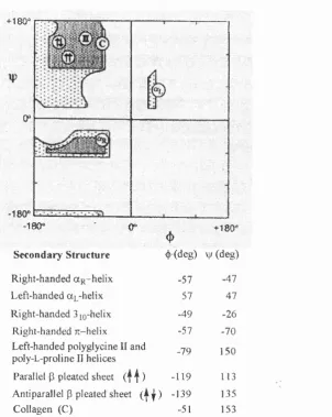
Figure 2: Local minimum energy bond lengths in A around which the structure can fluctuate...2 1 Figure 3: A Ramachandran plot o f sterically allowed ^ and \\i angles for poly-L-alanine...23
Figure 4: Hydrogen bonding patterns o f helical polypeptides... 25
Figure 5: The a-helix: residues 15-33 o f cytochrome c peroxidase... 24
Figure 6; The poIy-L-proline II helix: Pro-rich ligand for the SrcSH3 domain... 28
Figure 7: Tracing the 13-atom hydrogen bonded loop o f a piece o f an a - or 3.6 1 3-helix... 30
Figure 8: Kemp’s helical peptidyl-template molecule... 32
Figure 9: The hydrazone-ethylene based cyclic template attached to a polypeptide chain... 33
Figure 10: Dilactam-bridged four residue peptide...35
Figure 11: A single spaced lactam bridge in a segment o f an a-helical peptide... 35
Figure 12: Conformationally constrained hexapeptide... 36
Figure 13: A 22-membered lactam ring contained within a sequence o f hGRF, with the Gly spacer between Asp(8) and Om(12)...38
Figure 14: An f,(/+7) lactam bridge between L-Lys and (a) D-Glu, (b) but not always possible with L-Glu...38
Figure 15: A tripodal lactam-bridged model peptide... 39
unnatural D- or 2-amino acid...40
Figure 18: Ring closing metathesis (with a catalyst) o f terminal olefin side chains spaced /,(^+4) apart...41
Figure 19: Two overlapping bridges... 42
Figure 20: Model host peptides used for ‘host-guesf experiments to determine guest (X) 5-values... 43
Figure 21: (a) Control peptide (b) Test peptide with X-X as the synthetic constraint...46
Figure 22: The synthetic constraint: the ‘double-headed’ amino acid, (2R,9S)-2,9-diaminodecanedioic acid... 47
Figure 23: Modified sequence o f (1-22) hGRF with an f,(/+4) bridge...48
Figure 24: (a) Homocysteine bridge; (b) Leucine residue... 48
Figure 25: The protected form o f (2R,9S)-2,9-diaminodecanedioic acid (1) ready for solid phase peptide synthesis ...49
Figure 26: Common routes o f cyclisation on-resin... 51
Figure 27: The general solid phase synthesis o f a tripeptide amide... 53
Figure 28: Synthetic strategy for a branched cyclic peptide... 55
Figure 29: Cleavage o f a temporary N^-Fmoc protection group from a dipeptide...56
Figure 30: Cleavage o f the semi-permanent Dde protection group... 57
Figure 31 : Cleavage o f the semi-permanent Allyl Protection group on a Glu side chain... 57
Figure 32: Cleavage o f a permanent Boc protection group on a Lys side chain... 58
Figure 33: A tricyclic homodetic peptide prepared by a 3-dimensional orthogonal protection strategy... 59
Figure 34: The activated dipeptide undergoes intramolecular nucleophilic attack with loss o f Y, etc...60
Figure 36: An 0-acylisourea is formed from a nucleophilic attack o f a carboxylic
acid on DIC...62
Figure 37: An amide bond is formed with an activated ester which is generated from HOBt, etc... 62
Figure 38: The neighbouring group effect o f the 7-isomer o f HOAt...63
Figure 39: The HOCt coupling reagent... 64
Figure 40: The phosphonium based coupling reagents...65
Figure 41: The aminium based coupling reagents...66
Figure 42: T3P... 66
Figure 43: The attempted synthesis o f cyclo[Dab(2)-D-Glu(3)] with HBTU, etc...67
Figure 44: Other coupling reagents... 69
Figure 45: Synthesis o f Tyrocidine A on the oxime resin... 71
Figure 46: An end-to-end cyclisation o f an /,(/+4) segment o f human calcitonin analogue...72
Figure 47: The dashed lines represent the 4 attachments points used to synthesise the target decapeptide... 73
Figure 48: Attachment o f a Lys or Om side chain to an activated carbonate resin...75
Figure 49: Head-to-tail cyclisation with Ser side chain attachment... 76
Figure 50: Attachment o f a Tyr side chain to various resins via the Mitsunobu reaction...77
Figure 51: The BAL concept, etc... 78
Figure 52: Synthesis o f a cyclic hexapeptide, etc...79
Figure 53: Synthesis o f a Targe loop’ peptide... 80
Figure 54: Synthesis o f a thioether-bridged cyclic peptide... 81
Figure 55: (A) Cyclic peptide analogue, (b) N-protected ©-aminoalkylidene Gly building block... 82
Figure 57: The cyclic tripeptide analogue... 86
Figure 58: Solid phase synthesis o f resin bound conotoxin G1...87
Figure 59: Synthesis o f a cyclolathionine peptide by a novel thioether bridge formation...89
Figure 60: Connecting the A^-alkylated side chains in a peptide chain by olefin ring closing metathesis... 90
Figure 61: Grubbs olefin metathesis ruthenium catalyst...91
Figure 62: Introduction o f an alkene side chain to an amide nitrogen in solution or on solid support... 91
Figure 63: A retrosynthetic analysis o f the target residue, etc...93
Figure 64: The isopropyl moiety influences the direction o f attack, etc... 95
Figure 65: Literature synthesis o f the Schollkopf chiral auxiliary... 95
Figure 66: Synthesis o f compound (31) following the modified Bull methodology... 97
Figure 67: The bis-iminium cation... 98
Figure 68: The attempted alkylation o f a lithiated dihydropyrazine... 99
Figure 69: Cyanocuprate formation, etc...100
Figure 70: The Finkelstein reaction... 101
Figure 71: Chiral azidation o f an enolate-metal chelate... 102
Figure 72: Synthesis o f the oxazolidinone... 102
Figure 73: Addition o f the lithiated oxazolidinone, etc... 103
Figure 74: Chiral azidation o f compound 43... 104
Figure 75: Synthesis o f trisyl azide... 104
Figure 76: The proposed mechanism o f chiral azidation, etc... 104
Figure 77: Summary o f the route to the target ‘masked’ amino acid... 105
Figure 78: Cleavage o f the Evans auxiliary... 106
CHAPTER ONE
Introduction
1.1 Helices in Peptides
The secondary structure o f a protein is defined by the local conformation o f its peptide backbone. ^ Many aspects o f protein secondary structure are derived from the sequence o f amino acids contained in the polypeptide chain.^ X-ray studies o f amino acids and dipeptides by Pauling and Corey deduced the peptide group as a rigid planar structure (Figure 1) with the peptide bond having approximately 40% double-bond character because o f resonance interactions.^
O
9
1
I
Figure 1. Resonance structure o f the amide bond.
The N-C“ single bond is 0.13 A longer than the C-N bond, and the C = 0 bond is 0.02 A longer than that found in aldehydes or ketones (Figure 2). When the amide bond is planar, its 7t-bonding overlap is accentuated with a resonance energy value o f about 85 kJ/mol.
O
.
Three backbone dihedral angles per residue (N-C“ bond, C“ -C bond, \\f; C-N bond, co) and the dihedral angles (%i, %3 ) the side chain entirely describe the
local polypeptide conformation.^ However, as the partial double bond character o f the polypeptide keeps © close to 180° in the trans or extended form and near to 0° in the cis form, <|) and vj/ suffice for the main chain.
The two-dimensional Ramachandran plot utilises ^ and vj/ angles to indicate the sterically allowed backbone conformation for polypeptide secondary structures (Figure 3).^ Even though a polypeptide could have a highly coiled conformation, this is not by chance, instead restrictive conditions exist denoting allowed conformations in the proximal area o f a main chain residue. This is defined in terms o f contact criteria, allowing the distances between pairs o f atoms not to be less than the specified van der Waals minima.
Secondary structures o f polypeptides are generally classified according to their respective type o f regular conformation.^ The prominent conformations include a-helices and p-sheets, or nonrepetitive structures like turns and loops. Determining regular secondary structure is an important focal point when attempting to comprehend the structure o f a complex protein.
Regular polypeptide conformations are classified according to their repetitive ^ and
+180^ r r
y
- w ' c r +180^
S econdary S tru ctu re 4»(deg) V (d eg )
Right-handed aR -helix -57 -47
Left-handed UL-helix 57 47
Right-handed 3 jQ-helix -49 -26
Right-handed TC-helix -57 -70
Left-handed polyglycine II and
poly-L-proline II helices -79 150
Parallel P pleated sheet ( f j ) -119 113
Anti parallel P pleated sheet ( | | ) -139 135
Collagen (C) -51 153
Figure 3. A Ramachandran plot o f sterically allowed <}) and \\f angles for poly-L-alanine.^ The dark shadow areas are for the ‘normally allowed’ ({) and vj/ angles and the light shadow area represents conformations having ‘outer limit’ van der Waals distances.^
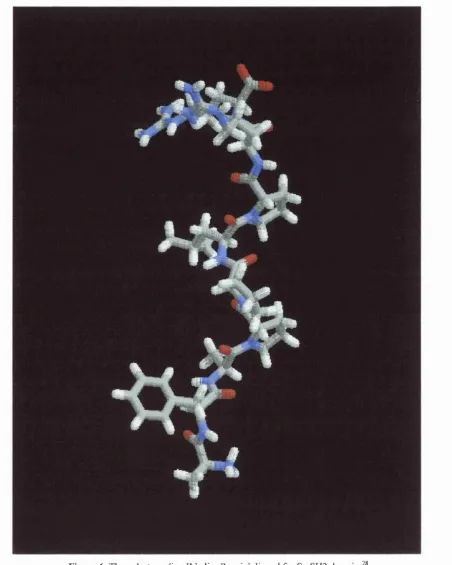
Hydrogen bonding patterns o f helical polypeptides occurs between the C = 0 of the rth residue and NH o f either the (/+3) or (/+4) and (/+5) residue (Figure 4).^ For the a-helix, carbonyl groups and amide protons are orientated in a ‘head-to-tail’ fashion enabling the hydrogen bonds to face the same direction on the helix surface. The characteristic /,(/+4) hydrogen-bonded a-helix has 3.6 residues per turn, and a pitch of 5.4 A (Figure 5).^ Apart from a-helices being one o f the most dominant forms of secondary structure in nature, they are predominately right-handed helices due to the chirality of the constituent T-amino a c i d s . T - a m i n o acids, if stacked in the
Figure 5. The a-helix: residues 15-33 o f cytochrome C peroxidase
left-handed form, would experience a sterically destabilising interaction between the carbonyl oxygen o f the rth residue and the p-carbon o f the (z+4)th residue.
o
o II
rth residue ^ 9
II
%■ |
I
Ï ' ' *^ I (/+3) residue
I
PI ^ 3 , „ - h e ü x f a-helix ( ' ^ « s i d u e
H H
MO .
) 71-hebx
Figure 4. Hydrogen bonding patterns o f helical polypeptides (atoms around C“ have been omitted for clarity).
The Sjo'helix has a pitch o f 6.0 A, and therefore rises steeper than the a-helix. Its (j) and \\j angles place it in a region o f conformations having ‘outer limit’ van der Waals distances, indicating that its R groups may experience some steric impediment. The close proximity o f (j) and vp conformational space between a - and 3^Q-helices may imply a gradual transformation into either helical form with a near-helix conformation maintained throughout.^® Studies utilising computer simulations to address the o/Sjo/coil equilibrium suggest that the S^Q-helix is a kinetic intermediate within the a-helix unfolding p a t h wa y . ^ 3 , o - He l i c e s have also been proposed as thermodynamic intermediates in helical peptides existing as a mixture o f a - and
3io-structures.^^
310-Helices were initially thought to be rare in globular proteins. However, a survey
3iQ-helix is less common than a-helix, it does have an important biochemical role in some cases. The interconversion o f the two helices has recently been implicated in the activity o f lactate dehydrogenase^^ and aspartate aminotransferase.^^
Distinguishing an a-helix from the S^Q-helix by NMR or CD spectroscopy is difficult. Discrimination between the two types o f helices in aqueous solution with peptide sequences similar to polyalanine helices studied by Marqusee and Baldwin was possible by employing double spin ESR spectroscopy. Analysing the distances between spin labels indicated the presence o f 3^Q-helicity at the C-terminus in the 16-residue peptide. Increasing the length by a further 5 residues converted the peptide largely to an a-helix. This indicates that the proportion o f a-helix and 3iQ-helix in polyalanine peptides is partly dependent on peptide length. Protein crystallographic data also implied that shorter helical stretches are likely to exist as 3io-helix^ and longer a-helices often have the final turn at the C-terminus as a
3^ 0-h elix.^ ^
In contrast to a - and 3 iQ-helices, 7i-helices are very rare.^ This type o f helix has been observed in protein crystal structures in catalase^^ and cytochrome P450.^^ The rarity o f the 7t-helix has been attributed to its energetically unfavourable dihedral angles^ and requirement for at least four residues to be in the correct orientation for /,(/+5) hydrogen bonds (Figure However, main chain hydrogen bonding was observed between glutamine side chains spaced /,(/+5) in a polyalanine peptide in a molecular dynamics simulation study.^^ Water molecules bridging the side chains helped stabilise these respective hydrogen bonds.
stnictures^^’^^ (Figure 6).^^ Proline residues were dominant, but, not exclusive in these helices. Poly-L-proline II helices occur on the protein surfaces, and therefore are exposed and hydrogen bonded to water molecules. The presence o f proline minimises the occurrence o f main chain hydrogen bonds, whilst hydrogen bonds with water molecules could stabilise poly-L-proline II helices.^^ The more mobile geometry o f the poly-L-proline II helix may permit it to interconvert into either a right-handed a - or S^Q-helix. Polyglycine II and poly-L-proline II helices are found in collagen molecules in a twisted coiled coil form, stabilised by interchain hydrogen bonds with water.^^
Another form o f polypeptide secondary structure has been identified from X-ray diffraction studies o f gramicidin These studies have also confirmed its function as a membrane channel structure. The molecule consists o f 15 alternating L- and D-amino acids with both ends blocked with formyl and ethanolamine groups. It exists as a head-to-head dimer in a p-helix form.^^ Despite the potential o f either a right- or left-handed helix forming, spectroscopic data has identified a right-handed helix with six to seven residues per tum.^^
A novel regular helical secondary structure has been observed in the crystal structure o f a-chymotrypsin.^^ Denoted as the e-helix (e refers to the extended nature o f the helix), this has ^ and \\) angles o f -93° and 146°, -2.7 residues p e r turn (left-handed helices have negative values) and a unit height o f 3.3 Â.
4
#
Figure 6. The poly-L-proline II helix: Pro-rich ligand for SrcSH3 domain28
require a-helical motifs. Key receptor-binding constituents o f peptide hormones such as calcitonin,^"^’^^ neuropeptide and human growth hormone releasing factor^^ are often a-helical. The basic-leucine zipper (bZIP) class o f proteins have an a-helical segment that is responsible for sequence-specific DNA recognition when binding occurs.^^ Apart from the suggestion that the a-helix plays a dominant role in protein folding initiation;^^ other key structural contributions include coiled coils in fibrous proteins,^® and 4-a-helical bundles in cytokines.^^
1.2 The i\(f+4) Hydrogen Bonded a-H elix and Stabilising Constraints
Designing intrinsically flexible short peptide segments to adopt well defined helical conformations in solution is important in the study o f biologically active molecules. Whilst nature uses constraints such as the cyclic amino acid proline and macrocycles formed by disulfide bonds to reduce the flexibility o f polypeptide c h a i n s , s e v e r a l synthetic approaches have been used to constrain peptides. In this review, stabilising a-helical polypeptides by reducing the entropie cost with strategically placed ‘constraints’ are considered.
1.2.1 Stabilising Hydrogen Bonding Interactions
The hydrogen bond in an a-helix can be thought o f as a closed loop o f 11 backbone atoms, plus a carbonyl O atom at one end and an amide H atom at the other end, hence, a total o f 13 atoms. (Figure 1 )? This is in contrast to the 3jQ-helix which has
I N H
0 = ^ ^ ^ 13-atom loop N H
S ^ N — H - ; O— f
^ H-bond f
Figure 7. Tracing the 13-atom hydrogen bonded loop in a piece o f a - or 3.6j3 -helix. (Only atoms directly involved in a single hydrogen bonded loop are shown for clarity).
Electron density maps o f a-helices indicate that the carbonyl oxygens point towards the C-termini and are slightly tilted outward away from the helix axis.*^^ An a-helix can be described as an array o f dipoles with an aggregate dipole directed along the helix axis, ending with a negative pole at the C-terminus, and a positive pole at the N-terminus.^^’^ Stabilising charge-dipole interactions o f positive charges with the C-terminus and negative charges with the N-terminus are possible.^^’"^^ This electric field generated by the polypeptide backbone could be a significant property responsible for binding o f charged substrates or useful as an ‘active site’ for enzyme catalysis.^^"* However the helix effect may be due only to a few localised dipoles and not the helix macrodipole.^^
presence o f residues with side chains able to form hydrogen bonds with the initial unpaired four NH donors and final CO acceptor groups in the main chain.^^’"^^ Residues such as Asn, Asp, Glu, Gin, Ser, Thr and His at the N-terminus and Asn, Arg, Gin, Lys and Ser at the C-terminus have been implicated."^^
The interaction o f distal residue side chains with exposed NH or CO main chain groups at the ends o f helices may also be favoured by the presence o f sterically unhindered Gly.^^ The contribution o f this interaction at the C-terminus to helix stability is not clear, partly because Gly is also observed to be a helix-breaker.^^’"^^ The presence o f Pro within a sequence is more helix destabilising than Gly,^®’^^’^^ however, at the N-terminus it may promote helix stabilisation because it restricts conformational freedom. The frequent occurrence o f Gly and Pro at helix termini provides strong evidence for these suggested capping mechanisms in peptides and even proteins.^^’^^
shown to stabilise a-helicity in aqueous solution in a specific order. The findings o f Forood et were in agreement with the previous study by Presta and Rose."^^ Hence, the helix-inducing ability o f various residues was deduced to be as follows: Asp>Arg>Ser>Glu>Gln>Ala at the amino terminus and Arg>Lys>Ala> at the carboxyl term in u s.R esid u es Pro and Gly might correctly orientate the polar side chains o f end residues such as Arg (at the C-terminus) and Asp (at the N-terminus) for hydrogen bonding to free CO and NH groups at the helix termini.
Introducing synthetic ‘capping’ templates at the helix termini is another method used to initiate and stabilise a-helicity in a peptide chain. Helix fraying at the termini is prevented by directing the otherwise free hydrogen-bonding groups o f the terminal residues to interact with the capping template. Novel use o f a rigid tricyclic proline-related template constrained by a S-CH2 bridge (Figure 8), with peptides
attached to its C-terminus, promotes hydrogen bonding between the correctly orientated CO and the relevant peptide NH groups. Induced helicity in short peptides with sequences o f poor helical preference was reported.
Peptide
A
A helix template with a hydrazone-ethylene link (-CO-N(R)-N=CH-CH2CH2-) as a
covalent replacement for the /,(/+4) hydrogen bond (-CO-NH—0=C(R)-NH-), maintains the 13-membered hydrogen bonded loop o f an a-helix (Figure 9). Attachment o f this template to the N-terminus o f a 10-residue peptide, promoted helix stability in aqueous solution.
HN.
O
Figure 9. The hydrazone-ethylene based cyclic template attached to a polypeptide chain.
1.2.2 Stabilising /,(i+4) Side Chain Interactions
Investigation o f the relative effectiveness o f Glu-Om, Glu-Lys and Glu-Arg ion pairs in stabilising alanine-based a-helical peptides also confirmed /,(/+4) spacing as ideal.^^ The highest helical content in the host peptide was with the latter Glu-Arg ion pairs at neutral pH.
The importance o f stabilising /,(/+4) hydrogen bonding interactions between guest ion pair residues, such as Gln-Asp;^ His-Asp;^^»^^ and Glu-Lys, Glu-His and Asp-Lys^^ in alanine-based host peptides has also been demonstrated. The shorter side chain His and Asp residues have stronger side chain interactions when compared to Lys, Glu or Arg. This is attributed to a lower loss o f side chain entropy when engaging in such an i n t e r a c t i o n . H o w e v e r , the stabilising effect o f His-Asp interactions seems ambiguous in the protein T4 lysozyme.^^ In another example, Lys or Arg to Glu salt bridges stabilised a-helicity in alanine-based peptides derived from smooth muscle caldesmon.^^
The strength o f side chain-side chain interactions between Glu-Lys^^ and Gln-Asn^® from modified forms o f the Lifson-Roig^^ theory for helix-coil transition has been estimated. The major contributor to these side chain interactions was established to be hydrogen bonding (by screening out electrostatic interactions at high salt NaCl concentrations), and a maximum free energy o f interaction around -1.9 kJ/mol for Glu"-Lys^ was det ermi ned. Li kewi se, -1.7 to -2.9 kJ/mol was found for Gln-Asn interactions.^®
between aromatic and charged residues are observed with the Trp-His"^ pair, and are helix stabilising in aqueous solution/^
1.2.3 Lactam Bridges between Side Chains
One o f the earliest cited examples about creating unnatural amide bonds between amino acid side chains involved adjacent Lys and Glu residues7^ This afforded the optimum size for incorporation o f dilactam rings and peptide backbone rigidity (Figure 10). Conformational analysis confirmed that the rigid peptide backbone maintained a p-tum structure.
|— NHCO— I I— nHCO— I
(CH2)4 (CH2)2 ( ^ 2)4 (CH2)2
MeCO— NHCHCO--- NHCHCO— NHCHCO— NHCHCO— NHMe
L-Lys Z-Glu D -L ys Z)-Glu
Figure 10. Dilactam-bridged four residue peptide.
However, for stabilising a-helical peptides, /,(/+4) spaced lactam bridges are r e q u i r e d . F o r example, studies with lactam-bridged Lys-Glu residues (Figure
1
.1)76,77
allowed comparisons to be made between the degree o f helicity observed inlinear and cyclic peptide analogues. Peptide assembly and cyclisation either on or o ff a solid support was found to yield stable helices in aqueous buffer and in TFE/water mixtures.
CD and NMR data confirmed that two overlapping lactam bridges induced a hexapeptide to adopt an extremely stable a-helical conformation in aqueous solution (Figure 12)7^ Lactam bridges between Lys and Asp are known to be more helix stabilising than Lys-Glu bridges/^
Boo — Lys 1 — Lys 2 — Ala 3 — Ala 4 — Asp 5 — Asp 6 — OPac
Figure 12. Conformationally constrained hexapeptide.
A direct comparison o f Glu-Lys salt bridges in a 14-residue linear peptide and Glu-Lys lactam bridges in a cyclic analogue, confirmed a higher helical content in the latter peptides from CD spectroscopy.^^
Lactam bridges have been used to stabilise a-helical conformations in a variety o f biologically active peptides. For example, CD data verified the ability o f these bridges to stabilise and enhance the dimérisation required for a 24-residue sequence derived from a zinc finger peptide library. This overall conformational stability allowed the peptide to maintain a two-stranded a-helical coiled-coil conformation in aqueous solution, even at low peptide concentrations.^^ In addition, the peptides lacking the lactam bridges between Glu and Lys, were less effective in inhibiting the binding o f the His-Phe-Val-Gln-His Zn finger peptide to the monoclonal antibody IgA in a dose dependent manner.
stabilisation with Lys-Asp residues /,(/+3) apart; however, the presence o f 3ig-helicity could not be ruled out. Lys-Asp Lactam bridges spaced /,(/+4) apart in the parathyroid hormone-related protein (7-34) amide, both stabilised a-helical structure in aqueous solution and helped elucidate the ‘bioactive conformation’. Analogues o f other peptide hormones such as neuropeptide human calcitonin^^ and human growth hormone releasing factor^^’^^ confirm the stabilising effects o f various /,(/+4) lactam bridges on their conformational and pharmacological properties.
HNyNHz
NH
(CH2)3
HO
O % H O V H _
Asp(8)y " °
-oc
Om(12)
-SPACER-Figure 13. A 22-membered lactam ring contained within a sequence o f hGRF, with the Gly spacer between Asp(8) and Om(I2).^^
In addition to f,(i+4) and /,(i+3) lactam bridges, these synthetic constraints also occur between side chains o f similar residues spaced f,(/+7) apart. Optimum side
chain-to-side chain cyclisation was observed between Z)-Glu and L-Lys spaced apart. Molecular modelling studies indicated that the orientation o f the Z-Glu side chain was not ideal for cyclisation and maintenance o f an a-helix structure (Figure 14). However, attempted cyclisation on the solid support yielded 46% o f the L-Glu cyclised peptide and 76% o f the D-Glu cyclised peptide. The cyclisation with 7,-Glu required almost six times the reaction time required for the latter cyclisation. CD spectra o f the T-Glu cyclised peptide indicated a random structure in aqueous solution and an a-helix with the D-Glu cyclised peptide.
I-Glu D-Glu
C = 0 NH
(a) (b)
A novel tripodal lactam-bridged peptide was synthesised from a trifimctional template such as (6)-1,3-diaminopropionic acid. This ‘bridge’ united an / spaced Lys residue with two Asp residues (z+3) and (z+7) away (Figure 15).^^ However, no conformational data o f the resultant peptide was provided.
=0
H-Lys-Leu-Lys-Glu-Leu-Asp-Gln-Lys-Leu-Asp-Glu-Leu-Lys-Gln-OH
Figure 15. A tripodal lactam-bridged model peptide.
1.2.4 Disulfide Bridges between Side Chains
Many native proteins do contain disulfide bonds; smaller biologically active compounds such as peptide hormones and neurotransmitters also possess disulfide bridges between oxidised cysteine residues. They take up a cyclic structure and are conformationally restricted allowing for extensive examination o f bioactivity relationships. Examples include the neurohypophyseal hormone oxytocin, vasopressin and somatostatin (Figure 16).^h92
^ ________ Oxytocin_________^
H-Cys 1 -Tyr 2-Ile 3-Gln 4-Asn 5-Cys 6-Pro 7-Leu 8-Gly 9-NH2
Vasopressin_______
f
H-Cys 1-Tyr 2-Phe 3-Gin 4-Asn 5-Cys 6-Pro 7-(Lys or Arg) 8-Gly 9-NH2
g _________________________Somatostatin___________________________ g
I I
H-Ala 1-Gly 2-Cys 3-Lys 4-Asn 5-Phe 6-Phe 7-Trp 8-Lys 9-Thr 10-Phe 11-Thr 12-Ser 13- Cys 14-OH
NMR studies showed that disulfide bridges stabilise a-helical motifs in an apamin-S peptide hybrid s e q u e n c e . T h e hybrid peptides containing the disulfide bridges folded into the same conformation as the native apamin peptide and possessed biological activity that complemented the S peptide. Hence, the helix-stabilising disulfide bridges did not interfere with its expected biological activity. In another example, short peptides with a two-turn a-helix conformation were stabilised by a disulfide bridge between i and (i+7) residues.^"* The 8- to 16-residue series o f peptides included the D- and Z-enantiomers o f fully deprotected 2-amino-6-mercaptohexanoic acid (Figure 17). The disulfide bond was made from the oxidation o f the free thiol moieties. CD data indicated a substantially higher degree o f helicity in the oxidised forms o f each peptide.
C OH
Fmoc-N N CH3
(a) (b)
Figure 17. (a) An /,(/+7) disulfide bridged peptide; (b) the fully protected form o f the unnatural D- or Z,-amino acid.
1.2.5 Other Covalent Linkages between Side Chains
A series o f cyclic helical heptapeptides incorporating an /,(/+4) carbon-carbon link between residue side chains were synthesised by ring closing metathesis utilising an olefin metathesis catalyst [(PCy))2Cl2Ru=CHPh] (Figure 18).^^ The linear peptides
comprised o f the sequence Boc-Val-X-Leu-Aib-Val-X-Leu-OMe, where X was the (olefin-containing) Z-Ser or L-homoserine (Hse) 0-allyl ether residues. The incorporation o f Aib residues into these peptides was justified by requiring amino acids known to stabilise and/or a-helical conformations in apolar polypeptides.^^ However, the CD spectra o f the peptide macrocycles did not indicate any significant conformational change to either helical conformation upon cyclisation. In addition, the type o f helical conformation present, that is, a - and/or S^g-helix remained unclear. There also appears to be uncertainty in the influence that the ‘ring closing olefin metathesis’ methodology has on the resultant biological activity o f synthetic conformationally rigid a-helical peptides.
Figure 18. Ring closing metathesis (with a catalyst) o f terminal olefin side chains spaced /,(/+4) apart.
Short peptides with a stable a-helical conformation in water were synthesised with
^-substituted benzene ring conformationally constrained a 14-residue peptide (Figure
J 9 ) 1 0 0
o ILN
o
Figure 19. Two overlapping i,{i+l) bridges.
In summary, conformationally constrained short a-helical peptides in solution are attainable either by manipulating hydrogen bonding and side chain interactions, or incorporating covalent bonds between residue side chains. The principal aim o f this project is to design a peptidomimetic with a stable a-helical conformation in aqueous solution. The appropriate model peptide and type o f constraint chosen are considered in the next section.
1.3 The Target Peptide and Synthetic Constraining Residue 1.3.1 Helix Formation in Short Peptides
Lifson-Roig^^ (LR) and Zimm-Bragg^®^ (ZB) statistical models. The LR and ZB formalisms considered helix formation in a peptide as a stringent two-state transition o f population o f molecules from random coil to helix conformation. This is an untypical situation in peptides; instead a transition occurs between populations o f mostly helical, with frayed ends, and molecules that are almost o f random coil conformation. 10 h 105,106 xhese theoretical models also ignore the stabilising effects o f sequence- and position-dependent residue side chains.
A major advance in the study o f helical propensities o f amino acids in short peptides came from the findings o f Marqusee and Baldwin. They discovered that the significant entropie cost associated with helix formation in 16 or 17-residue alanine-based peptides could be offset by intrachain /,(/+4) hydrogen bonding between neutral or charged ion paired residues, and by the high helix propensity o f Ala. These studies questioned the original ‘host-guesf experiments and their suggestions that short peptides (<20 amino acids) could not form stable helices in an aqueous environment.^®^ Consequently, further studies by the Stellwagen,^®^ Kallenbach^^^’^^^ and Baldwin^®^»^®^’^^®»*^^"^^^ groups using a range o f host peptides (Figure 20) have established the 5-values o f guest amino acids and effects o f their sequence positioning.
HOST PEPTIDE PEPTIDE SEQUENCE REFERENCE
AEK AC-Y-EAAAK-EAXAK-EAAAK-A-NH2 (61,108)
AK AC-Y-KAAXA-KAAXA-KAAXA-K-NH2 (18,106,109,
110)
E4K4 succinyl-YS-EEEE-KKKK-XXX-EEEE-KKKK-NH2 (111,112)
AQ A c-A AQ A A-AAQ AA-AAQ AA-Y-N H 2 (69,113,114,
115)
AXA succinyl-YS-EEEE-KAKK-AXA-EEAE-KKKK-NH2 (109) Figure 20. Model host peptides used for ‘host-guest’ experiments to determine guest (X) s-values.^^^
Marqusee and Baldwin determined the helix content o f a 16-residue alanine-based peptide solubilised by incorporating Lys residues in aqueous solution. Compared to earlier work,^^ the novelty o f their findings indicated the highly helix promoting capacity o f alanine in the absence o f side chain interactions. The design o f the peptides eliminated ion pair formation or charged group-helix dipole interactions. Charged residues were strategically placed in the peptide sequence to spiral around the helix and avoid amphiphilicity and help solubilise the entire peptide. Helix formation was monitored by CD at 1°C, at pH 3-12, in both 1.0 and 0.01 M NaCl. The extent o f helix formation could be easily monitored by measuring the minimum o f the CD spectra at 222 nm; referred to as the molar ellipticity -[0]2i2 They established helix formation as an enthalpy driven process; increasing the temperature was accompanied by unfolding o f the helical p e p t i d e s . T h e peptides remained monomeric and had the highest helicity at neutral pH with three or four Lys residues at PC and 1.0 M NaCl. At low salt concentration, enhanced helicity was observed with an increasing pH.
the effects o f interactions between the bulky host side chains and the guest residues can not be ruled out, implying that alanine-based peptides are best suited to determining ^-values. 1 ^21
Vila et al^^^ suggested that alanine-based peptides, with three or four Lys residues inserted for water solubility, are highly helical because o f interference o f these Lys amino acids with the hydration o f the peptide backbone in the nonhelical conformation. This would imply that Lys has a higher 5-value than Ala, however, the opposite is observed. Kemp also challenged the helix stability o f de novo designed peptides, including alanine-based peptides and suggested the intrinsic helix propensity o f Ala is accentuated in the presence o f long side chains such as Lys, Gin or Glu.^^^’^^^ Recently both these suggestions have been challenged with the observed high helical content o f an alanine-based peptide flanked by pairs o f charged residues such as Om or Dpr.^^^ These residues have short side chains which are unable to directly interact with neighbouring Ala residues. In addition, the spacing o f the charged residues disputes the solvation effect o f Vila et The findings o f Kemp et 23,124 ^lay be attributed to stabilising interactions between Lys and the template itself. Increased helicity is observed in alanine-based peptides with one, two or three Lys residue substitutions^H ow ever, the contribution o f this increased solubility from an escalating charge to its apparent enhanced helicity is not clear.
1.3.2 The Model Peptide
The model peptide chosen for this project is a variant o f the Marqusee and Baldwin^ ^ type peptides (Figure 21). Hence, this is a 16-residue alanine-based peptide with four Lys residues incorporated /,(z+5) apart, for solubility and to minimise aggregation o f the peptide in solution. Lys Residues have the optimum side chain length for basic amino acids to minimise helix destabilising interactions with the peptide backbone. The peptide would have an acetyl group at the N-terminus and an amide group at the C-terminus to avoid helix destabilising interactions o f the terminal a-NHj''" and a-COO" with the helix dipole.^ ^ A synthetic conformational constraint would then be placed near the supposedly least helical segment o f the peptide chain and direct comparison o f helicity between the unsubstituted and substituted peptides in solution could then be made (refer to chapter 4). This would provide information about the effects o f conformationally constraining this short peptide in aqueous and non-aqueous environments.
AC-KAAAAKAAAAKAAAAK-CONH
2(a)
AC-KAAAAKAAXAKAXAAK-CONH
2(b)
Figure 21. (a) Control peptide (b) Test peptide with X-X as the synthetic constraint.
1.3.3 The Synthetic Residue Constraint
The region near the C-terminus o f the control/unsubstituted model peptide has been reported to be less helical than the N-terminus; CD and NMR data indicated the greatest a-helical content in Marqusee and Baldwin^® type peptides between the middle o f the 16-residue peptide and its N - t e r m i n u s . E v e n though initially Marqusee and B a l d w i n t y p e peptides were thought to have some 3iQ-helicity at the C-termini, the role o f the imposed flexible spin labels used in these ESR experiments do question the accuracy o f their findings. The N terminal acetyl blocking group could promote helix stability by establishing capping interactions o f its otherwise free C = 0 moiety with unpaired amide backbone groups and minimise f r a y i n g . H e n c e , the synthetic constraint is to be incorporated near the C-terminus in the model peptide (Figure 21).
The constraint replaces two Ala residues o f /,(/+4) spacing in an attempt to maintain the hydrogen bonding pattern o f an a-helical peptide b a c k b o n e . T h i s constraint is a covalently bonded ‘scaffold’ requiring four sites within it that can be manipulated. The first site o f this synthetic residue needs to be incorporated whilst chain extension continues with the second site, then the third site needs to intramolecularly cyclise on to a bridging Ala residue, and the final site needs to continue the required peptide chain extension (Figure 22).^^^ This novel type o f synthetic constraint is able to generate a cyclic peptide by a unique synthetic route (refer to chapter 2 and 4).
Final Site nHo
= First Site
,COOH
IS HOOC''
X
X
Third Site NHj
Second Site
The length o f this 8-carbon aliphatic bridge has been developed from initial molecular modelling s t u d i e s ^ w i t h an idealised a-helical segment o f a modified sequence o f human growth hormone releasing factor using the SYBYL (Tripos) program (Figure 23).^^
H2
N-Tyr-Ala-Asp-Ala-Ile-Phe-Thr-Asn^-Xaa^-Tyr-Arg-Lys^^-Xaa^^-Leu-Ala^^-Glu-Leu-Ser-Ala-Arg-Lys-Leu-C02H
Figure 23. Modified sequence o f (1-22) human growth hormone releasing factor with an /,(/+4) bridge. (Xaa refers to the substituted amino acids).
Initial studies reported in the literature, on the effects o f covalent bridges upon helix stability within this sequence (Figure 23), involved a lactam bridge between residues 8 and 12.^^ In addition to the lactam bridge, an Ala residue replacing a Gly at position 15 did correspondingly increase the stability o f the helix significantly in aqueous solution. However, the covalent bridge in this modelling study was a disulfide link between substituted D- and L- homocysteine residues at positions 9 and 13 (Figure 24). The 8-atom bridge was replaced with carbons atoms only and the energy o f helix formation o f the peptide determined by using the electrostatic parameters o f Leu. It appeared that the bridged sequence had a lower energy than the segment lacking the bridge. (Refer to Appendix A for the photograph o f the computer generated molecular model).
2 V s V 7
8
HzN'^^COgH HjN^^COsH
(a)
Figure 24. (a) Homocysteine bridge; (b) Leucine residue.
In this project, the aliphatic bridge was obtained by the incorporated (2R,
9*S)-2,9-diaminodecanedioic acid. The synthesis o f (2/?, 95^-2,9-diaminodecanedioic acid required the use o f two separate enantiospecific methodologies to generate a residue with two chiral centres for use in peptide synthesis (refer to chapter 3). This unnatural bifunctional amino acid is used in solid phase peptide synthesis^^^ (refer to chapter 4) in its fully protected form (Figure 25).
NH
OH
NH
Figure 25. The protected form o f (2/?, 95)-2,9-diaminodecanedioic acid (1) ready for solid phase peptide synthesis.
CHAPTER TWO
Solid Phase Synthesis of Cyclic Peptides
2.1 Introduction
Interest in cyclic peptides continues over the decades due to the occurrence o f such structures in nature as antibiotics or toxins. Some o f the best known examples are the cyclic antibiotic decapeptide gramicidin S which was discovered about 50 years ago,^^^ tyrocidins/"^^ and valinomycin.^"^^ Many small peptides regulate numerous biological functions, acting as hormones, inhibitors or neurotransmitters. 131,142-144
Hence, the efforts in drug design are concerned with establishing highly specific agonists or antagonists o f these peptides with minimal toxicity. The design o f such analogues requires distinct information about the bioactive conformation for specific interactions with the many available receptors. Linear peptides built from natural Z-amino acids are generally less suitable for studying specific structure-activity relationships between a ‘bioactive molecule’ and its respective receptor. Apart from their inherent flexibility in solution, problems o f bioavailability and enzyme degradation are also unavoidable. Incorporation o f a constraint could lock the active conformation by reducing its conformational entropy, resulting in an increased affinity. These conformationally restricted molecules are ideal lead compounds for drug design combining high receptor affinity and reduced side effects.
homodetic i f lactam linkages connect the constituent amino acids, and heterodetic if the linkages are disulfide, thioether, ether or ester moieties.
Traditionally, both types o f cyclic peptides were prepared entirely in solution; alternatively the linear precursor was initially assembled on solid support, cleaved from the resin and finally cyclised in solution, Classical ‘cyclisation in solution’ methodologies were limited by cyclodimérisations and cyclo-oligomerisations even under conditions o f high dilution. Cyclisation o f the linear peptide whilst still attached to the resin may be more beneficial due to the ‘pseudodilution’ effect. This is a kinetic occurrence where intramolecular reactions dominate over intermolecular side reactions. The solid phase approach may also drive the cyclisation o f the peptide to completion by allowing the use o f excess reagents which can be easily removed with washing and filtration.
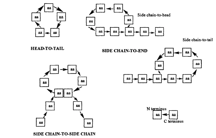
Three main routes to cyclisation o f peptides on-resin have been described (Figure
2 6 ). 145,146
y Side chain-to-head
aa aa
Side chain-to-tail
HEAD-TO-TAIL
SIDE CHAIN-TO-END
y B ^ E y
B
y
-*"| aa aa [-*-j aa [
N t s m i a u s C term inus
SIDE CHAIN-TO-SIDE CHAIN
Ring closure between the terminal amino and carboxyl groups is referred to as head-to-tail or end-to-end mode o f cyclisation. Closure is also possible between an amino group o f a Lys/Om residue side chain and the carboxyl terminus, or an Asp/Glu side chain carboxy group and the amino terminus; these closures are referred to as the side chain-to-end or side chain-to-backbone mode o f cyclisation. Finally, homodetic and heterodetic cyclic peptides are generated from side chain-to-side chain cyclisations involving lactam bridges formed between functional groups on residue side chains or disulfide linkages between two oxidised cystine residues.
2.1.1 Solid Phase Synthesis o f Linear Peptides
The pioneering work by Merrifield^^^ in the early 1960’s developed into a novel method for the automated synthesis o f polypeptides. This process involves the stepwise assembly o f a linear peptide chain whilst still attached to a solid support (Figure 27). The solid support consists o f polymer resin beads attached to a linker. The first amino acid is attached to the solid support directly via this linker. The linear peptide is built up from individually coupled N^-temporarily protected amino acids (with permanent side chain protection if necessary).
reagents to optimise reaction conditions, and the easier work-up between amino acid couplings.
Fmoc-HN-
HoN-X .
Fmoc-HN CONH
Fmoc-HN
CONH-_ Polvmer resin bead
LINKER
D eprotect terminal amine from linker (or handle)
LINKER
Couple amino a cid 1
LINKER
1. Deprotect N^-Fmoc
2. Couple amino a c id 2
LINKER
1. Deprotect hN-Fmoc
2. Couple amino a cid 3
3. D eprotect N -terminal Fmoc group
4. Cleave tripeptide fro m a cid labile linker
R'
H sN ^ ^ C O H N '-^ C O H N K '^ C O —Ii2i\ u w n ix V/wniT uu—NH2
Figure 27. The general solid phase synthesis o f a tripeptide amide. (R^-R^ are arbitrary amino acid side chain groups.)
When the desired linear peptide sequence has been assembled, the final peptide can be cleaved from the solid support using conditions that are dependent on the type o f linker used. The N-terminal protecting group could be removed before cleavage or left on the final target peptide. The choice o f linker (or handle) permits either a peptide acid or amide to be generated. The remainder o f this chapter discusses examples concerning the solid phase synthesis o f cyclic peptides.