Wong, TY; Cheng, CY; Davila, S; Hibberd, M (2018) Human pharyn-geal microbiota in age-related macular degeneration. PloS one, 13 (8).
e0201768. ISSN 1932-6203 DOI: https://doi.org/10.1371/journal.pone.0201768
Downloaded from: http://researchonline.lshtm.ac.uk/4649166/ DOI:10.1371/journal.pone.0201768
Usage Guidelines
Please refer to usage guidelines at http://researchonline.lshtm.ac.uk/policies.html or alterna-tively contactresearchonline@lshtm.ac.uk.
Human pharyngeal microbiota in age-related
macular degeneration
Eliza Xin Pei Ho1☯
*, Chui Ming Gemmy Cheung2☯
, Shuzhen Sim1☯
, Collins Wenhan Chu1,
Andreas Wilm1, Clarabelle Bitong Lin1, Ranjana Mathur2, Doric Wong2, Choi Mun Chan2, Mayuri Bhagarva3, Augustinus Laude4, Tock Han Lim4, Tien Yin Wong2, Ching Yu Cheng2,
Sonia Davila1, Martin Hibberd1,5
1 Genome Institute of Singapore,Singapore, Singapore, 2 Singapore Eye Research Institute, Singapore National Eye Center, Duke-NUS Medical School, National University of Singapore, Singapore, Singapore, 3 Department of Ophthalmology, National University of Singapore and National University Health System, Singapore, Singapore, 4 National Healthcare Group Eye Institute, Tan Tock Seng Hospital, Singapore, Singapore, 5 Faculty of Infectious and Tropical Diseases, London School of Hygiene and Tropical Medicine, London, United Kingdom
☯These authors contributed equally to this work. *hoxpe@gis.a-star.edu.sg
Abstract
Background
While the aetiology of age-related macular degeneration (AMD)—a major blinding disease —remains unknown, the disease is strongly associated with variants in the complement fac-tor H (CFH) gene. CFH variants also confer susceptibility to invasive infection with several bacterial colonizers of the nasopharyngeal mucosa. This shared susceptibility locus impli-cates complement deregulation as a common disease mechanism, and suggests the possi-bility that microbial interactions with host complement may trigger AMD. In this study, we address this possibility by testing the hypothesis that AMD is associated with specific micro-bial colonization of the human nasopharynx.
Results
High-throughput Illumina sequencing of the V3-V6 region of the microbial 16S ribosomal RNA gene was used to comprehensively and accurately describe the human pharyngeal microbiome, at genus level, in 245 AMD patients and 386 controls. Based on mean and dif-ferential microbial abundance analyses, we determined an overview of the pharyngeal microbiota, as well as candidate genera (Prevotella and Gemella) suggesting an association towards AMD health and disease conditions.
Conclusions
Utilizing an extensive study population from Singapore, our results provided an accurate description of the pharyngeal microbiota profiles in AMD health and disease conditions. Through identification of candidate genera that are different between conditions, we provide preliminary evidence for the existence of microbial triggers for AMD.
a1111111111 a1111111111 a1111111111 a1111111111 a1111111111 OPEN ACCESS
Citation: Ho EXP, Cheung CMG, Sim S, Chu CW,
Wilm A, Lin CB, et al. (2018) Human pharyngeal microbiota in age-related macular degeneration.
PLoS ONE 13(8): e0201768.https://doi.org/
10.1371/journal.pone.0201768
Editor: Simon J. Clark, University of Manchester,
UNITED KINGDOM
Received: April 20, 2018 Accepted: July 20, 2018 Published: August 8, 2018
Copyright:©2018 Ho et al. This is an open access
article distributed under the terms of theCreative Commons Attribution License, which permits unrestricted use, distribution, and reproduction in any medium, provided the original author and source are credited.
Data Availability Statement: Raw sequence data
are available from ENA, study accession number PRJEB15613.
Funding: This project is funded by grant number
10/1/35/19/671 from the Biomedical Research Council, Singapore. The funders had no role in study design, data collection, analysis, and interpretation, manuscript preparation, or decision to publish.
Competing interests: The authors have declared
Ethical approval for this study was obtained through the Singapore Health Clinical Institu-tional Review Board, reference numbers R799/63/2010 and 2010/585/A.
Introduction
The human throat harbors a complex bacterial community located at the intersection of the digestive and respiratory tracts. This pharyngeal microbiome is dominated by the phyla Firmi-cutes and Bacteroidetes [1] and is thought to protect against respiratory tract infections and inva-sive disease by preventing the outgrowth of potentially pathogenic bacteria (reviewed in [2]).
The human complement system is a major innate immune defense against meningococcal disease. Briefly, the binding of complement proteins to antigen-antibody complexes (classical pathway) or to the pathogen surface (alternative pathway) activates a triggered-enzyme cas-cade, resulting in an amplified response that brings about pathogen lysis, pathogen opsoniza-tion, and the recruitment and activation of phagocytes.
To prevent damage to host cells, this potent response is kept under regulatory control. This host cell protection mechanism nonetheless can be exploited by microbes developing comple-ment evasion mechanisms that aid in their pathogenicity [3]. These pathogens include Neis-seria meningitidisandStreptococcus pneumoniae, which are carried asymptomatically in the nasopharynx [4,5]. These microbes exploit this dampening mechanism by factor H sequestra-tion, via binding to domains 6–7 or through carboxyl-terminal domains 19–20 on factor H [5]. The complement factor H (CFH) gene encodes a negative regulator of the alternative com-plement pathway. Individuals homozygous for risk variants ofCFH, when exposed to Chla-mydia pneumoniae, were found to face an increased risk of age-related macular degeneration (AMD) progression [6]. Similarly, variants inCFHwere found to show strong associations with susceptibility to meningococcal disease [7].
Intriguingly, theCFHgene is also associated with susceptibility to AMD [8–9], a blinding disease that is an important cause of vision loss in the elderly. It is thought that the disease will afflict 288 million people globally by 2040 [10].
Early-stage AMD, which presents minimal symptoms and is usually detected through rou-tine eye examinations, is characterized by the presence of drusen deposits within the ocular sub-retinal space. Most vision loss occurs during advanced stage disease, which takes two main forms: geographic atrophy ("dry" AMD), characterized by degeneration of large, conflu-ent regions of the retinal pigmconflu-ented epithelium, and neovascular ("wet") AMD, characterized by invasion of the retina by leaky choroidal blood vessels and macrophages (reviewed in [11]).
Despite extensive research, the etiology of AMD is unknown. It is well known, however, thatCFHrisk variants encode proteins that bind with lower affinity to Bruch’s membrane (the multilayered extracellular matrix separating the retina from choroidal blood vessels) and to other complement pathway components, potentially resulting in an impaired inhibitory effect [12–14]. In addition, increased alternative pathway activity has been reported for several AMD-associated risk variants in other complement pathway components and regulators, with protective variants having the opposite effect [15]. Thus, one hypothesis for AMD etiopatho-genesis is complement over-activation in response to injury and debris in the retina, rendering increased exposure to complement-mediated damage to host cells. There are postulations that interactions between microbial colonizers and host complement could provide a trigger for AMD [6,16].
In this study, we used 16S rRNA sequencing to comprehensively characterize the pharyn-geal microbiomes of 260 AMD cases and 386 controls, thus allowing us to investigate possible roles for specific microbial genera in the pathogenesis of AMD.
Materials and methods
Study population and sample collection
Following protocols described previously, we recruited 311 AMD cases at three clinical sites: Singapore National Eye Centre, Tan Tock Seng Hospital and National University Hospital [17]. Ethical Approval was obtained through the Singapore Health Clinical Institutional Review Board (R799/63/2010; 2010/585/A), with written informed consent from participants. AMD diagnosis was made according to standard definitions based on ophthalmic examina-tions, including dilated fundus photography, fluorescein angiography, indocyanine green angiography and optical coherence tomography. Grading of fluorescein angiograms for pres-ence of choroidal neovascularization was done using a modification from the Macular Photo-coagulation Study [18]. Indocyanine green angiography was done to diagnose definitive polypoidal choroidal vasculopathy (PCV), using the Japanese Study Group guidelines [19]. Similarly, 421 healthy participants from the Singapore Chinese Eye Study without AMD nor carrying risk factors such as hypertension, hyperopic refraction and chronic kidney disease, were recruited as control subjects [17,20]. Throat swabs were stored immediately at -80˚C upon collection until deoxyribonucleic acid (DNA) template extraction [21].
Nucleic acid extraction, 16S rRNA amplification and DNA sequencing
Extraction of total DNA from throat swabs was performed via a combination of mechanical and chemical lysis using FastPrep Instrument (MP Biomedicals) and Qiagen DNeasy EZ1 Advanced XL system (Qiagen). Extraction of samples was conducted without differentiating between Case/Control statuses. An approximate 750bp region (encompassing V3-V6) on pro-karyotic 16S rRNA was amplified from total DNA as described previously [22,23]. In sum-mary, we purified DNA amplicons from 35 cycles of PCR using Qiagen Hotstar (Germany), which yielded approximately 1μg of nucleic acids. Amplicons were sheared and constructed into sequencing libraries using GeneRead DNA Library I Core Kit (Qiagen) according to the manufacturer’s protocol. DNA libraries were multiplexed using Illumina 48-plex barcodes and 76bp paired-end sequenced on the Illumina HiSeq 2500. After demultiplexing (with the Illu-mina bcl2fastq 2.17.1.14 software) and removal of reads that failed IlluIllu-mina’s purity filters (PF = 0), reads were converted to FASTQ format. Reads were then trimmed by removal of trailing bases with quality score2. Read pairs containing reads shorter than 60bp were also removed [24].Reconstruction and classification of 16S rRNA amplicon sequences
To reconstruct the original amplicon (V3-V6) sequences, we followed the workflow described previously [22,23], which is largely based on EMIRGE [24], an expectation maximization method not only reconstructs amplicons but also provides estimates of relative taxon abun-dances. In brief, quality-trimmed reads (see above) were input into EMIRGE (GIT version 98787b5). EMIRGE performs template-guided “assembly” based on a modified SILVA SSU (version 102) database that contains sequences between 1200 – 1900bp, using an expectation-maximization algorithm to iterate, align, and assign reads to candidate 16S sequences [24]. This iterative mapping of paired-end reads also prevents chimeric sequences from mapping. The EMIRGE-based reconstruction methodology has been evaluated and benchmarked
against modQIIME and RTAX, and was found to be comparatively more robust and to pro-duce highly concordant estimates of taxonomic abundance [22], thus clearly demonstrates its utility for profiling microbiomes in a precise, high-resolution manner.
To reduce computational requirements and runtime, we applied EMIRGE to the top (in terms of average quality) 500,000 reads in each sample [22]. We found this number to be robust enough to accurately reflect the 16S composition in each sample (S1 FigandS1 Table.) [22]. De facto outputs indicate OTU abundance; where sequences with relative abundance below 0.1% were removed for data quality control.
EMIRGE-reconstructed sequences were trimmed to the primer-amplified section and searched using BLAST against the chimera-checked Greengenes 16S rRNA database (curren-t_GREENGENES_gg16S_unaligned.fasta) [25]. BLAST hits were sorted by (in consecutive order) smallest E-value, highest bit score, highest percent identity, and longest alignment length; only the top hit after this sorting was used for classification. Minimum percent identity levels were set at 80%, 90%, and 95% for the phylum, family, and genus levels respectively; hits below these percentages were dropped and not considered for classification purposes.
Characterization of microbial community composition
EMIRGE assigns abundance estimates to reconstructed sequences, allowing us to directly use these results to characterize community composition at various taxonomic levels. A relative abundance profile of OTUs was generated for each sample, excluding those that failed to meet the minimum percent identity levels after BLAST (laid out above).
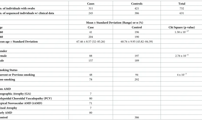
Throat microbiome profiles were successfully obtained (i.e. sufficient 16S PCR product for library building, and>500,000 high quality reads such that EMIRGE reconstruction suc-ceeded) from 245 out of 311 AMD-positive throat swabs, and from 386 out of 421 control throat swabs. Out of the 245 cases, 80 were classified as early-stage AMD and 165 as late-stage AMD (Table 1). Smoking status was available for 126/245 cases and 386/386 controls; only these microbiomes were used for the analysis of the impact of smoking on the pharyngeal microbiome.
Data visualization and statistical analyses
Abundance estimates from EMIRGE were converted to relative abundances at the genus level for each sample. Clinical data, OTU relative abundance tables, Shannon’s and Simpson’sα -diversity indices were processed and calculated using customized R script in R version 3.3.2. OTU relative abundance tables were input for Principal Component Analyses (PCA). The R package “DESeq2” was used to determine differential abundance for case/control conditions (in all samples), and early/late AMD conditions (in case samples). Age and gender were included as covariates. Only taxa found to be significant (P<0.05) were reported.
Generalised Linear Mixed Model (GLMM) was performed to determine genera with signif-icantly different relative abundances between conditions. P-values were corrected for multiple testing using Benjamini-Hochberg correction.
We used “SVA” R package to examine batch effects across samples. Additionally, Principal Coordinate Analyses (PCoA) and Guided Principal Component Analyses (gPCA) [26] were performed to visually inspect for batch effects using “stats” R package.
Quantitative-PCR (qPCR) of total bacteria and selected microbial genera
Absolute gene counts of totalPrevotella,GemellaandStreptococcuswere determined with quantitative PCR (Q-PCR). Twenty samples were randomly selected from among the AMD case population for 16S qPCR gene copy determination; this was matched by an equal numberof randomly selected control samples. To quantify total bacteria, primers were selected that amplify a uniform 16S region in these genera (S2 Table) [27].PrevotellaandStreptococcus
primers were sourced from Matsuki et al. and Picard et al. respectively [28,29], whileGemella
primers were established in-house via multiple sequence alignments ofGemella16S sequences obtained from the NCBInrrepository. Primers were tested to ensure specificity to correspond-ing genera.
Reactions were carried out in 384-well qPCR plates with duplicate 10μL reaction volumes containing the KAPA SYBR FAST qPCR Master Mix (2X) for LightCycler 480 (Sigma-Aldrich, Inc.), the primers (15μM) and 2.0μL of sample DNA. The LightCycler1480 Instrument II (Roche) was used for amplification and detection with the following thermocycling parame-ters: 1 cycle of 95˚C for 20s followed by amplification at 95˚C for 15s, 60˚C for 20s for 40 cycles and 1 cycle of 95˚C for 15s, 60˚C for 15s and 95˚C for 15s with readings collected at final step for melting curve analysis. Ct values were recalculated for log copy number/ul DNA from raw data exported into Excel.
Calibration standards for converting Ct values to bacterial 16S rRNA gene copy numbers were generated as follows. Double-stranded DNA oligomers were synthesised (gBLOCK, Inte-grated DNA Technologies, Inc.) to span respective regions covered by0020corresponding Pre-votella/Gemella/Streptococcusforward and reverse primers. A 10 ng/μL stock solution was prepared from the lyophilised oligomers as per manufacturer’s instruction. Copy number/μL of stock solution were determined from calculations (DNA concentration / (fragment length x weight of base pair)) and serially diluted to 6 standards with final concentrations spanning 1.0 Table 1. Clinical parameters of study subjects.
Cases Controls Total
No. of individuals with swabs 311 421 732
No. of sequenced individuals w/ clinical data 245 386 631
Mean±Standard Deviation (Range) or n (%)
Age Case Control Chi Square (p-value)
<60 41 196 1.58 x 10⁻¹⁷
>60 204 190
Mean age±Standard Deviation 67.46±8.57 (32–85.26) 60.76±9.95 (45.82–84.39)
Gender
Female 88 197 2.76 x 10⁻⁴
Male 157 189
Smoking Status
Current or Previous smoking 48 94 4 x 10⁻³
Non-smoking 78 292
Late AMD
Geographic Atrophy (GA) 7
Polypoidal Choroidal Vasculopathy (PCV) 80
Typical Neovascular AMD (tAMD) 71
Mixed Atrophy 7
Early AMD 80
Control 386
x 103to 1.0 x 10⁸copy numbers. A plot of Ct versus log10(copy number) produced linear cali-bration curves with typical R2values of 0.99.
Copy numbers for each genera were expressed as percentage of total microbial copy num-ber. Pearson’s correlation coefficient between qPCR and 16S sequencing techniques was deter-mined using Microsoft Excel 2013.
Results
Pharyngeal microbiome structure in AMD cases and controls
We successfully obtained bacterial 16S rRNA sequence data from 631 out of 732 individual throat swabs. Of these, 245 were derived from AMD cases, and 386 from controls. Clinical parameters, including age, gender, smoking status, and disease stage are shown inTable 1. With the exception of disease-type variable for AMD cases, pairwise statistical comparisons (PERMANOVA) of microbial community abundance among case and control samples revealed no obvious stratification of pharyngeal microbiome profiles by gender and age vari-ables (S3 Table). No significant surrogate variables were identified from “SVA” analysis (data not shown). Visual inspection for batch effects using Principal Coordinate Analysis (PCoA), similarly indicated an absence of batch effects among samples (S2 Fig). Finally, application of Guided Principal Components Analysis (gPCA) revealed that proportion of variance due to batch effects was not significantly greater than would be expected (p<0.05) (S3 Fig).
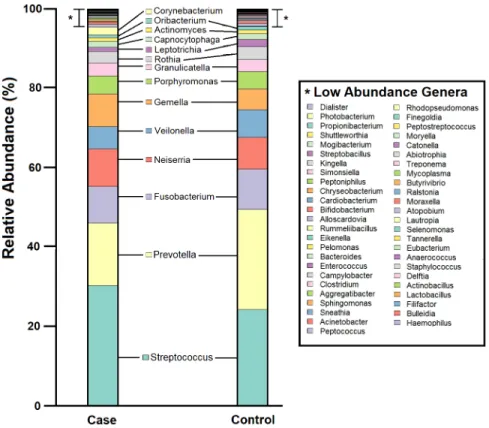
In total, nine phyla and 63 genera were detected across all 631 subjects. The mean number of identified genera were 17 (SD= 6) and 18 (SD= 5) in 245 cases and 386 controls respec-tively. This reflects some degree of similarity in the number of identified genera from samples collected from a specific anatomical niche, albeit with varied disease status. Consistent with previous reports [1], two dominant phyla—Firmicutes and Bacteroidetes—accounted for more than half of all OTUs. Members of the microbial community in case and control samples were similar, however, relative abundances of each member differed between case (n = 245) and control (n = 386) groups (Fig 1). In terms of genera prevalence among study population,
Streptococcus,Prevotella,VeilonellaandGemellawere most prevalent in both case (n = 245) and control (n = 386) samples, being detected in>85% of all subjects (S4 Fig). Shannon and Simpson diversity indices did not differ significantly between cases and controls (p<0.05, Mann-Whitney U test) (S5 Fig).
Principal Components Analysis (PCA) revealed the lack of clustering across conditions, implying that maximum abundance variances are insufficient to distinguish between condi-tions. It also suggests that genera relative abundances between conditions are subtle (S3 Fig).
Association of specific microbial genera with AMD
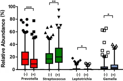
245 case and 386 control samples were used for DESeq2 to evaluate differential abundance of bacterial genera. DESeq2 analyses found the case samples to be enriched forGemella(Adj-p= 3.15 x 10⁻⁵), whilePrevotellahad reduced relative abundance (Adj-p= 1.89 x 10⁻⁵) (Fig 2). While, in the comparisons between early versus late AMD cases, no genera was detected to be significantly different in relative abundance among the groups. Likewise was in the smoking status comparisons (Adj-p<0.05). Using Generalised Linear Mixed Model analysis, we observed the robust associations ofPrevotellaandGemellawith case/control status, as similarly shown in the DESeq2 analysis. In addition, we detectedStreptococcusandLeptotrichiato asso-ciate with case/control status as well (Fig 3andS4 Table).StreptococcusandPrevotella, the two most abundant genera, showed an opposite trend:Streptococcuswas present at a significantly higher abundance in cases than in controls (mean relative abundance of 23.4% vs 18.6%;Adj-p
Adj-p= 6.95 x 10⁻⁵). In addition,Gemellawas significantly more abundant in cases than in controls (6.0% vs 4.0%;Adj-p= 0.007).Leptotrichiawhich were present at low relative abundance (<1%), also differed significantly (Adj-p= 0.007) between cases and controls (Fig 3andS4 Table). Analysing the>60 years subset revealed onlyPrevotella,LeptotrichiaandStreptococcus
to be significantly associated (Streptococcus Adj-p= 1.85 x 10⁻⁵;Leptotrichia Adj-p= 0.005;
Streptococcus Adj-p= 0.035) among cases and controls (S5 Table).
Stratifying the AMD cases by disease into early and late stages,PrevotellaandLeptotrichia
relative abundance was significantly lower in late AMD samples than controls (Fig 4andS6 Table). Conversely, late AMD samples were revealed to have greater relative abundances of
StreptococcusandGemella(Streptococcus Adj-p =1.19 x 10⁻⁴;Gemella Adj-p =0.028) (Fig 4). Comparing early AMD samples to controls did not reveal significant differences in genera rel-ative abundances between both conditions. Further analysis to compare microbiomes between subgroups of late AMD disease (as described inTable 1) was under powered by reduced num-bers of individuals, but remains an area of future interest.
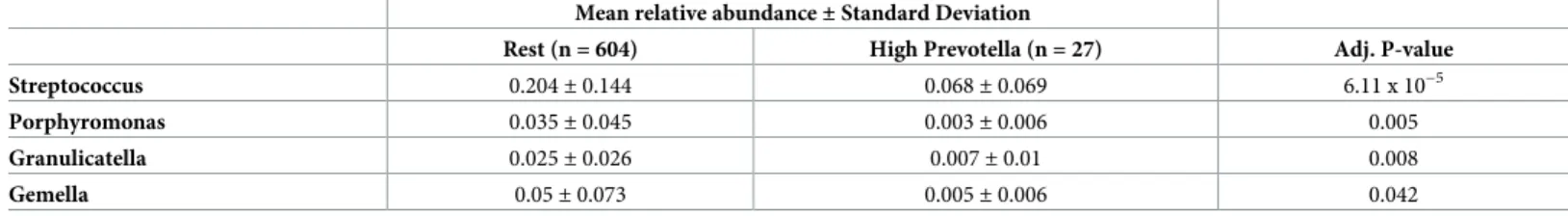
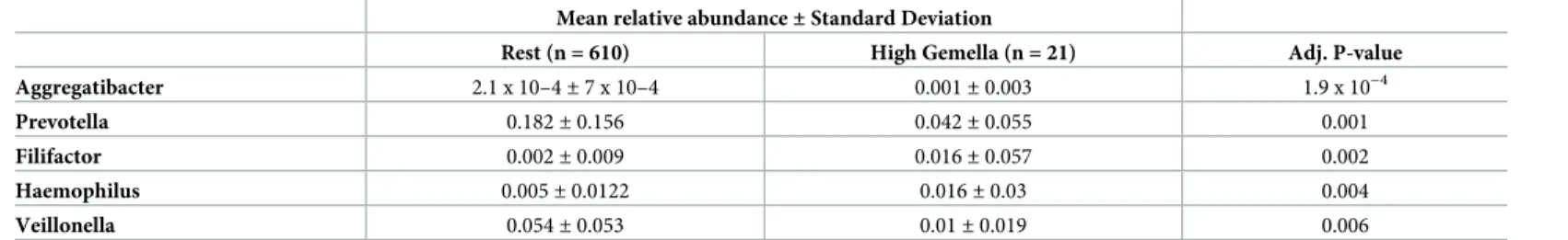
Sampling rarely yields individual microbes in isolation; instead, microbes exist in commu-nities together with other bacterial genera. To understand bacterial commucommu-nities associated withPrevotellaandGemella, we identified subjects with high relative abundance of each genus, defined as relative abundance greater than two standard deviations above the population mean. Comparing the microbiota of these high abundance groups with the rest of the study population revealed distinct subsets of bacterial genera that are associated with each genus (Tables2and3).
Fig 1. Pharyngeal microbiome profile of the study population, at genus level. Cumulative relative abundance of
each of the 63 bacterial genera in case and control samples. Microbial community members with relative abundance <0.1% are listed as Low Abundance Genera (indicated by “”).
HighPrevotellaabundance was uniquely associated with reducedGemella,Granulicatella,
Porphyromonas and Streptococcusabundance, and highGemellaabundance was uniquely asso-ciated with higherFilifactor,Haemophilus and Aggregatibacterabundances. Using PCA to map Fig 2. DESeq2 differential abundance analysis expressed as Log2FC comparison of AMD-positive samples and control samples. Negative fold change scores (log2) indicate genera with decreased abundance in AMD-positive
samples, and positive fold change scores indicate genera with increased abundance in AMD-positive samples. Each point represents a genus. Genera detected to have significant difference in abundance (Adj-p<0.05) are shown.
https://doi.org/10.1371/journal.pone.0201768.g002
Fig 3. Relative abundances of significant genera between 245 case and 386 control samples. AMD-positive and
control samples are denoted by (+) and (-) respectively. Statistical significance is indicated by (Adj-p<0.05), (Adj-p
<0.005) or (Adj-p<0.0005). Mean relative abundances, standard deviations and P-values are presented inS4
Table.
samples harbouring either highPrevotellaor highGemellaabundances, a distinct cluster of 27 samples was observed with highPrevotellaabundance, of which six were case samples (S6 Fig).
Quantification-PCR (qPCR) validation of selected genera
To test the ability of our 16S sequencing to detect differences in abundance, we utilised qPCR assays as a validation platform to quantify microbial loads of three highly prevalent genera exhibiting narrow difference in mean abundance among controls and cases. Pearson’s correla-tion showed both techniques were positively correlated to some extent (R = 0.322). Microbial loads reported by qPCR demonstrated a similar trend as relative abundance by sequencing for case/control status. Using qPCR,Streptococcuswas significantly enriched in case samples and
Prevotellain controls.Gemellawas increased in case samples, however, this difference was not significant by qPCR (Fig 5). While we found that 16S sequencing and qPCR are consistent in capturing overall microbial loads, the choice of different technology will result in different out-puts and data formats. In addition to abundance estimates, 16S sequencing is able to identify members of a microbial community and their respective quantities in relation to other members.
Fig 4. Relative abundances of significant genera between 386 controls and 165 individuals with late AMD. (-) and
L-AMD denote controls and late AMD samples respectively. Statistical significance is indicated by (Adj-p<0.05),
(Adj-p<0.005) or (Adj-p<0.0005). Mean relative abundances, standard deviations and P-values are presented
inS6 Table.
https://doi.org/10.1371/journal.pone.0201768.g004
Table 2. Microbial genera significantly associated with highPrevotellarelative abundance.
Mean relative abundance±Standard Deviation
Rest (n = 604) High Prevotella (n = 27) Adj. P-value
Streptococcus 0.204±0.144 0.068±0.069 6.11 x 10⁻⁵
Porphyromonas 0.035±0.045 0.003±0.006 0.005
Granulicatella 0.025±0.026 0.007±0.01 0.008
Gemella 0.05±0.073 0.005±0.006 0.042
Discussion
We have comprehensively and accurately described the human pharyngeal microbiome, at genus level in this a large case-control study of 631 AMD subjects, by shotgun sequencing the V3-V6 region of the microbial 16S rRNA gene. Consistent with previous studies [1,30–33], pharyngeal microbiomes in our study population were dominated by relatively few bacterial genera, of whichStreptococcusandPrevotellawere most abundant and prevalent. These "core" genera may play roles in maintaining the stability and complexity of individual microbiomes over time, properties thought to be associated with health [1].
Investigating community relative abundances using PCA revealed that the microbial driv-ers in the pharyngeal community are not vastly different between cases and controls. Rather, they suggest that the pharyngeal community is overall stable in terms of community genera members and their respective relative abundances across both disease and health conditions. Overall, it is interesting to note that pharyngeal community relative abundances in early AMD were more similar to healthy controls, while late AMD condition revealed significant dissimi-larity in community relative abundance when compared to controls, which potentially suggests some dysbiosis related to disease progression. AMD is a progressive disease, and this observa-tion suggests that alteraobserva-tions in the pharyngeal microbiota becomes more evident as the dis-ease progresses. Current treatment of “wet” AMD is based on suppression of vascular
endothelial growth factor (VEGF) with intraocular injections of anti-VEGF agents. While this has significantly improved the outcomes in the management of AMD, this treatment does not Table 3. Microbial genera significantly associated with highGemellarelative abundance.
Mean relative abundance±Standard Deviation
Rest (n = 610) High Gemella (n = 21) Adj. P-value Aggregatibacter 2.1 x 10⁻⁴±7 x 10⁻⁴ 0.001±0.003 1.9 x 10⁻⁴ Prevotella 0.182±0.156 0.042±0.055 0.001 Filifactor 0.002±0.009 0.016±0.057 0.002 Haemophilus 0.005±0.0122 0.016±0.03 0.004 Veillonella 0.054±0.053 0.01±0.019 0.006 https://doi.org/10.1371/journal.pone.0201768.t003
Fig 5. Microbial loads from qPCR. qPCR-derived copy numbers of each genera were expressed as a percentage of
total microbial copy number within each sample. Twenty samples were randomly selected for each disease status. Statistical significance is indicated by (P<0.01) or (P<0.001) as determined by Mann-Whitney U test.
reverse damage related to AMD, is invasive, expensive, and incurs significant healthcare resources. Thus, a search for the underlying cause of AMD with strategies to prevent its devel-opment and progression remains a key research priority.
GemellaandPrevotellawere detected to have significant differential abundance between case and control conditions. Difference in mean relative abundance of both genera in disease and health conditions also reached statistical significance. This could either suggest that health/disease conditions could have an impact on community structure, or community abun-dances play a role towards disease propagation. Our findings provide preliminary insight into the composition of pharyngeal microbiota, as well as candidate genera with their abundance alterations in AMD health and disease conditions. Metagenomic sequencing and translational studies can be performed in future to provide more robust evidence to elucidate relevant bio-logical factors leading to this observation.
We were also able to investigate potential roles for pharyngeal microbiome members in the initiation and pathogenesis of AMD. While overall diversity and composition of pharyngeal microbiomes from AMD cases and controls were relatively similar, we observed clear differ-ences in specific bacterial genera between the two groups.GemellaandPrevotella(also the two most abundant genera), showed opposite trends, withGemellabeing more abundant in cases andPrevotellamore so in controls.Gemellais a dominant member of the laryngeal mucosal community and has been identified to be present at higher abundances in patients with laryn-geal squamous cell carcinoma than healthy subjects [34,35].Prevotellaspecies are not known to be protective; some such asPrevotella intermediaare known to cause periodontal disease [36], andPrevotella copriin the gut has also been associated with rheumatoid arthritis [37]. While there is a possibility that the protective effect we observed here for AMD is due to corre-sponding lower levels ofGemellain individuals with highPrevotellaloads, nevertheless it begets the question of why some community members show differential abundance in health or disease conditions.
To better understand the composition of communities harbouring highPrevotellaand
Gemellaloads, we looked for community members associated with highPrevotellaand Gemella. Most associations were negative, i.e. high driver abundance correlated with reduced abundance of the genus in question. This could reflect the fact that the drivers were the most abundant genera (such that other genera are correspondingly low), or alternatively could indi-cate the existence of competitive mechanisms. Conversely, positive associations (for example, highGemellawas associated with highHaemophilus,FilifactorandAggregatibacter) could indi-cate cooperative or symbiotic interactions. These patterns suggest that intra-community inter-actions are likely to affect the overall contribution of the individual microbiota to AMD pathogenesis. PCA clustering revealed highPrevotellasamples (n = 27) to form an exclusive cluster, suggesting the highPrevotellasubset to possess unique microbial profile against the rest of the samples. Among this subset were six case samples, indicating that while increased
Prevotellaabundance is associated with control status, it is not the sole scenario. It suggests greater complexity between association of the pharyngeal microbiota and AMD to be beyond the sheer abundance of a single genus.
Host genetic differences between cases and controls may also influence pharyngeal micro-biome composition and colonization efficiency. A recent study revealed the rs3006458-T allele (associated with lower abundance ofAerococcusandMicrococcaceain the nasopharynx) [38] was associated with increased PGLYRP4 expression in mucosal tissues supporting antimicro-bial innate immunity [39]. These data suggest that host genotype regulates PGLYRP4 expres-sion on the respiratory mucosa that leads to modulatory effect on bacterial abundance [38]. A number of pathogenic bacteria, includingNeisseria meningitidis,Streptococcus pneumoniae,
CFH, thus mediating evasion of complement-mediated killing [40,41]. Genetic variants in CFH associated with increased AMD risk have been shown to alter binding toS.pyogenes sur-face proteins, and consequently impact complement activation, opsonization, and phagocyto-sis [42,43]. In addition, individuals carrying the Tyr402His CFH risk variant who also harbored high levels of antibodies againstChlamydia pneumoniaeshowed an increased risk of AMD progression [6]. Our work provides further evidence that these factors play a role in AMD disease and progression.
Conclusions
Through 16S sequencing of an extensive population cohort, this study has provided accurate representation of the population as well as considerable confidence in a role for the pharyngeal microbiota profile in AMD health and disease conditions. Pharyngeal microbiota members and their relative abundances are highly stable among populations. Subtle differences were identified in relative abundances ofPrevotella,Gemella,StreptococcusandLeptotrichiain the pharyngeal composition of AMD cases when compared to controls, a phenomenon similarly observed in comparisons between controls and patients with late-stage AMD. Prospective studies investigating pharyngeal microbiomes prior to disease, longitudinal studies tracking microbiomes over the course of disease progression, and analysis of genetic factors influencing microbiome composition will be necessary to fully characterize microbial triggers of AMD.
Supporting information
S1 Fig. Rarefaction curves indicating the number of genera detected with less than
<500,000 reconstructed reads in 30 randomly picked samples. Samples are labelled with “PHT” prefix and corresponding sample number.
(DOCX)
S2 Fig. Visual inspection for batch effects using Principal Coordinate Analysis (PCoA) based on relative abundance at genus level. Each point represents an individual.
(DOCX)
S3 Fig. Guided Principal Component Analysis (gPCA) based on relative abundance on (A) Gender, (B) Disease status (C) Age and (D) Disease progression factors. Each point rep-resents an individual. Case samples has a similar community composition similar to that of control samples. Additionally, microbial community composition is highly similar among early/late AMD status.
(DOCX)
S4 Fig. Prevalence (percentage of individuals in which the genus was detected) of each microbial genus in case and control samples.
(DOCX)
S5 Fig. Measures of pharyngeal microbiome community diversity. Shannon and Simpson diversity indices for pharyngeal microbiomes in AMD cases and controls. Each data point rep-resents an individual pharyngeal microbiome; boxes indicate the mean and 25th and 75th per-centiles; whiskers indicate interquartile ranges. Shannon and Simpson diversity indices did not differ significantly between cases and controls (p<0.05, Mann-Whitney U test). (DOCX)
S6 Fig. PCA plots colored by samples harbouring (A) highPrevotella(n = 27) and (B) high
(A). (DOCX)
S1 Table. Comparison of identified genera in 30 samples using 500,000 and<500,000 reconstructed reads.
(DOCX)
S2 Table. Quantitative-PCR (qPCR) primers and their corresponding amplicon sizes. (DOCX)
S3 Table. Gender, age and disease-type Pairwise Statistical Comparisons (PERMANOVA) of microbial community abundance among case and control samples.
(DOCX)
S4 Table. Association between microbial genera and AMD status. Genera with significantly different relative abundances in case/control conditions are shown.
(DOCX)
S5 Table. Association between microbial genera and AMD status in individuals>60 years. Genera with significantly different relative abundances in case/control conditions are shown. (DOCX)
S6 Table. Association between microbial genera with control and Late AMD status. Genera with significantly different relative abundances in control and Late AMD conditions are shown.
(DOCX)
Acknowledgments
We thank all patients and their families for their participation in this study, as well as hospital staff who assisted in patient enrolment and sample collection. We also thank the GERMS Microbial Genomics platform at the Genome Institute of Singapore for sequencing and bioin-formatics expertise.
Author Contributions
Conceptualization: Tien Yin Wong, Ching Yu Cheng, Sonia Davila, Martin Hibberd. Data curation: Eliza Xin Pei Ho, Ching Yu Cheng.
Formal analysis: Eliza Xin Pei Ho, Collins Wenhan Chu. Funding acquisition: Tien Yin Wong, Martin Hibberd.
Investigation: Eliza Xin Pei Ho, Tien Yin Wong, Ching Yu Cheng, Martin Hibberd. Methodology: Eliza Xin Pei Ho, Martin Hibberd.
Project administration: Eliza Xin Pei Ho, Chui Ming Gemmy Cheung, Ranjana Mathur, Doric Wong, Choi Mun Chan, Mayuri Bhagarva, Augustinus Laude, Tock Han Lim, Sonia Davila.
Resources: Chui Ming Gemmy Cheung, Andreas Wilm, Clarabelle Bitong Lin, Ranjana Mathur, Doric Wong, Choi Mun Chan, Mayuri Bhagarva, Augustinus Laude, Tock Han Lim, Tien Yin Wong, Sonia Davila.
Supervision: Chui Ming Gemmy Cheung, Andreas Wilm, Sonia Davila. Validation: Eliza Xin Pei Ho, Clarabelle Bitong Lin.
Writing – original draft: Eliza Xin Pei Ho, Shuzhen Sim.
Writing – review & editing: Shuzhen Sim, Collins Wenhan Chu, Andreas Wilm, Sonia Davila, Martin Hibberd.
References
1. Consortium THMP. Structure, function and diversity of the healthy human microbiome. Nature. 2012 Jun 14; 486(7402):207–14.https://doi.org/10.1038/nature11234PMID:22699609
2. Gao Z, Kang Y, Yu J, Ren L. Human Pharyngeal Microbiome May Play A Protective Role in Respiratory Tract Infections. Genomics Proteomics Bioinformatics. 2014 Jun; 12(3):144–50.https://doi.org/10. 1016/j.gpb.2014.06.001PMID:24953866
3. Freeley S, Kemper C, Le Friec G. The “ins and outs” of complement-driven immune responses. Immu-nol Rev. 2016 Nov 1; 274(1):16–32.https://doi.org/10.1111/imr.12472PMID:27782335
4. Yazdankhah SP and Caugant DA. Neisseria meningitidis: an overview of the carriage state. J Med Microbiol. 2004 Sep; 53(Pt 9):821–32.https://doi.org/10.1099/jmm.0.45529-0PMID:15314188
5. Meri T, Amdahl H, Lehtinen MJ, Hyva¨rinen S, McDowell JV, Bhattacharjee A, et al. Microbes Bind Com-plement Inhibitor Factor H via a Common Site. PLOS Pathog. 2013 Apr 18; 9(4):e1003308.https://doi. org/10.1371/journal.ppat.1003308PMID:23637600
6. Baird PN, Robman LD, Richardson AJ, Dimitrov PN, Tikellis G, McCarty CA, et al. Gene-environment interaction in progression of AMD: the CFH gene, smoking and exposure to chronic infection. Hum Mol Genet. 2008 May 1; 17(9):1299–305.https://doi.org/10.1093/hmg/ddn018PMID:18203751
7. Davila S, Wright VJ, Khor CC, Sim KS, Binder A, Breunis WB, et al. Genome-wide association study identifies variants in the CFH region associated with host susceptibility to meningococcal disease. Nat Genet. 2010 Sep; 42(9):772–6.https://doi.org/10.1038/ng.640PMID:20694013
8. Klein RJ, Zeiss C, Chew EY, Tsai J-Y, Sackler RS, Haynes C, et al. Complement Factor H Polymor-phism in Age-Related Macular Degeneration. Science. 2005 Apr 15; 308(5720):385–9.https://doi.org/ 10.1126/science.1109557PMID:15761122
9. Edwards AO, Ritter R, Abel KJ, Manning A, Panhuysen C, Farrer LA. Complement factor H polymor-phism and age-related macular degeneration. Science. 2005 Apr 15; 308(5720):421–4.https://doi.org/ 10.1126/science.1110189PMID:15761121
10. Wong WL, Su X, Li X, Cheung CMG, Klein R, Cheng C-Y, et al. Global prevalence of age-related macu-lar degeneration and disease burden projection for 2020 and 2040: a systematic review and meta analy-sis. Lancet Glob Health. 2014 Feb; 2(2):e106–116.https://doi.org/10.1016/S2214-109X(13)70145-1
PMID:25104651
11. Ambati J, Atkinson JP, Gelfand BD. Immunology of age-related macular degeneration. Nat Rev Immu-nol. 2013 Jun; 13(6):438–51.https://doi.org/10.1038/nri3459PMID:23702979
12. Laine M, Jarva H, Seitsonen S, Haapasalo K, Lehtinen MJ, Lindeman N, et al. Y402H Polymorphism of Complement Factor H Affects Binding Affinity to C-Reactive Protein. J Immunol. 2007 Mar 15; 178 (6):3831–6. PMID:17339482
13. Clark SJ, Perveen R, Hakobyan S, Morgan BP, Sim RB, Bishop PN, et al. Impaired Binding of the Age-related Macular Degeneration-associated Complement Factor H 402H Allotype to Bruch’s Membrane in Human Retina. J Biol Chem. 2010 Sep 24; 285(39):30192–202.https://doi.org/10.1074/jbc.M110. 103986PMID:20660596
14. Sjo¨berg AP, Trouw LA, Clark SJ, Sjo¨lander J, Heinegård D, Sim RB, et al. The Factor H Variant Associ-ated with Age-relAssoci-ated Macular Degeneration (His-384) and the Non-disease-associAssoci-ated Form Bind Dif-ferentially to C-reactive Protein, Fibromodulin, DNA, and Necrotic Cells. J Biol Chem. 2007 Apr 13; 282 (15):10894–900.https://doi.org/10.1074/jbc.M610256200PMID:17293598
15. Tortajada A, Montes T, Martinez-Barricarte R, Morgan BP, Harris CL, de Co´rdoba SR. The disease pro-tective complement factor H allotypic variant Ile62 shows increased binding affinity for C3b and enhanced cofactor activity. Hum Mol Genet. 2009 Sep 15; 18(18):3452–61.https://doi.org/10.1093/ hmg/ddp289PMID:19549636
16. Khor CC, Hibberd ML. Host–pathogen interactions revealed by human genome-wide surveys. Trends Genet. 2012 May; 28(5):233–43.https://doi.org/10.1016/j.tig.2012.02.001PMID:22445588
17. Cheung CMG, Li X, Cheng C-Y, Zheng Y, Mitchell P, Wang JJ, et al. Prevalence, Racial Variations, and Risk Factors of Age-Related Macular Degeneration in Singaporean Chinese, Indians, and Malays.
Ophthalmology. 2014 Aug; 121(8):1598–603.https://doi.org/10.1016/j.ophtha.2014.02.004PMID:
24661862
18. Laser photocoagulation of subfoveal recurrent neovascular lesions in age-related macular degenera-tion. Results of a randomized clinical trial. Macular Photocoagulation Study Group. Arch Ophthalmol Chic Ill 1960. 1991 Sep; 109(9):1232–41.
19. Japanese Study Group of Polypoidal Choroidal Vasculopathy. [Criteria for diagnosis of polypoidal cho-roidal vasculopathy]. Nippon Ganka Gakkai Zasshi. 2005 Jul; 109(7):417–27. PMID:16050460
20. Lavanya R, Jeganathan VSE, Zheng Y, Raju P, Cheung N, Tai ES, et al. Methodology of the Singapore Indian Chinese Cohort (SICC) eye study: quantifying ethnic variations in the epidemiology of eye dis-eases in Asians. Ophthalmic Epidemiol. 2009 Dec; 16(6):325–36.https://doi.org/10.3109/
09286580903144738PMID:19995197
21. Hang J, Desai V, Zavaljevski N, Yang Y, Lin X, Satya RV, et al. 16S rRNA gene pyrosequencing of ref-erence and clinical samples and investigation of the temperature stability of microbiome profiles. Micro-biome. 2014 Sep 16; 2:31.https://doi.org/10.1186/2049-2618-2-31PMID:25228989
22. Ong SH, Kukkillaya VU, Wilm A, Lay C, Ho EXP, Low L, et al. Species Identification and Profiling of Complex Microbial Communities Using Shotgun Illumina Sequencing of 16S rRNA Amplicon
Sequences. PLoS ONE. 2013 Apr 8; 8(4):e60811.https://doi.org/10.1371/journal.pone.0060811PMID:
23579286
23. The HC, Florez de Sessions P, Jie S, Pham Thanh D, Thompson CN, Nguyen Ngoc Minh C, et al. Assessing gut microbiota perturbations during the early phase of infectious diarrhea in Vietnamese chil-dren. Gut Microbes. 2017 Aug 2;0.
24. Miller CS, Baker BJ, Thomas BC, Singer SW, Banfield JF. EMIRGE: reconstruction of full-length ribo-somal genes from microbial community short read sequencing data. Genome Biol. 2011; 12(5):R44.
https://doi.org/10.1186/gb-2011-12-5-r44PMID:21595876
25. DeSantis TZ, Hugenholtz P, Larsen N, Rojas M, Brodie EL, Keller K, et al. Greengenes, a Chimera Checked 16S rRNA Gene Database and Workbench Compatible with ARB. Appl Environ Microbiol. 2006 Jul 1; 72(7):5069–72.https://doi.org/10.1128/AEM.03006-05PMID:16820507
26. Reese SE, Archer KJ, Therneau TM, Atkinson EJ, Vachon CM, de Andrade M, et al. A new statistic for identifying batch effects in high-throughput genomic data that uses guided principal component analy-sis. Bioinforma Oxf Engl. 2013 Nov 15; 29(22):2877–83.
27. Chua MC, Ben-Amor K, Lay C, Neo AGE, Chiang WC, Rao R, et al. Effect of Synbiotic on the Gut Micro-biota of Caesarean Delivered Infants: A Randomized, Double-Blind, Multicenter Study. J Pediatr Gas-troenterol Nutr. 2017 May 3;
28. Picard FJ, Ke D, Boudreau DK, Boissinot M, Huletsky A, Richard D, et al. Use of tuf sequences for genus-specific PCR detection and phylogenetic analysis of 28 streptococcal species. J Clin Microbiol. 2004 Aug; 42(8):3686–95.https://doi.org/10.1128/JCM.42.8.3686-3695.2004PMID:15297518
29. Matsuki T, Watanabe K, Fujimoto J, Takada T, Tanaka R. Use of 16S rRNA gene-targeted group-spe-cific primers for real-time PCR analysis of predominant bacteria in human feces. Appl Environ Microbiol. 2004 Dec; 70(12):7220–8.https://doi.org/10.1128/AEM.70.12.7220-7228.2004PMID:15574920
30. Boutin S, Graeber SY, Weitnauer M, Panitz J, Stahl M, Clausznitzer D, et al. Comparison of Micro-biomes from Different Niches of Upper and Lower Airways in Children and Adolescents with Cystic Fibrosis. PLoS ONE. 2015 Jan 28; 10(1):e0116029.https://doi.org/10.1371/journal.pone.0116029
PMID:25629612
31. Bogaert D, Keijser B, Huse S, Rossen J, Veenhoven R, van Gils E, et al. Variability and Diversity of Nasopharyngeal Microbiota in Children: A Metagenomic Analysis. PLoS ONE. 2011 Feb 28; 6(2): e17035.https://doi.org/10.1371/journal.pone.0017035PMID:21386965
32. Charlson ES, Chen J, Custers-Allen R, Bittinger K, Li H, Sinha R, et al. Disordered Microbial Communi-ties in the Upper Respiratory Tract of Cigarette Smokers. PLoS ONE. 2010 Dec 20; 5(12):e15216.
https://doi.org/10.1371/journal.pone.0015216PMID:21188149
33. Morris A, Beck JM, Schloss PD, Campbell TB, Crothers K, Curtis JL, et al. Comparison of the Respira-tory Microbiome in Healthy Nonsmokers and Smokers. Am J Respir Crit Care Med. 2013 May15; 187 (10):1067–75.https://doi.org/10.1164/rccm.201210-1913OCPMID:23491408
34. Gong H, Shi Y, Zhou X, Wu C, Cao P, Xu C, et al. Microbiota in the Throat and Risk Factors for Laryn-geal Carcinoma. Appl Environ Microbiol. 2014 Dec; 80(23):7356–63.https://doi.org/10.1128/AEM. 02329-14PMID:25239901
35. Gong H-L, Shi Y, Zhou L, Wu C-P, Cao P-Y, Tao L, et al. The Composition of Microbiome in Larynx and the Throat Biodiversity between Laryngeal Squamous Cell Carcinoma Patients and Control Population. PLoS ONE [Internet]. 2013 Jun 18 [cited 2015 Jun 15]; 8(6).
36. Potempa M, Potempa J, Kantyka T, Nguyen K-A, Wawrzonek K, Manandhar SP, et al. Interpain A, a Cysteine Proteinase from Prevotella intermedia, Inhibits Complement by Degrading Complement Fac-tor C3. PLoS Pathog. 2009 Feb 27; 5(2):e1000316.https://doi.org/10.1371/journal.ppat.1000316
PMID:19247445
37. Scher JU, Sczesnak A, Longman RS, Segata N, Ubeda C, Bielski C, et al. Expansion of intestinal Pre-votella copri correlates with enhanced susceptibility to arthritis. eLife. 2013 Nov 5; 2:e01202.https://doi. org/10.7554/eLife.01202PMID:24192039
38. Igartua C, Davenport ER, Gilad Y, Nicolae DL, Pinto J, Ober C. Host genetic variation in mucosal immu-nity pathways influences the upper airway microbiome. Microbiome. 2017 Feb 1; 5:16.https://doi.org/ 10.1186/s40168-016-0227-5PMID:28143570
39. Consortium TGte. The Genotype-Tissue Expression (GTEx) pilot analysis: Multitissue gene regulation in humans. Science. 2015 May 8; 348(6235):648–60.https://doi.org/10.1126/science.1262110PMID:
25954001
40. Schneider MC, Prosser BE, Caesar JJE, Kugelberg E, Li S, Zhang Q, et al. Neisseria meningitidis recruits factor H using protein mimicry of host carbohydrates. Nature. 2009 Apr 16; 458(7240):890–3.
https://doi.org/10.1038/nature07769PMID:19225461
41. Jo´zsi M, Zipfel PF. Factor H family proteins and human diseases. Trends Immunol. 2008 Aug; 29 (8):380–7.https://doi.org/10.1016/j.it.2008.04.008PMID:18602340
42. Haapasalo K, Vuopio J, Syrja¨nen J, Suvilehto J, Massinen S, Karppelin M, et al. Acquisition of Comple-ment Factor H Is Important for Pathogenesis of Streptococcus pyogenes Infections: Evidence from Bac-terial In Vitro Survival and Human Genetic Association. J Immunol. 2012 Jan 1; 188(1):426–35.https:// doi.org/10.4049/jimmunol.1102545PMID:22140259
43. Haapasalo K, Jarva H, Siljander T, Tewodros W, Vuopio-Varkila J, Jokiranta TS. Complement factor H allotype 402H is associated with increased C3b opsonization and phagocytosis of Streptococcus pyo-genes. Mol Microbiol. 2008 Nov; 70(3):583–94.https://doi.org/10.1111/j.1365-2958.2008.06347.x