IMMUNITY IN HODGKIN'S DISEASE: STATUS AFTER 5 YEARS'
REMISSION
B. W. HANCOCK*, L. BRUCE*, M. D. WHITHAM*, I. R.
DUNSMORET
A. MILFORD
WARD$
AND J. RICHMOND*From the *Departments of Medicine, tProbability and Statistics and
tImmunology,
Royal HallamshireHospital and University ofSheffield, Sheffield
Received13March 1982 Accepted24May 1982
Summary.-Immunological indices have been reassessedin27 patientsinremission fromHodgkin's disease for 5 years after treatment and the findings correlatedwith initial treatment and splenectomy status. Neutrophil counts were lower and lymphocyte and monocyte counts higher at 5 years' remission than at presentation; the increases in lymphocyte count were mainly a feature of the splenectomized patients. Neutrophil function (nitro-blue tetrazolium) was unchanged in remission but cellular immunity (leucocyte migration inhibition and lymphocyte transforma-tion) was depressed at 5 years and progressive falls in serumimmunoglobulinswere noted. Low values of IgG and
IgM
were particularly found in patients who had splenectomy and chemotherapy; there was however no excess of infections in this smallgroup.THE LONG-TERM EFFECTS of radio-therapy, chemotherapy and splenectomy in Hodgkin's disease are not yet fully established. Cellular immunity may remain depressed for years after
chemo-therapy (Fisher et al., 1980) and after radiotherapy long-lived defects in cellular immunity may also be found (Fuks et al., 1976; Bjorkholm et al., 1977b), though Kun & Johnson (1975) found normal
delayed hypersensitivity skin-test responses 5 years after radical
radio-therapy. Impaired humoral immunity has been observed in treated Hodgkin's dis-ease (Weitzman et al., 1977); however, other reports (Fisher et al., 1980; Kun &
Johnson 1975) suggest that in the long-term humoral immunity is not depressed. In 1977 we reported our early follow-up studies on the immune status of patients with Hodgkin's disease after splenectomy and treatment (Hancock et al., 1977). Cellular immunityas judged byskin tests andleucocyte migration inhibition showed little evidence of disturbance after one
year but serum IgG and IgM levels fell with intensive chemotherapy in splenec-tomized patients; and IgA and IgMlevels were lower, irrespective of splenectomyor therapy status, after 1 year's remission than either at presentation or after treatment. Twenty-seven of the patients assessed in that study have been reasses-sed 5 years after completion of therapy. All patients entered complete remission withtreatment and remained disease-free at reassessment. The findings have been correlated with initial treatment and splenectomystatus.
METHODS
Peripheral blood counts
Total lymphocyte, neutrophil and
mono-cyte counts were performedat each stage of
theassessment. Neutrophilfunction
Thenitro-blue tetrazolium (NBT)testused
wasthe unstimulatedsemi-quantitative
histo-chemical technique of Park et al. (1968). Correspondence to: Dr B. W. Hancock, Dept ofMedicine, RoyalHallamshire Hospital, GlossopRoad,
B. W.HANCOCKETAL. Heparinized blood was incubated with
buf-fered0.1% NBTsolution. Smearsweremade
onglass slides and stained. The percentage of neutrophils containing formazan deposits were counted. Normally less than 10% of neutrophils showareduction.
Cellular immunity
Leucocyte
migration
inhibition test. he leucocyte migration inhibitiontestwas modi-fied from the method ofS0berg & Bendixen (1976). Separated peripheral leucocytes were allowedtomigrate from microcapillarytubes in wells containing (a) purified protein derivative, (b) Candida albicans extract and(c) control medium. Migration areas with antigensineachexperiment werestatistically compared with control migration areas (Student's ttest) andsignificantinhibition of migration was said to have occurred if P<0-05. If significant migration inhibition occurred with one or both antigens the patient was considered immunocompetent in
thistest. Thistest wasrepeatedateachstage
ofassessment.
Lymphocytetransformation
The lymphocyte transformation was modi-fied from that of Schellekens & Eijsvoogel
(1968). Lymphocytes were separated from whole blood by a Ficoll-based centrifugation method and distributed into triplicate cul-tures containing culture medium, autologous plasma and phytohaemaglutinin. Control healthy lymphocyte cultures werealso set up
and all cultures were incubated at 37°C for 72h. Tomeasurelymphocytetransformation byestimation of DNA synthesis the
lympho-cytes were labelled with tritiated thymidine for the last hour of incubation. Synthesis was
then arrested by cooling to 40Cand the cells wereharvested. Radioactivitywascounted to
give a final result in disintegrations per minute (d.p.m.). Each patient was assessed
against age and sex-matched control ranges.
Values below these ranges were interpreted as subnormal. Lymphocyte transformation
assessments were made before treatment and 5yearsaftertreatment.
T-cellcounts
A spontaneous E-rosette-formation tech-nique basedon that ofSteel et al. (1974) was
used. Washed, fresh, defibrinated, preserva-tive-free sheep red cells (Gibco Bio-Cult Ltd)
were added to washed, separated
lympho-cytesin culturemedium, centrifuged at 200 g
for 5minandincubated at 4°C for 1 h. After
re-suspension the number and proportion of
rosette-forminglymphocyteswascounted in a
haemocytometer. The normal range in our
laboratory for this technique is 40-80%. T-cell studies were performed at presentation
and at 5years. Humoralimmunity
Immunoglobulins.
-Serum immunoglobu-lins were determined by automatedimmuno-precipitation (the local standard preparation being calibrated in relationship to the mass equivalentof theIFCCpreparation74/1). Patients
Twenty-seven patients with Hodgkin's disease attaining complete remission were
assessed at presentation, immediately after radiotherapy or before the third or fourth
courseof intensivechemotherapy and after 1
and5years' remissiotn.
During the period of study itwasthe policy of theLymphoma Group in Sheffield, U.K.,to
select patients for laparotomy and
splenect-omy on the basis of clinical staging and histological type. Those patients with Stage
IAand IIA disease of lymphocyte
predomin-antand nodularsclerosinghistology and with Stage IIIB, IVA, IVB (all histologicaltypes) did not have laparotomy; all other patients did. Patients with Stage I-IIIA disease had radical radiotherapy (mantle, inverted Y or
total nodal irradiation); patients with stage
IIIB-IV disease had intensive chemotherapy (modified MOPP, seeappendix).
Of the 27 patients, 15 had undergone splenectomy and of these7 weresubsequently treated with chemotherapy and 8 with
radiotherapy. Of the 12 patients not under-goingsplenectomy, 7hadradiotherapy and5
hadchemotherapy. The clinical detailsof the
patientsaccordingtotheir
treatment/splenec-tomy status areshowninTableI. Stati.stical analysis
To reduce the effects of variations in the
basic levels of each quantitative variable (neutrophils, lympocytes, immunoglobulins)
between the patients the changes at each stage ofassessment foreach ofthe variables wereconsidered.As asimple first step changes within the group as a whole were analysed
HODGKIN'S DISEASE: IMMUNITY AT5YEARS 01m c> p: m t Co ; c'~) Is o pq~~~~~~C oCO¢ C .= ~ .0
~~~~~~~~
0~~~~~~~ bCD 4 ~~ ~ ~ ~ ~ zm* ¢_ GQ 595B.W.HANCOCKET AL.
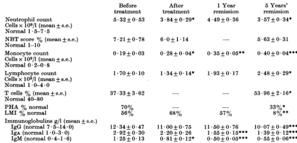
TABLE II.-Follow-up assessments ofimmunestatus inpatients with
Hodgkin's
disease in remissionNeutrophil count Cells x109/1 (mean+s.e.)
Normal 1-5-7-5
NBTscore % (mean+ s.e.) Normal 1-10
Monocyte count Cellsx109/1 (mean+s.e.) Normal0 2-0 8 Lymphocyte count Cellsx109/1 (mean+s.e.) Normal 10* 4-0 Tcells % (mean+s.e.)
Normal 40-80 PHA % normal LMI % normal
Immunoglobulins g/l (mean+s.e.)
IgG (normal 7-5-14-0) IgA(normal 10-3 0) IgM(normal 0.4-1-6) Before treatment 5-32+ 0-53 After treatment 3-84+0-29* 7-21+0-78 6-0+1-14 1 Year remission 4-49+0-36 5Years' remission 3-57+0-34* 5-63+0-31 0-19+0-03 0-28+0-04* 0.35+0.05** 0.40+0.04*** 1-70+0-10 1-34+0-14* 1-93+0-17 2-48+0-29* 37-33+3-62 70% 56% 12-34+0-47 2-92+0-30 1-25 + 0-13 - 53-96+2-16* 68% 11-00+0-75 2-20+0-26 0-81+0 12* 57% 11-50+0 76 1.55+0.15*** 0.50+0.05*** 33%* 8%** 10-07+0-49*** 1-39+0.12*** 0-55+0-06***
***P <0001; **P> 0 * 01;*P < 0 * 05(compared with pretreatment values). separately using paired Student's t and x2
tests (Table II). The patients were then assessed to relate the relativechangesinlevels of eachofthevariablesto the 2 mainfactors, i.e. radiotherapy/chemotherapy and splenec-tomy/no splenectomy. A 2-way analysis of variance with interaction withunequal num-bersofobservations in the cells(Scheff6, 1960) wasused.Inspectionof the datarevealedthat the underlying assumption of normality seemedreasonable.
RESULTS Leucocyte counts
Neutrophil
counts (Fig. 1) in the group asawhole fell significantly after treatment(P<
0.05),
increased by 1 year (P<0-05) butwerestillsignificantlylowerat 5years' remission than at presentation (P < 0-05). This trend was not evident for thosepatients who had splenectomy and
radio-therapy (P<
0.01).
Lymphocyte counts (Fig. 1) overall fell after treatment(P<
0.05)
and increased 1 year and at 5 years after treatment (P< 0-01 and 0 001 respectively). This increase was accounted for by counts in those patients who underwent splenectomy (P <001). Mono-cyte counts (Table II) increased aftertreatment (P<0.05) at 1 year (P<0.01) and at 5 years (P < 0-001) compared with pretreatment values. Individual groups didnotshow significantlydifferent values atany stage.
Neutrophilfunction
There was no significant change in neutrophil function as assessed by NBT scores (Table II) aftertreatmentorafter5 years' remission.
Cellularimmunity
Leucocyte migration inhibition studies showed no significant changes after treat-mentand after 1year'sremission butwere
significantly lower (P<0-01) at 5 years (Table II). All groups of patients showed this depression of reactivity. PHA lym-phocytetransformation wasassessed only
at presentation and in remission. The numberofpatients with normal responses washowever significantlylower (P<0.05)
at 5 years than at presentation. T-cell
population studies were also assessed at presentation and after 5years. Therewas an overall increase in both the percentage and the number ofT cells (P< 0 01). This 596
Neutrophil counts x109/I -2.0 --1.0 - 0-1.0
Post lyr 5yr
Lymphocyte counts
RT/S
All
.A
---CT/NS
Post l-yr
5rT/NS
Post
lyr
5yr
FIG. 1.-Alean clhanges in leucocyte;counts from before treatment to following treatment (post) and to 1,year and 5 year assessments. Key: RT, radiotherapy; S, splenectomy; CT,
chemo-thlerapy;NS,nosplenectomy. IgG 1gA g/l -1.0, .40, ~~CTAIS All RT/S 0-1.0o 2.0
-Post lyr 5yr
Ak
\T/NRT/SCT/NS
Post lyr 5yr
1gM g/l -0.4 -0.2 - 0-0.2 -0.4 -0.6 -0.8 - 1.0- 1.2-RT/NS CT/NS All RT/S CT/S
Post lyr 5yr
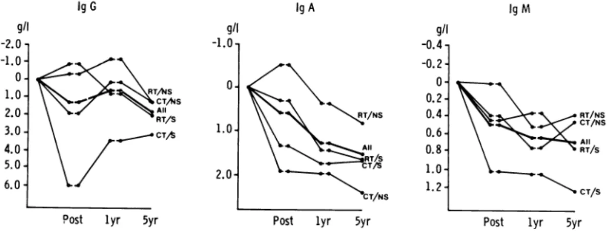
FIG. 2. MAeanchlangesin serumimmunoglobulinlevels frombeforetreatment tofollowingtreatment
(post)and to 1 year and5 yearassessments. Key: RT, radiotherapy;S, splenectomy; CT,
chemo-therapy; NS,nosplenectomy.
was a particular feature of the
splenec-tomized patients though there were no
significant differences between individual
groups.
Humioral irnintuity
Allimmunoglobulin classes showed falls during the period of study (Fig. 2). At presentation, values (particularly IgA) for the group as a whole were not
unexpec-tedly near the top of the normal ranges.
Individual patients had values above the normal range (3 IgG, 9IgA and 5 IgM). All fell to normal with remission though 2/5 patients with initially raised IgM had subnormalvalues at 5years. Inall, atthis stage there were 3, 5 and 7 patients with
subnormal IgG, IgA and 1gM values
respectively. With IgG the 5-year
assess-ment was significantly lower than
pre-treatment and 1-yearlevels(P<0 001 and
005 respectively). Lowvaluesthroughout
x109/1
-2.0 --1.0 -1.0 2.0 -3.0 -4.0 -g/l -2.0 -1.0* 0-1.0. 2.0. 3.0. 4.0-5.0.6.0-B.W. HANCOCK ET AL.
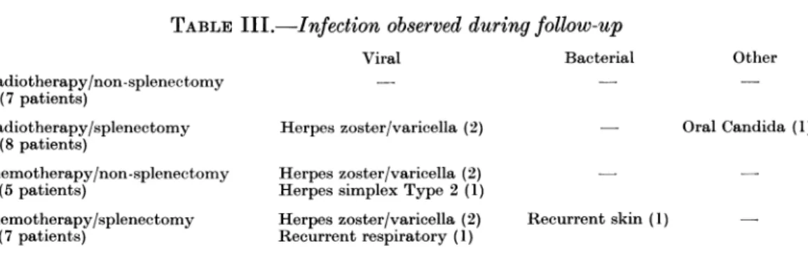
TABLE III.-Infection observed during follow-up
Viral Bacterial Radiotherapy/non-splenectomy (7 patients) Radiotherapy/splenectomy (8 patients) Chemotherapy/non-splenectomy (5 patients) Chemotherapy/splenectomy (7 patients) Herpes zoster/varicella (2) Herpeszoster/varicella (2) Herpessimplex Type 2 (1) Herpes zoster/varicella (2) Recurrentrespiratory (1)
Oral Candida(1)
Recurrent skin(1)
follow-up were seen more frequently in
those patientswhohadchemotherapyand splenectomy. IgA levels fell significantly compared with pretreatment values by 1 year(P<001) andthis fall wassustained
at 5 years (P<0-001). IgM levels fell
significantly withtreatment (P<0*05)and
valuesat1 and5years'remissionwerealso
significantly lower than at presentation (P<0-001); low values throughout follow-up were a particular feature of the
chemotherapy/splenectomygroup.
Correlation withinfection (Table III) The list of infections is not exhaustive,
as trivial nonspecific respiratory and
mucosal infections were not documented.
The only group which stands out is the
radiotherapy/non-splenectomy group, in
which there were no major infections.
Viralinfectionswerenotuncommoninthe
other groups (particularly with Herpes
zoster/varicella).
DISCUSSION
Changes in the immunological status of patients with Hodgkin's disease in the follow-up period after radiotherapy or
chemotherapy are variable. Undoubtedly
radiotherapy and chemotherapy depress immunity (particularly cellular immunity) during and for some time after the
conclusion of treatment. Immunity may
return to normal as the patients' general
condition improves after treatment. Kun & Johnson (1975) were able to show no
evidence of residual haematological or
immunological depression in 71
consecu-tive patients treated successfully for Hodgkin's disease by radiotherapy 5years
previously. In particular the quantitative immunoglobulin levels were normal and
delayed hypersensitivity reactions were
intact. Fuks et al. (1976), however, in a
study of26 patientsin completeremission
12-111 months after radiation therapy showed T-cell lymphocytopenia and sig-nificant impairment of in vitro lympho-cyte-transformation responses persisting
foraslongas 10yearsaftertreatmentwith radiotherapy; most of these patients had had laparotomy with splenectomy. Bjorkholm et al. (1977a, b) have also demonstrated persistent defects 15-18 months after radiotherapy and particu-larlyinagroupof9 curedpatients (10-28 yearsaftertreatment).
It is generally accepted that serious infections (e.g. septicaemia) occur more
frequently aftersplenectomy, particularly where patientshave had aggressive
treat-ment of the underlying lymphoma and Weitzman et al. (1977) showed that treatment with combined radiotherapy and chemotherapy impaired humoral de-fence mechanisms against Haemophilus
influenzae
TypeB andthatserumlevels ofIgMwere significantlyreduced inpatients
having chemotherapy and prior splenec-tomy. Significant reductions in E-rosette and mitogen-induced proliferation were
also observedin47 long-term survivors of Hodgkin's disease who had been
success-fully treated with MOPP chemotherapy (Fisheretal., 1980); the defects continued foras long as 11 years.
In our study absolute lymphocyte and
monocyte counts had risen 5 years after
treatment; the T-cell population had also increased. However,
leucocyte-migration-Other
inhibition and lymphocyte-transformation asssessments were significantly lower at 5 yearsthan at presentation, when responses were already subnormal. Since all these patients were in clinical remission when retested, these findings invalidate our previous suggestion (Hancock et al., 1977) that deteriorating cellular immunity may be a useful indicator of relapse. Our patients also showed falls in all the immunoglobulin classes (G, A and M) moni-tored over the period of study. When splenectomy and therapy status were taken into account there were no differ-encesbetween groups in respect of cellular immunity but low values of IgG and IgM were a particular feature of the
chemo-therapy/splenectomygroup.
The clinical significance of the depressed in vitro responses is difficult to evaluate sincetherehave been no large-scale clinico-immunological correlative studies. In our own small study there were no serious
infections in those patients attaining remission. Herpes/varicellainfections were commonin that 6/27 patients (22%) were
affected (invariably within 2 years of presentation). In the initial stages of the study3patientswho had had splenectomy died of septicaemia (Hancock etal., 1976), but since then no further patients have had major problems with sepsis though it is recognized that infections may arise several years after splenectomy and the relevance of the persistently low IgM levels inour patientsremains to be seen.
Splenectomy has been claimed to have
"protective" effects on peripheral leu-cocyte counts during therapy and our earlier study (Hancock et al., 1977) confirmed this. At 5 years, however, the
neutrophil count was unchanged only in theradiotherapy/splenectomy group
com-pared with the decreased level in other patients. However, total lymphocyte counts at 5yearswere significantly higher inboth splenectomygroups.
The discrepancies between the increased
total-lymphocyteand T-lymphocytelevels
comparedwith depressed in vitro lympho-cyte function in remission is difficult to
explain and this is further complicated by reports of normal skin-test reactivity in other series (Kun & Johnson, 1975; Fisher et al., 1980). However, the non-concord-ance of the various methods ofassessment of immunity is well recognized and it may well be that different aspects of the immunological system are variably affec-ted by the therapeutic regimes being used or that such immune defects are peculiar to patients with Hodgkin's disease. It is known, for example, that prolonged defects in immunity are notusuallyfound inpatientswith non-Hodgkin'slymphoma
having similar chemotherapeutic regimes (Fisher et al., 1980).
It has been suggested that certain aspects of T-cell function, rather than
beingdepressed bythe presence of soluble
factors, may in fact be affected by either suppressorT cellsorsuppressormonocytes and certainly increased sensitivity to normal monocyte suppressor cells regu-lating mixedlymphocyte culture responses has been observed (Fisheret al., 1981). In this context it is interesting to note the significant increase in the monocyte count over the period of remission in our patients.
It seemsthen that persistent defects in both cellular and humoral immune sys-tems are seenduring the 5 yearsfollowing
treatment of Hodgkin's disease. Such
findings,particularly when takentogether
with the American (Fuks et al., 1976; Fisher et al., 1980) and Swedish
(Bjorkholm et al., 1977a,b) studies show-ingprolonged abnormalities in
T-lympho-cyte function, favour the hypothesis of a
constitutional, rather than just a disease
and/or treatment-mediated defect. Hum-oraldefects are more afeature ofpatients
having splenectomy and chemotherapy. However, there has been no excess of infections in this small group and the clinical relevance ofour findings remains tobedetermined.
Wearegratefultotheconsultantmedical staff of Weston Park Hospital whose patients have been
studied, and to the Cancer Research Campaign (Yorkshire Branch) for financialassistance.
600 B. W.HANCOCK ET AL.
APPENDIX
INTENSIVECYCLICAL CHEMOTHERAPY Modified MOPPregime
Mustine 6 mg/m2i.v. )
Vincristine(Oncovin) - Days 1 and8
1-4mg/M2 i.v. J
Oral procarbazine 100 mg/2 Days1and 14 Oralprednisolone 40 mg/day j
Six courses beginning at 28-day intervals. Then 4 further courses at3-monthly intervals.
REFERENCES
BJORKHOLM, M., HOLM,G.&MELLSTEDT, H. (1977a)
Persisting lymphocyte deficiencies during
remis-sion in Hodgkin's disease. Clin. Exp. Immunol., 28, 389.
BJ6RKHOLM, M., HOLM,G.&MELLSTEDT,H.(1977b)
Immunologic profile of patients with cured
Hodgkin'sdisease. Scand. J.Haematol.,18,361.
FISHER, R. I., DEVITA, V.T., BOSTIK, F.& 4others
(1980) Persistent immunologic abnormalities in long-term survivors of advanced Hodgkin's disease.Ann. Int.Med.,92, 595.
FISHER,R.I., VANHAELAN, C.&BOSTIK, F. (1981) Increased sensitivity to normal adherent sup-pressor cells in untreated advanced Hodgkin's disease.Blood, 57, 830.
FUKS, Z., STROBER, S., BOBROVE, A. M., SASAZUKI, T., MCMICHAEL, A. &KAPLAN, H. S. (1976) Long
term effects of radiation on T and Blymphocytes in peripheral blood of patients with Hodgkin's disease. J. Olin.Invest., 58, 803.
HANCOCK, B. W.,BRUCE,L., MILFORD WARD,A. &
RICHMOND,J. (1976)Changes in immunestatusin patients undergoing splenectomy for the staging of Hodgkin'sdisease. Br.Med.J., 1,313. HANCOCK, B. W., BRUCE, L., DUNSMORE, I. R.,
MILFORD WARD, A. & RICHMOND, J. (1977)
Follow-up studies on the immune status of patients with Hodgkin'sdisease aftersplenectomy and treatment, in relapse and remission. Br. J. Cancer,36, 347.
KUN, L. E. & JOHNSON, R. E. (1975) Hematologic
and immunologic status in Hodgkin's disease 5 years after radical radiotherapy. Cancer, 36,
1912.
PARK, B. H., FIKRIG, S. M. & SMITHWICK, E. M.
(1968) Infection and nitro-blue tetrazolium
reduction byneutrophils.Lancet,ii, 532.
SCHEFFA, H. (1960)Analysis of Variance. New York: Wiley.p. 112.
SCHELLEKENS, P.T. A. &EIJSVOOGEL,F. P. (1968) Lymphocyte transformation in vitro. Clin. Exp. Immunol.,3, 571.
S0BERG, M. & BENDIXEN, G. (1976) Human lympho-cytemigrationas aparameterof hypersensitivity. ActaMed.Scand.,181,247.
STEEL, C. M., EVANS, J. & SMITH, M.A. (1974)The sheep cellrosette test onhumanperipheral blood lymphocytes: An analysis of some variable factors inthetechnique. Br. J. Haematol., 28,245.
WEITZMAN, S. A., AISENBERG, A. C., SIBER,G. R. &
SMITH, D.H. (1977)Impaired humoralimmunity in treated Hodgkin's disease. N. Engl. J. Med.,