University of Zurich Main Library Strickhofstrasse 39 CH-8057 Zurich www.zora.uzh.ch Year: 2019
Periodontal bacterial supernatants modify differentiation, migration and
inflammatory cytokine expression in human periodontal ligament stem cells
Ramenzoni, Liza L ; Russo, Giancarlo ; Moccia, Maria D ; Attin, Thomas ; Schmidlin, Patrick R
Abstract: Periodontal ligament stem cells (PDLSC) play an important role in periodontal tissue home-ostasis/turnover and could be applied in cell-based periodontal regenerative therapy. Bacterial super-natants secreted from diverse periodontal bacteria induce the production of cytokines that contribute to local periodontal tissue destruction. However, little is known about the impact of whole bacterial toxins on the biological behavior of PDLSC. Therefore this study investigated whether proliferation, migration, inflammatory cytokines expression and transcriptional profile would be affected by exposure to endotoxins from bacterial species found in the subgingival plaque. PDLSC were cultured with the following bacterial supernatants: S. mutans, S. anginosus, P. intermedia, F. nucleatum, P. gingivalis and T. denticola. These supernatants were prepared in dilutions of 1:1000, 1:500, 1:300 and 1:50. Using quantitative RT-PCR, gene expression of selected inflammatory cytokines (IL-6, IL-8 and IL-1�) and cell-surface receptors (TLR2, TLR4) showed upregulation of �2.0- to 3.0-fold, when exposed to P. intermedia, F. nucleatum, P. gingivalis and T. denticola. However, supernatants did not affect proliferation (MTT) and migration (wound scratch assays) of PDLSC. Next generation RNA sequencing confirmed modified lineage commitment of PDLSC by stimulating chondrogenesis, adipogenesis and inhibition of osteogenesis under P. gingivalis supernatant treatment compared to control. Taken together, this study shows stem cell immunomodulatory response to different periodontal bacteria supernatant and suggests that stem cell transcriptional capacity, migration/proliferation and osteogenesis may differ in the presence of those pathogens. These results bring into question stem cell contribution to periodontal tissue regeneration and onset of inflammation.
DOI: https://doi.org/10.1371/journal.pone.0219181
Posted at the Zurich Open Repository and Archive, University of Zurich ZORA URL: https://doi.org/10.5167/uzh-181380
Journal Article Published Version
The following work is licensed under a Creative Commons: Attribution 4.0 International (CC BY 4.0) License.
Originally published at:
Ramenzoni, Liza L; Russo, Giancarlo; Moccia, Maria D; Attin, Thomas; Schmidlin, Patrick R (2019). Pe-riodontal bacterial supernatants modify differentiation, migration and inflammatory cytokine expression in human periodontal ligament stem cells. PLoS ONE, 14(7):e0219181.
Periodontal bacterial supernatants modify
differentiation, migration and inflammatory
cytokine expression in human periodontal
ligament stem cells
Liza L. RamenzoniID1,2, Giancarlo Russo3, Maria D. Moccia3, Thomas Attin1, Patrick
R. Schmidlin1,2
*
1 Clinic of Conservative and Preventive Dentistry, Center of Dental Medicine, University of Zurich, Zurich,
Switzerland, 2 Laboratory of Applied Periodontal and Peri-implantitis Sciences, Clinic of Conservative and Preventive Dentistry, Center of Dental Medicine, University of Zurich, Zurich, Switzerland, 3 Functional Genomics Center Zurich, ETH, University of Zurich, Zurich, Switzerland
*patrick.schmidlin@zzm.uzh.ch
Abstract
Periodontal ligament stem cells (PDLSC) play an important role in periodontal tissue homeostasis/turnover and could be applied in cell-based periodontal regenerative therapy. Bacterial supernatants secreted from diverse periodontal bacteria induce the production of cytokines that contribute to local periodontal tissue destruction. However, little is known about the impact of whole bacterial toxins on the biological behavior of PDLSC. Therefore this study investigated whether proliferation, migration, inflammatory cytokines expression and transcriptional profile would be affected by exposure to endotoxins from bacterial spe-cies found in the subgingival plaque. PDLSC were cultured with the following bacterial supernatants: S. mutans, S. anginosus, P. intermedia, F. nucleatum, P. gingivalis and T.
denticola. These supernatants were prepared in dilutions of 1:1000, 1:500, 1:300 and 1:50.
Using quantitative RT-PCR, gene expression of selected inflammatory cytokines (IL-6, IL-8 and IL-1β) and cell-surface receptors (TLR2, TLR4) showed upregulation of�2.0- to 3.0-fold, when exposed to P. intermedia, F. nucleatum, P. gingivalis and T. denticola. However, supernatants did not affect proliferation (MTT) and migration (wound scratch assays) of PDLSC. Next generation RNA sequencing confirmed modified lineage commitment of PDLSC by stimulating chondrogenesis, adipogenesis and inhibition of osteogenesis under
P. gingivalis supernatant treatment compared to control. Taken together, this study shows
stem cell immunomodulatory response to different periodontal bacteria supernatant and suggests that stem cell transcriptional capacity, migration/proliferation and osteogenesis may differ in the presence of those pathogens. These results bring into question stem cell contribution to periodontal tissue regeneration and onset of inflammation.
a1111111111 a1111111111 a1111111111 a1111111111 a1111111111 OPEN ACCESS
Citation: Ramenzoni LL, Russo G, Moccia MD,
Attin T, Schmidlin PR (2019) Periodontal bacterial supernatants modify differentiation, migration and inflammatory cytokine expression in human periodontal ligament stem cells. PLoS ONE 14(7): e0219181.https://doi.org/10.1371/journal. pone.0219181
Editor: Alain Haziot, INSERM, FRANCE Received: September 28, 2018 Accepted: June 18, 2019 Published: July 3, 2019
Copyright:©2019 Ramenzoni et al. This is an open access article distributed under the terms of the Creative Commons Attribution License, which permits unrestricted use, distribution, and reproduction in any medium, provided the original author and source are credited.
Data Availability Statement: All relevant data are
within the manuscript and its Supporting Information files.
Funding: The authors received no specific funding
for this work.
Competing interests: The authors have declared
Introduction
Periodontitis is a chronic inflammatory disease that leads to the destruction of periodontal
tis-sues and subsequent tooth loss, mainly due to subgingival bacterial infection [1,2]. Endotoxins
secreted from pathogenic bacteria are crucial virulence factors involved in the initiation and establishment of periodontitis, stimulating proinflammatory cytokine production, monocyte/
lymphocyte infiltration and bone resorption [3,4]. In both healthy and pathological
condi-tions, periodontal ligament stem cells (PDLSCs) play an important role in maintaining homeo-stasis and inducing tissue regeneration owing to their ability to differentiate into major cellular types, such as osteoblasts, fibroblasts and cementoblasts [5–7]. More specifically, studies have demonstrated that due to their osteogenic potential, PDLSCs have the capacity not only to regenerate and repair alveolar bone tissues [8], but also to modulate differentiation and immu-nomodulatory characteristics of the periodontal tissue [9]. Endotoxins produced by periodon-tal pathogens may interact with progenitor periodonperiodon-tal stem cells, which could either result in tissue homeostasis and resolution of inflammation, or further progression of periodontitis.
To better understand the impact of inflammatory bacterial endotoxins on PDLSCs differen-tiation, more knowledge is required. Some reports have revealed stimulating [10] or inhibiting effects of purified lipopolysaccharides (LPS) on the differentiation potential of PDLSCs and changing cell fate by enhancing the production of IL-6 and IL-8 [11]. Moreover, it has been
suggested thatP.gingivalisLPS could induce the expression of pro-inflammatory cytokines in
periodontal ligament cells, and thus lead to disturbance of the differentiation of dental stem cells [11–14]. Even though PDLSCs may play a role in the immune inflammatory response [10,15], the interaction of different periodontopathogenic endotoxins with PDLSCs has never been fully investigated. Toll-like receptors -2 (TLR2) and -4 (TLR4) have been identified as sig-naling receptors of bacterial endotoxins [12] but the PDLSCs capacity to express TLRs for endotoxin still need further investigation. Additionally, potential migration of stem cells and their possible innate immune response against bacterial endotoxins could play an important role in their possible therapeutic use on periodontal treatments [16–18]. Therefore, the success of regenerative periodontal treatment could be improved by understanding the migration and cytokine secretion of PDLSCs when exposed to periodontal bacterial endotoxins.
Accordingly, the present study was undertaken to provide new insight into the impact of periodontal bacterial endotoxins from different periodontal bacteria on the pro-inflammatory cytokine production and functional properties of PDLSCs. PLDSCs immunoresponse and dif-ferentiation capacity in the presence of culture supernatants collected from different periodon-tal bacteria species was analyzed. Additionally, cell proliferation and migratory efficacy of endotoxin-treated PDLSC was examined.
Materials and methods
Isolation, cultivation and phenotype analysis
Disease free impacted third molars were obtained from 3 adult donors patients undergoing tooth extraction for orthodontic or therapeutic reasons at Department of Oral Surgery of the Center of Dental Medicine, University of Zurich. Approval to conduct this study was granted by the Swiss ethics committees for research involving humans (2016–00243) and informed consent obtained from the donors, in accordance with the Declaration of Helsinki. PDLSCs were isolated and cultured following previous study methodology [11]. Periodontal ligament cells were scraped from these third molars and then enzymatically digested for 30 min at 37˚C in a solution of collagenase/dispase (Sigma, St. Louis, MO, USA). Cells were cultured in Dul-becco’s modified Eagle’s medium (DMEM), supplemented with 10% fetal bovine serum (FBS),
streptomycin (50μg/ml) and penicillin (100 U/ml) at 37˚C in a 5% CO2humidified
atmo-sphere. The cells were then seeded onto T75 culture dishes (Falcon BD) and incubated at 37˚C
in 5% CO2. Cells at passage P3–P6 were used in the experiments. For the phenotype analysis,
primary PDLSC were stained with monoclonal antibodies STRO-1, CD105, CD166 and CD90-biotin IgM antibody (eBiosciences, San Diego, CA, USA; 2 lg/ml) for 1 h on ice. A goat anti-human IgM FITC secondary antibody (Molecular Probes, Eugene, Carlsbad, CA, USA; 2 lg/ml) was incubated for 1 h. Cell surface-marker STRO-1, CD105, CD166 and CD90 positive cells (Table 1) were analyzed and sorted by fluorescence-activated cell sorting in a flow cytom-etry facility (FACSCalibur, Becton Dickinson, CA, USA).
Bacterial strains and supernatants
Six bacterial species were used in this study:Streptococcus mutansUAB159 (OMZ 918),
Strep-tococcus anginosusATCC9895T (OMZ 871),Prevotella intermediaATCC25611T (OMZ 278),
Fusobacterium nucleatumKP-F8 (OMZ 598),Porphyromonas gingivalisATCC33277T (OMZ
308) andTreponema denticolaATCC35405T (OMZ 661).Treponema denticolawas cultivated
under anaerobic conditions in 10 mL of spirochetes medium OMIZ-W68 and cultivated anaerobically at 37˚C. The other strains were cultured on Columbia Blood Agar (CBA) plates under the same anaerobic conditions. Liquid pre-cultures were prepared by inoculation of the bacterial colonies from the Columbia Blood Agar (CBA) plates into modified fluid universal medium (mFUM). The growth was obtained for 3 to 4 days at 37˚C, followed by further anaer-obic sub-culturing for 2 to 3 days at 37˚C in Brain Heart Infusion broth, containing 0.5% hemin and 0.2% menadione. After incubation, the total of bacterial counts per bacterial species
were determined using a stereomicroscope at 16×magnification (Zeiss, Oberkochen,
Ger-many). The bacterial cultures were grown to the same density (approximately 5×10⁷CFU/ml,
supporting informationS1 Fig) and were adjusted to OD550 nm = 1.0. Correspondence was
established between OD 550 nm and bacteria counted in microscopy. Then, they were
centri-fuged at 10.000×g for 10 min at 4˚C. The supernatants were finally filter–sterilized (0.2μm
fil-ter) to free them from bacterial debri/cells and stored at−80˚C. The concentration of protein
content of all supernatants was measured with Qubit Protein Assay Kit (Life Technologies). Finally, for stimulation experiments, the bacterial supernatants were directly diluted in the cell culture medium of the PDLSCs to provide distinct working solutions with final dilution of 1:1000, 1:500, 1:300 and 1:50 [19] for comparison among each bacterial species for 24 h. Cells treated with phosphate-buffered saline (PBS 1X) were used as negative control (untreated cells). Based on the collection method used here and other studies [19–21], the expected viru-lence factors present in the bacterial supernatants in the present study are as follows: LPS, fim-briae, lipd-A associated proteins, hemagglutinin, hemolysins, peptidoglycan, DNA, cystein proteases (gingipains), trypsin-like protease, outer membrane vesicles, sialidade and metallo-proteases. However, supernatant components were not specifically identified or quantified for the series of experiments in this study.
Table 1. Flow cytometric analysis of cell surface expression of STRO-1, CD105, CD166 and CD90. One million
PDL cells were analyzed with antibodies against the human antigens STRO-1, CD105, CD166 and CD90 by flow cytometry. Positive expression was defined as the level of fluorescence greater than 99% of the corresponding isotype-matched control antibodies. Data represent (%) mean±standard deviation (SD) (n = 3).
Marker Percentage positivity
STRO-1 92.1±33.5%
CD105 96.5±10.8%
CD166 67.2±19.1%
CD90 84.5±35%
Cell viability and proliferation
PDLSCs viability was determined by thiazolyl blue tetrazolium (MTT; Sigma–Aldrich) dye
reduction assay (5 mg/ml in phosphate buffered saline). 5×10³cells/well were seeded in
96-well plates. At 24, 48 and 72 h after exposure to the 1:1000, 1:500, 1:300 and 1:50 dilutions
ofP.gingivalisandT.denticola, 500 ml of MTT was added to each well and incubated for 4 h
at 37˚C in the dark. In the next step, MTT was removed by aspiration from the wells and iso-propanol was added (200 ml; 1 N HCl) to solubilize the MTT formazan crystals formed. The untreated control was used as the positive control and as reference for the sample cell viability. Absorbance was measured in optical density at a wavelength of 570 nm with an ELISA plate spectrophotometer reader (Quant Bio-Tek Instruments Inc., Winooski, USA). The following formula was used to calculate cell viability: Cell viability (%) = (OD of sample / OD of positive control) x 100. For estimation of PDLSCs proliferation, cells were seeded at concentration
2×10⁴cells/well in 24-well plates and cultivated in standard conditions. After 24 h seeding,
cells were treated with 1:1000, 1:500, 1:300 and 1:50 of supernatant dilutions for further 24, 48 and 72 h. For cell proliferation assays, cell numbers were counted using flow cytometry at
dif-ferent times post-treatment. Cells were centrifuged and re-suspended in 100μl of ice-cold PBS
containing flow cytometry buffer. Afterwards, 20μl of fluorescein isothiocyanate
(FITC)-con-jugated monoclonal anti-human STRO-1 (eBiosciences, San Diego, CA, USA) were added to this cell suspension and incubated on ice in the dark for 30 min. The cells were washed twice after staining, finally re-suspended in 0.2 ml of flow cytometry buffer and measured by flow cytometry (FACSCalibur, Becton Dickinson, CA, USA). Each sample was acquired in tripli-cate. For data analyses the software CellQuest 3.3 (Becton Dickinson, Franklin Lakes, NJ, USA) was used.
Cell migration assay
Cell migration was evaluated by a scratch wound assay where PDLSC were seeded at
concen-tration of 2×10⁴cells/well in 24-well plates and cultivated under serum starvation to maximal
60% of confluence. The scratch was produced 16 hours after the beginning of serum starvation of the cells, to halt proliferative response during wound closure. Next, each well was wounded
by scratching with a 10μl pipette tip. In each well, 3 different scratches were performed and
images from all scratches were considered on the results. Following PBS washes to remove
cell debris, the cultures were exposed toP.gingivalisandT.denticolasupernatant dilutions
(1:1000, 1:500, 1:300 and 1:50). Cell counting was also performed before and after the closure to assure low change in cell number between the start and end of the experiment. Digital images were captured using a camera-equipped, inverted microscope (Carl Zeiss, Inc., Thor-wood, NY, USA) using the LAS-X Leica image acquisition software (Leica Microsystems, Wet-zlar, Germany) and wound width measurements were subtracted from wound width at time zero to obtain the net wound closure. The images of the migration assay were obtained from the exact same position within the assay well. The distance between edges of the injured mono-layer was measured by Image J software (Software 1.48q, Rayne Rasband, National Institutes of Health, USA) in pixels and wound closure areas were expressed as the difference in width at 0 h, 12 h and 24 h after wound simulation by subtracting the total amount of greyscale pixel counted in the cell-free area remaining after 24 h from the initial wound area (wound closure area). Since the scratch width varied to some extent from one wound to the other, a “relative wound closure” (RWC) area was calculated by normalizing the measured wound closure area (in pixels) to the total area of the image, which is covered in pixels (RWC [%] = wound closure
area [pixel]×100 [%]/×[pixel]. Assays were performed three times on independent days using
Quantitative real time PCR
Gene expression analysis was conducted using PDLSCs that were seeded at concentration of
2×10⁴cells/well in 24-well plates with medium described above, then treated with collected
bacterial supernatants in dilutions (control [untreated], 1:1000, 1:500, 1:300 and 1:50) for 24 h. Total RNA was extracted from the cells by Trizol Reagent (Gibco, Life Technologies, Carlsbad, CA, USA). DNase I (AM2238, Invitrogen, Carlsbad, USA) was added to RNA samples and incubated at 37˚C for 30 min to degrade DNA in the presence of RNA. Then, RNA was quanti-fied using NanoDrop ND-1000 (Thermo-Fisher Scientific, Wohlen, Switzerland). cDNA was synthesized using an iScript kit (Bio-Rad, Hercules, CA, USA). Quantitative real time PCR (RT-PCR) reactions were carried out on a CFX96 real-time PCR system (Bio-Rad) by initial incubations of 2 min at 50˚C and 10 min at 95˚C, followed by 40 cycles of 15 s at 95˚C and 1
min at 60˚C and run in a total reaction volume of 15μL, containing 7.5μL of SYBRGreen PCR
Master Mix (LifeTechnologies, Zug, Switzerland), 6μL of sample (1ng) and 1.5μL of primer
solution of 1μM (mixture of forward and reverse primers). Gene expression levels were
deter-mined by RT-PCR for innate/adaptive immunity response for the following genes:TLR2,
TLR4,IL-6,IL-8andIL-1β. Three independent experiments were performed for all genes for
humanGAPDH(forward primer: 5-AATCCCATCACCATCTTCCA-3, reverse primer: 5’-TG
GACTCCACGACGTACTCA-3’,IL-6(foward primer: 5’-GGTACATCCTCGACGGCATC
T-3’, reverse primer: 5’-GTGCCTCTTTGCTGCTTTCAC-3’),IL-8(foward primer: 5’-ATGA
CTTCCAAGCTGGCCGTGGCT-3’, reverse primer: 5’- TCTCAGCCCTCTTCAAAAACTTC
TC-3’),IL-1β(foward primer: 5’- ACAGATGAAGTGCTCCTTCCA-3’, reverse primer: 5’- G
TCGGAGATTCGTAGCTGGAT -3’),TLR2(foward primer: 5’-GCTCTCTGCTCCTCCCTG
TT-3’, reverse primer: 5’- CACACCGACCTTCACCATCT-3’),TLR4(foward primer: 5’- CA
GAGTTGCTTTCAATGGCATC-3’, reverse primer: 5’- AGACTGTAATCAAGAACCTGGA GG-3’). From the Cq values obtained with the qPCR, the expression levels of transcripts were
calculated by using the comparative Ct method (2−ΔΔCTformula) after normalization to the
internal reference gene (GAPDH). The results are presented in means±standard deviations.
Enzyme-linked immunosorbent assay (ELISA)
PDLSC linked immunosorbent assay (ELISA) read after 24 h in 96-well flat-bottomed plates in medium described above. Cells were grown until confluent monolayers, followed by
stimula-tion with dilustimula-tions of 1:500 and 1:50 ofP.gingivalisbacterial supernatant for further 24 h.
Untreated cells were considered as control. The supernatants collected were centrifuged for 10 min at 1000g (4˚C) to remove cell debris. The concentration of the cytokines was measured
using commercially available ELISA kits for IL-1β(ab46052) and IL-6 (ab46027) (Abcam,
Cambridge, UK) according to the instructions provided by the manufacturer. Absorbance was measured at a wavelength of 450 nm using a microplate reader (Bio-Rad Laboratories). Values were calculated on the basis of a standard curve constructed for each assay.
Western blot analysis
PDLSCs were seeded at concentration of 2×10⁴cells/well in 24-well plates and cultured with
dilutions of 1:500 and 1:50 ofP.gingivalissupernatant for 24 h. Untreated standard media was
used as control. Then, cells were solubilized with radioimmunoprecipitation assay lysis buffer (Thermo Fisher Scientific, Inc.) and protein concentration was measured using a BCA kit
(Thermo Fisher Scientific, Inc.). Approximately 50μg of protein from each sample was
sepa-rated by 12% SDS-PAGE and transferred to a polyvinylidene fluoride membrane (EMD Milli-pore, Burlington, MA, USA). Following blocking with 5% bovine serum albumin (1:100; Sigma-Aldrich; Merck KGaA) in Tris-buffered saline with Tween for 2 h at room temperature,
membranes were incubated with TLR2 (ab191458) and TLR4 (ab13556) primary antibodies (Abcam, Cambridge, UK) at a dilution of 1:1000 overnight at 4˚C followed by incubation with anti-TLR4 antibody (Biotin) (ab183459) and anti-TLR2 antibody (ab191458) (at a dilution of 1:2000; Abcam) for 1 h at room temperature. GAPDH (ab80863); dilution 1:2000; Abcam) was used as an internal control. Protein blots were visualized and analyzed using a chemilumines-cence system (Bio-Rad Laboratories, Inc., Hercules, CA, USA) and autoradiography films (Kodak Image Station 440; Kodak, Rochester, NY, USA). Relative concentration of proteins was quantitated using the Image J Software (National Institutes of Health, Bethesda, USA).
Alizarin red staining
The PDLSCs osteogenic differentiation was assessed using Alizarin red S (Sigma-Aldrich) stain-ing as a biochemical mineralization assay. Cells were seeded in 24-well culture plates at a density
of 2×10⁴cells/well to confluence in normal medium. Osteogenic medium wasα-MEM
supple-mented with 10% FBS, 100 nM dexamethasone, 5 mMβ-glycerophosphate and 50μg/ml
L-ascorbic acid (Sigma-Aldrich). Experimental group cells were cultured for 21 days in induction
medium additionally supplemented with dilutions of 1:500 and 1:50 ofP.gingivalis. The
induc-tion medium was changed every three days. Cell fixainduc-tion was performed using 4% paraformal-dehyde for 10 min at 4˚C. Before the staining, fixed cells were washed twice with deionized water. Then, 1 ml of 1% w/v Alizarin Red S solution (Sigma-Aldrich) was added to each well for 10–15 min, followed by washing with deionized water before imaging. For Alizarin Red S stain-ing quantification, stained cells were washed with deionized water and incubated with 200 ml of destaining solution (20% methanol, 10% acetic acid in deionized water) during 15–20 min before measuring the absorbance of the solution at 405 nm in the microplate reader.
RNA sequencing analysis
PDLSCs were seeded at concentration of 2×10⁴cells/well in 24-well plates and cultured in
DMEM containing 10% FCS/1% antibiotics with or without dilutions of 1:500 and 1:50 ofP.
gingivalissupernatants for 24 h. For RNA sequencing (RNAseq), 3 samples with 1:500
treat-ments, 3 samples with 1:50 treatment and 3 control samples (without supernatant treatment) underwent total RNA isolation using a RNA Cell Miniprep System (Promega, USA). TruSeq RNA Access library kit (Illumina, Inc., San Diego, CA, USA) was applied to make RNA sequencing libraries and samples were analyzed at the Functional Genomics Center Zurich (FGCZ), ETH Zurich, University of Zurich. The quality and quantity of the enriched libraries were validated using Qubit (1.0) Fluorometer and the Caliper GX LabChip GX (Caliper Life Sciences, Inc., USA). The product was a smear with an average fragment size of approximately 260 bp. Sequencing were performed on the Illumina HiSeq 4000 single end 125 bp using the TruSeq SBS Kit HS4000 (Illumina, Inc, California, USA) according to standard protocols used
at the FGCZ. To identify the lineage commitment ofP.gingivalistreated PDLSC, focus was
placed on identifying any variations in the expression of genes involved in inflammation
(con-firmation of RT-PCR results:TLR2,TLR4,IL-6,IL-8andIL-1β), osteogenesis, chondrogenesis
and adipogenesis. Differentially expressed genes were identified using the R package edgeR from Bioconductor Version 3.0. Gene Ontology (GO) clustering was analyzed by using the online Database for Annotation, Visualization and Integration Discovery (DAVID) bioinfor-matics resources, version 6.7 (http://david.abcc.ncifcrf.gov).
Statistical analysis
Statistical analysis was completed using IBM SPSS software (IBM SPSS Statistics for Windows, version 23.0; IBM Corp., Armonk, NY). For the RT-PCR analysis, linear mixed model statistics
were performed to simultaneously explore the effect of the different bacterial supernatants, the different dilutions and the genes examined, as simultaneous models usually increase the power of the statistical analysis compared to a large series of pairwise comparisons. The group differ-ences were identified using post hoc tests with a Bonferroni correction for multiple compari-sons. It was assumed that repeated measurements taken from the same bacterial supernatant with the same dilution are correlated. Quantitative RT-PCR data was presented together with the significance of paired-tests between the different configurations of genes, bacterial
super-natants and dilution. The quantitative data is presented as mean±standard deviation (SD);
p�0.05 was considered to be statistically significant. For cell viability, proliferation and
migration, analysis of variance (ANOVA) was used to show significant differences in results followed by the post hoc Fisher least significant difference (LSD) test.
Results
Bacterial supernatants stimulated expression of immune response genes
and impairs the osteogenic potential in PDLSC
The effects of different dilutions (untreated, 1:1000, 1:500, 1:300 and 1:50) of bacterial
superna-tants (S.mutans,S.anginosus,P.intermedia,F.nucleatum,P.gingivalis and T.denticola) on the
immunomodulatory related gene expression levels (IL-1β,IL-6,IL-8,TLR2andTLR4) in PDLSCs
were investigated. RT-PCR analysis determined that 24 h treatment with 1:1000 and 1:500 of all bacterial species had no impact on constitutive gene expression in PDLSCs (Fig 1). Expression
augmentation forIL-6,IL-8andIL-1βwas found up to 3.5-fold higher when in cells exposed to
1:300 and 1:50 dilutions of supernatants derived specially fromP.intermedia,F.nucleatum,P.
gin-givalisandT.denticola(Fig 1). Whereas no significant expression was observed forS.mutansand
S anginosus. Expression ofTLR2andTLR4was found to be maximum 2-fold compared to the
control (Fig 1), also when in cells exposed to 1:300 and 1:50 dilutions of supernatants derived
fromP.intermedia,F.nucleatum,P.gingivalisandT.denticola. Values of PDLSC cultivated in
absence of supernatants (control) were set as 1.�p<0.05. Mean±S.D. The statistical linear
mixed models analysis confirmed the explorative and pairwise comparison results. In addition,
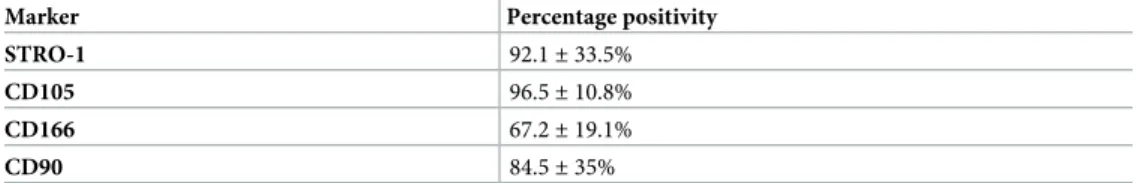
PDLSCs culture supernatants were analyzed after 24 hours of treatment for IL-1β, IL-6 and IL-8
production using ELISA. A significant increase in IL-1β, IL-6 and IL-8 protein levels was detected
withP.gingivalis1:50 dilution treatment compared toP.gingivalis1:500 dilution and the control
(Fig 2,�p<0.05). To ascertain the role ofP.gingivalisbacterial supernatant in the activation of
the TLR2 and TLR4 of PDLSCs, we stimulated PDLSCs byP.gingivalisbacterial supernatants
(1:500 and 1:50) for 24 h. Western blot analysis showed that the expression of both TLR2 and
TLR4 in PDLSCs was not altered byP.gingivalis1:500 dilution treatment. However,P.gingivalis
1:50 dilution treatment increased TLR2 and TLR4 protein concentration in 2 folds compared to
control (Fig 2,�p<0.05). To verify the role ofP.gingivalisbacterial supernatant in regulating the
PDLSCs osteogenic differentiation,P.gingivalissupernatants (1:500 and 1:50) were added to the
osteogenic induction medium. Alizarin red S staining showed thatP.gingivalissupernatant 1:500
dilution impaired the osteogenic differentiation ability of PDLSC, which was demonstrated by the decreased formation number of calcified nodules stained with Alizarin Red compared with the
control group after 21 days induction (Fig 2, day 21,�p<0.05).
Effect of
P
.
gingivalis
and
T
.
denticola
supernatants on PDLSC viability and
proliferation
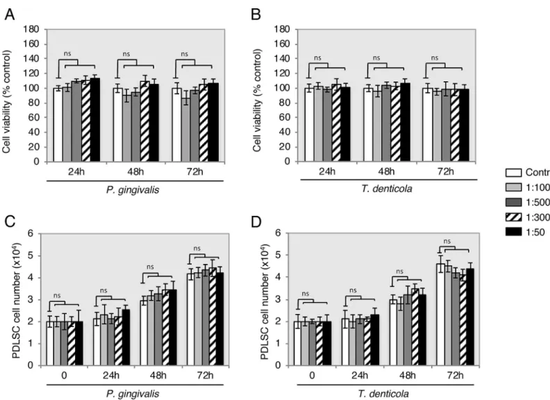
Cell viability and proliferation were performed using onlyP.gingivalisandT.denticolaspecies
Fig 1. Bacterial supernatants modified expression of immune-related molecules in PDLSC. Gene expression levels determined by RT-PCR
analysis showed upregulation ofIL-1β(A)IL-8(B),IL-6(C),TLR2(D) andTLR4(E) in PDLSCs. Representative blots of at least three independent experiments are shown. Statistically significant differences presented as fold change relative to the untreated negative control (control, open columns)
�p<0.05. Mean±S.D.
https://doi.org/10.1371/journal.pone.0219181.g001
Fig 2.P.gingivalissupernatant impairs the osteogenic potential of PDLSC. (A) IL-1β, IL-6 and IL-8 production was measured using ELISA after PDLSCs were treated withP.gingivalis(P.g.) supernatant’s dilutions (1:500 and 1:50), or untreated cells as control simultaneously for 24 h. IL-1β, IL-6 and IL-8 is expressed in pg/ml (±standard deviation). (B) Western blot analysis showed the protein expression of TLR2, TLR4 and GAPDH was used as the internal control. (C) Confluent PDLSC were stained with Alizarin Red after 21 days of cultivation in osteogenic medium containingP.
gingivalissupernatant’s dilutions (1:500 and 1:50) and Alizarin Red was then extracted and measured for light absorbance at 405 nm.�p<0.05 as determined by unpaired two-tailed t tests. Data from three biologically independent replicates. https://doi.org/10.1371/journal.pone.0219181.g002
showed that all supernatants dilutions ofP.gingivalisandT.denticola(Fig 3) had no signifi-cant effect on cell viability at 24 h, 48 h and 72 h compared with the control. The PDLSCs showed proliferation as expected by approximately doubling of the cell number throughout the observation periods of 24 h and 72 h. However, the proliferation rate compared to the con-trols within each individual period of time had no statistically significant alteration. Bacterial supernatants did not have an effect on proliferation in each time periods (Fig 3).
Involvement of
P
.
gingivalis
and
T
.
denticola
supernatants in PDLSC
migration
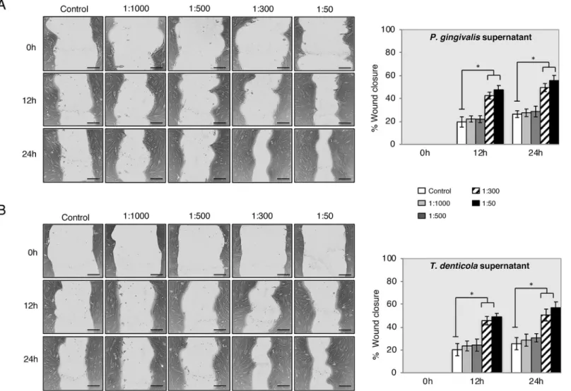
Following cell viability and proliferation, the migration capacity of PDLSC in the presence of
P.gingivalisandT.denticolasupernatants in different dilutions with the in vitro scratch
wound healing assay was determined. Images were acquired at 0 h, 12 h and 24 h after expos-ing the cells to bacterial supernatants (Fig 4). After 24 h, PDLSC migrated and covered
approx-imately 60% of the wound area observed at time zero when exposed toP.gingivalisandT.
denticolaat dilutions of 1:300 and 1:50.
Fig 3. Viability and proliferation of PDLSC exposure toP.gingivalisandT.denticola. Upon supernatant treatment (untreated, 1:1000, 1:500, 1:300 and 1:50),
PLDSC viability and proliferation percentages did not alter throughout periods of time (24 h, 48 h and 72 h) when compared with untreated controls (open columns). ns: non-significant. Data are shown as the mean±S.D of 3 samples (3 wells each) from one representative experiment.
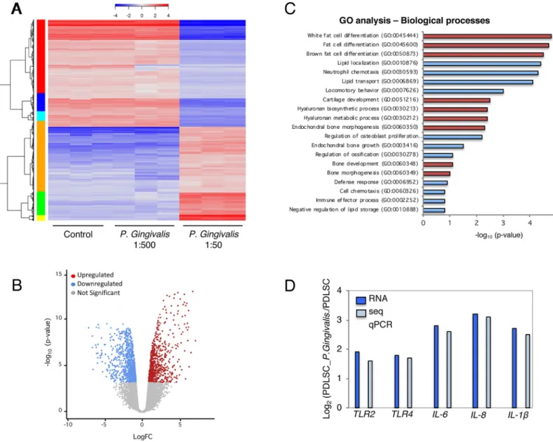
Transcriptional profile of PLDSC response to supernatants dilutions
Using the RNAseq method, the whole transcriptional profile of PLDSC in response to 1:500
and 1:50 dilutions ofP.gingivalissupernatant (24 h cell exposure) was screened. All
signifi-cantly changed genes were analyzed in heat map (Fig 5), which indicated more upregulated
genes induced byP.gingivalissupernatant at 1:50 supernatant dilution. After RNAseq data
processing, normalized and transformation, significantly changed genes of PLDSC with 1:50
dilution ofP.gingivalistreatment compared to the untreated control were shown in the
vol-cano Fig (Fold change>2.0, p<0.05 and False Discovery Rate<0.05 were considered as
sig-nificant) (Fig 5). The volcano analysis showed higher number of upregulated genes than
downregulated genes. The total of 502 genes were found significantly changed after 1:50P.
gin-givalissupernatant stimulation, of which 289 were upregulated, whereas 213 genes were
down-regulated. The analysis using KEGG pathway included 29 cytokines (10.9%), 32 signaling
transducers (e.g NF-κB) genes (9.7%), 17 metabolism genes (6.1%), and 211 other functional
genes (73.3%). To identify the key biological processes and pathways that are affected when
Fig 4. PDLSC migration was increased with supernatants ofP.gingivalisandT.denticolaby scratch wound healing assay. Representative images are shown from 3
independent experiments and light gray area define the areas lacking cells (Scale bar 120μm). Images were analyzed using ImageJ software to calculate wound area. Data is expressed as the mean values of percentage wound closure relative to the corresponding 0 h time point and represent the mean percentage closure±SEM (n = 3):
�p<0.01 vs. time-matched treated control for each time-point. https://doi.org/10.1371/journal.pone.0219181.g004
the PDLSCs are treated with 1:50 dilution ofP.gingivalsupernatant, gene ontology (GO) enrichment analysis performed by DAVID software allowed the listing of gene groups involved in the same biological processes. GO analyses of the common differentially expressed genes revealed that a large proportion of genes was involved in white and brown fat cell differ-entiation, lipid transport and lipid localization (Fig 5). PDLSCs 1:50-supernatant dilution
treatment showed significant enrichment for 1,116 GO terms (p<0.05), including 856 for
biological processes. We then narrowed down key GO terms based on high significance (p
value) to highlight the widespread effect ofP.gingivalissupernatant on PDLSCs. The key GO
terms significantly enriched in biological processes were white fat cell differentiation (GO:
Fig 5. Transcriptomic analysis elucidates PDLSC response to bacterial supernatant-induced bioactivity. (A) Bacterial supernatant treated groups (1:500 and 1:50
dilutions) and untreated group (Ctrl) were marked. Heatmap showed significant gene profile for RNAseq data. Normalized log2 scale shown. (B) Volcano analysis was performed for significantly changed genes in the dilution of 1:50 supernatant treated group (red dots represented upregulated genes and blue dots represented downregulated genes, Fold change>2.0, p<0.05). (C) Significant GO terms of associated biological processes from differentially regulated genes from a ranking by enrichment scores (p<0.05). Adipogenesis and chondrogenesis processes identified by red columns. Enrichment scores were calculated as−log10(P-value), (D)
Comparison between RNAseq and qPCR gene expression showed similar values.
0045444), white fat cell differentiation (GO: 0045444) and brown fat cell differentiation (GO: 0050873). The analysis also indicated positive regulation of cartilage development
(GO:0051216), hyaluronan biosynthetic process (GO:0030213) and hyaluronan biosynthetic process (GO:0030213) (Fig 5). Terms related to biological process indicate strong biophysical
interactions between PDLSCs andP.gingivalissupernatant. Also, in additional comparison,
the expression of important genes participating in inflammation, which were significantly higher in PDLSCs treated with 1:50 dilution than in untreated controls was further validated by RT-PCR (Fig 5).
Discussion
Considering the key role of PDLSC in periodontal tissue homeostasis and regeneration [10, 15,16], the purpose of this study was to ascertain the effect of different periodontal bacterial supernatants on PDLSC immune response and cell differentiation potential. The results showed that periodontal bacteria supernatants could not only inhibit osteogenesis, but also increase the migration and immune response of PDLSC. In addition, this study also showed that these changes in biological process were most likely dependent upon increases in peri-odontal bacteria virulence.
Other previous in vitro studies using oral stem cells predominantly demonstrate the influ-ence of LPS and fail to address the important effect of other virulent factors secreted or bacte-rial-derived molecules on a variety of cell responses. In this study, we sought to address the contributions made by bacterial-derived molecules other than just the role of LPS on stimulat-ing PDLSCs inflammatory response and cell fate. Many pathogenicity determinants, which cause damage in a host organism, can be found in bacterial supernatants such as the ones employed in this study. For instance, several outer membrane proteins (lipd-A associated
pro-teins/LAP) have been shown inP.gingivalisto be potent inducers of cytokine synthesis [22]
with lowest concentration stimulation of only 10 ng/ml, whereas LPS fromP.gingivalisis 1 log
unit less active in this respect [22]. Several studies also have demonstrated the ability of
fim-brial protein to stimulate cytokine synthesis in various cell types.P.gingivalisfimbriae (1μg/
ml) stimulated human gingival fibroblasts to release IL-1β[23] and P. intermedia (100 ng/ml)
stimulated release of IL-1α, IL-1β, IL-6, and IL-8 [24]. Other virulent factors presents in
super-natants are cell surface cysteine proteinases, such asP.gingivalisgingipains that can also be
present in secreted soluble form on supernatants [25]. The gingipains have immunomodula-tory multiple effects by enhancing cell-mediated immune response in oral tissues [26–29].
Many components of the supernatants collected for this study have increased cell response and may be associated to the virulence potency of the bacterial species. In previous animal
studies on secreted determinants of pathogen virulence, the exopolysaccharide produced byP.
intermediaandP.gingivaliswas considered to be powerful menace for inflammation and
abscess formation in mice [30]. Fimbriae and other microbial virulence proteins were also pointed to augment virulence in many pathogenic bacteria [23]. Moreover, microbial viru-lence proteins have evolved to interact with and possibly exploit TLRs and the cell pattern
rec-ognition system, in ways that increase bacterial pathogenicity [31].T.denticola
membrane-associated lipoproteins/lipooligosaccharides were also found to induce production of inflam-matory mediators by mouse macrophages in a dose-dependent manner [32]. We believe that all those pathogenicity determinants from bacterial species used in this study may have played an important role on intensifying the observed PDLSCs response.
The literature suggests that pathogenic bacterial endotoxin concentrations in the subgingi-val biofilm vary greatly depending on the local micro-environmental conditions [33]. Follow-ing those reports, we decided to use bacterial supernatant dilutions, which could resemble the
endotoxins concentrations likely to be found in the subgingival plaque of periodontal pockets. In those studies, for the single stimulation experiments as performed here, the bacterial
super-natants were also directly diluted in the cell culture medium [20,21]. The bacterial strains
cho-sen for this study reprecho-sent some important members of the oral biofilm.P.intermedia,F.
nucleatum,P.gingivalisandT.denticolawere used as species representative of the pathogenic
subgingival biofilm related with periodontitis.S.mutansandS.anginosuswere used as
con-trols, to define the specificity of the results regarding the pathogenicity of the other gram-nega-tive bacterial species.
Changes in PDLSCs lineage fate, caused by bacterial endotoxins, were observed in recent studies, which implicate bacterial endotoxins as inhibitors of PDLSCs osteogenic
differentia-tion [11,12,34]. Accordingly, the transcriptional profile results presented here suggest that
inflammation may be associated with inhibition of PDLSCs osteogenic capability. In compari-son to literature assessing the effect of LPS on PDLSCs, adipogenesis and chondrogenesis in
bacterialP.gingivalissupernatant treated PDLSCs observed in the current study also presented
increased levels [35,36]. Even though adipogenesis and chondrogenesis differentiation of
PDLSCs have been questioned in vivo [37], bacterial endotoxins are still a pivot for change in PDLSCs differentiation into undesirable patterns of periodontal ligament tissue maintenance; directions associated with some additional pathologic changes. For example, the small size increments of neutral lipid droplets in cytosol correlation to various metabolic changes in cells, which appears during cellular senescence process and LPS-treated dental pulp stem cells
oxidative stress [38,39]. It may be speculated that PDLSC adipogenic differentiation is related
to in vitro cell culture conditions and changes in energy metabolism. In addition, an
inflam-matory microenvironment may modulate immunomodulatory PDLSC properties [13,14],
which supports the findings of the current study where those inflammatory cytokines (IL-6,
IL-8,IL1β) and transmembrane cell-surface receptors (TLR2andTLR4) were significantly
enhanced (Figs1and2). This was further confirmed by the whole transcriptional profile
RNA-seq analysis. The increase in gene expression was dependent on the supernatant dilution and the bacterial virulence, as the expression was higher in cells exposed to higher dilutions of
superna-tants (i.e. 1:300–1:50) derived from strongly pathogenic bacteria (P.intermedia,F.nucleatum,P.
gingivalisandT.denticola). These results may also support the hypothesis of the immune cell
like function of periodontal ligament cells [37]. In contrast, the RT-PCR analysis showed lower
augmentation ofTLR2andTLR4expression under the influence of bacteria supernatant
treat-ment. Possible explanations of this lower-expression could be due to cells adaptation mechanism to bacteria endotoxins exposure, or that TLR2 mostly identifies lipo-proteins, lipo-peptides or other virulence factors rather than LPS, whereas TLR4 recognizes LPS [39]. In fact, the family of the Toll-like receptors is involved in recognition of bacterial cell wall components [40].
More-over, the treatment of PDLSC with bacterial supernatant ofP.gingivalisandT.denticoladid not
increase cell viability and proliferation compared to the controls within each individual period of time analyzed (Fig 3), although previous studies suggested that migratory properties of stem cells enhanced by LPS treatment on murine odontoblasts-like cells, dental follicle periodontal
cells, bone marrow mesenchymal stem cells [41,42] and periodontal ligament cells [43,44]. In
contrast to the proliferation results, PDLSC presented significantly higher motility activity when
challenged withP.gingivalisandT.denticolaat 1:300 and 1:50 dilutions (Fig 4), whereas
dilu-tions of 1:1000 and 1:500 did not elicit migration. The unchanged migration upon lower super-natant dilution treatment could be explained by lower concentration of endotoxins content, which reduces supernatant stimulation potency. And it indicates that there is possibly a thresh-old for PDLSCs responsiveness to bacterial endotoxins. However, the data obtained from
dilu-tions of 1:300 and 1:500 supernatant stimulation verifies the stimulatory impact ofP.gingivalis
periodontal tissue repair process. This theory was also confirmed in previous studies which showed that LPS stimulated contractility of PDLSCs with elevated myofibroblast marker
expres-sion ofα-SMA, TGF-β, and FN proteins [34,45] specially found in periodontal ligament
fibro-blasts in response to chemical stress signals [46].
This study has provided further evidence of the broader role played by different periodontal bacterial species as to their influence on PDLSCs cell fate and differentiation properties. Also, the increase in gene expression related to adipogenic or chondrogenic noted in the RNA sequencing (Fig 5) and inhibition on osteogenesis (Fig 2) in this study suggests the negative effect of bacterial endotoxins on periodontal regeneration. For instance, by TLR4 mediation, LPS was found to activate the host cells in the periodontium, including polymorphonuclear
leukocytes, macrophages, fibroblasts and the epithelium [47,48]. And inhibition of TLR4 or
NF-κB pathway impaired alveolar bone loss caused by LPS treatment [12]. Nevertheless, the
detailed mechanism still needs further investigation, as the mechanism of LPS induced peri-odontitis is complicated and not completely understood. Aside from endotoxin modification of PDLSC differentiation, LPS can induce alveolar bone loss by stimulating the secretion of
proinflammatory cytokines (IL-1β, TNF-αor IL-6). LPS can also induce the secretion of
matrix metalloproteinases, formation of osteoclasts stimulation, causing direct damage to peri-odontal tissues [20]. An important question in cellular susceptibility to inflammation is to what extent the variability in pathogen species could induce higher or lower immune response. Strains of bacterial species are not equally pathogenic due to dissimilarity in virulence factor expression. Following this idea, the contribution of the pathogenic bacteria on the cytokine production was shown in our previous study [19], in which the same in vitro methodology used in the present study enabled the determination of cell-type specific response to different periodontal bacterial species according to its pathogenicity. Nevertheless, the immunomodula-tory activity of PDLSC still need to be assessed by additional studies to clarify which particles other that LPS can govern inflammation or whether it is mediated through suppression of other downstream signaling pathways.
In summary, this study demonstrates that different bacterial stimulation induces immunor-esponse, cell motility and most importantly the differentiation potential of PDLSC. Stem cell response to periodontal bacteria raises questions on the multipotent cells contribution to both outbreak of inflammation and periodontal tissue regeneration. Immunomodulation of stem cells through bacterial stimulation is a matter of great interest, as it may allow the development of therapeutic interventions for inflammatory diseases such as periodontitis.
Supporting information
S1 Fig. Growth curves of different bacteria species at the optical density measurements. Correspondence was established between OD 550 nm measured from 0 to 20h and bacteria
counted in microscopy. Colonies ofS.mutans,S.anginosus,P.intermedia,F.nucleatum,P.
gingivalisandT.denticolawere cultured under anaerobically conditions (6 h, 37˚C, 10% CO2)
in 5 mL brain heart infusion broth (Merck, Darmstadt, Germany). Cells from the logarithmic growth phase were used for the study after overnight standardization of bacterial cultures in 5
mL PBS to achieve a bacterial cell concentration of 106CFU/mL. The density of the resulting
inoculum was measured using a MicroSpeak densitometer and confirmed by single colony counting after 24-hour growth on brain heart infusion broth medium under the above-speci-fied conditions. The number of grown bacterial colonies (CFU/mL) was calculated. The results are means of three experiments and error bars represent standard deviations determined from at least three replicates.
S1 File. Minimal data set. (DOCX)
Acknowledgments
We thank Dr. Giancarlo Russo, Genomics Center Zurich (FGCZ), ETH Zurich, University of Zurich, for RNA sequencing and technical support. And we thank Prof. T. Mitsiadis and the Orofacial Development and Regeneration for steady support on the methodology develop-ment on this study. We acknowledge the University of Zurich Flow Cytometry Facility for assistance with the generation of flow cytometry data. Authors declare no conflict of interests in this study and received no specific funding for this work.
Author Contributions
Conceptualization: Liza L. Ramenzoni. Data curation: Giancarlo Russo. Investigation: Liza L. Ramenzoni. Methodology: Liza L. Ramenzoni.
Project administration: Patrick R. Schmidlin. Software: Giancarlo Russo, Maria D. Moccia. Supervision: Thomas Attin.
Validation: Giancarlo Russo.
Writing – original draft: Liza L. Ramenzoni.
References
1. Aas JA, Paster BJ, Stokes LN, Olsen I, Dewhirst FE. Defining the normal bacterial flora of the oral cav-ity. J Clin Microbiol. 2005; 43: 5721–5732.https://doi.org/10.1128/JCM.43.11.5721-5732.2005PMID: 16272510
2. Page RC & Kornman KS. The pathogenesis of human periodontitis: an introduction. Periodontology 2000. 1997; 14: 9–11. PMID:9567963
3. Graves DT, Oates T, Garlet GP. Review of osteoimmunology and the host response in endodontic and periodontal lesions. J Oral Microbiol. 2011; 3:1–15.
4. Chatzivasileiou K, Kriebel K, Steinhoff G, Kreikemeyer B, Lang H. Do oral bacteria alter the regenera-tive potential of stem cells? A concise review. J Cell Mol Med. 2015; 19: 2067–2074.https://doi.org/10. 1111/jcmm.12613PMID:26058313
5. Nagatomo K, Komaki M, Sekiya I, Sakaguchi Y, Noguchi K, Oda S, et al. Stem cell properties of human periodontal ligament cells. J Periodontal Res. 2006; 41: 303.https://doi.org/10.1111/j.1600-0765.2006. 00870.xPMID:16827724
6. Zhu W, Liang M. Periodontal ligament stem cells: current status, concerns, and future prospects. Stem Cells Int. 2015; 2015: 972313.https://doi.org/10.1155/2015/972313PMID:25861283
7. Bassir SH, Wisitrasameewong W, Raanan J, Ghaffarigarakani S, Chung J, Freire M, Intini G. Potential for stem cell-based periodontal therapy. J Cell Physiol. 2016; 231: 50–61.https://doi.org/10.1002/jcp. 25067PMID:26058394
8. Choi S, Cho TJ, Kwon SK, Lee G, Cho J. Chondrogenesis of periodontal ligament stem cells by trans-forming growth factor-β3 and bone morphogenetic protein-6 in a normal healthy impacted third molar. Int J Oral Sci. 2013; 5: 7–13.https://doi.org/10.1038/ijos.2013.19PMID:23579467
9. Tang HN, Xia Y, Yu Y, Wu RX, Gao LN, Chen FM. Stem cells derived from “inflamed”and healthy peri-odontal ligament tissues and their sheet functionalities: A patient-matched comparison. J Clin Periodon-tol. 2016; 43: 72–84.https://doi.org/10.1111/jcpe.12501PMID:26719165
10. Albiero ML, Amorim BR, Martins L, Casati MZ, Sallum EA, Nociti FH Jr, Silve´rio KG (2015). Exposure of periodontal ligament progenitor cells to lipopolysaccharide from Escherichia coli changes osteoblast dif-ferentiation pattern. J Appl Oral Sci. 2015; 23: 145–152.https://doi.org/10.1590/1678-775720140334 PMID:26018305
11. Kato H, Taguchi Y, Tominaga K, Umeda M, Tanaka A. Porphyromonas gingivalis LPS inhibits osteo-blastic differentiation and promotes pro-inflammatory cytokine production in human periodontal liga-ment stem cells. Arch Oral Biol. 2014; 59: 167–175.https://doi.org/10.1016/j.archoralbio.2013.11.008 PMID:24370188
12. Li C, Li B, Dong Z, Gao L, He X, Liao L, Hu C, Wang Q, Jin Y. Lipopolysaccharide differentially affects the osteogenic differentiation of periodontal ligament stem cells and bone marrow mesenchymal stem cells through Toll-like receptor 4 mediated nuclear factorκB pathway. Stem Cell Res Ther. 2014; 5: 67. https://doi.org/10.1186/scrt456PMID:24887697
13. Seo T, Cha S, Kim TI, Lee JS, Woo KM. Porphyromonas gingivalis-derived lipopolysaccharide-medi-ated activation of MAPK signaling regulates inflammatory response and differentiation in human peri-odontal ligament fibroblasts. J Microbiol. 2012; 50: 311–319. https://doi.org/10.1007/s12275-012-2146-xPMID:22538661
14. Bandow K, Maeda A, Kakimoto K, Kusuyama J, Shamoto M, Ohnishi T. Molecular mechanisms of the inhibitory effect of lipopolysaccharide (LPS) on osteoblast differentiation. Biochem Biophys Res Com-mun. 2010; 402: 755–761.https://doi.org/10.1016/j.bbrc.2010.10.103PMID:21036155
15. Andrukhov O, Andrukhova O, O¨ zdemir B, Haririan H, Mu¨ller-Kern M, Moritz A, Rausch-Fan X. Soluble CD14 Enhances the Response of Periodontal Ligament Stem Cells to P. gingivalis Lipopolysaccharide. PLoS One. 2016; 11:e0160848.https://doi.org/10.1371/journal.pone.0160848PMID:27504628
16. Chen FM, Gao LN, Tian BM, Zhang XY, Zhang YJ, Dong GY, Lu H, Chu Q, Xu J, Yu Y, Wu RX, Yin Y, Shi S, Jin Y. Treatment of periodontal intrabony defects using autologous periodontal ligament stem cells: a randomized clinical trial. Stem Cell Res Ther. 2016; 19: 7–33.
17. Liu Y, Zheng Y, Ding G, Fang D, Zhang C, Bartold PM, et al. Periodontal ligament stem cell-mediated treatment for periodontitis in miniature swine. Stem Cells. 2008; 26: 1065–73.https://doi.org/10.1634/ stemcells.2007-0734PMID:18238856
18. Tsumanuma Y, Iwata T, Washio K, Yoshida T, Yamada A, Takagi R, et al. Comparison of different tis-sue-derived stem cell sheets for periodontal regeneration in a canine 1-wall defect model. Biomaterials. 2011; 32: 5819–5825.https://doi.org/10.1016/j.biomaterials.2011.04.071PMID:21605900
19. Ramenzoni LL, Zuellig RA, Hussain A, Lehmann R, Heumann C, Attin T, Schmidlin PR. Bacterial super-natants elevate glucose-dependent insulin secretion in rat pancreatic INS-1 line and isletβ-cells via PI3K/AKT signaling. Mol Cell Biochem. 2019; 452: 17–27.https://doi.org/10.1007/s11010-018-3408-7 PMID:30039349
20. Bostanci N, Allaker RP, Belibasakis GN, Rangarajan M, Curtis MA, Hughes FJ, McKay IJ. Porphyromo-nas gingivalis antagonises Campylobacter rectus induced cytokine production by human monocytes. Cytokine. 2007; 39: 147–156.https://doi.org/10.1016/j.cyto.2007.07.002PMID:17709256
21. Yin L, Swanson B, An J, Hacker BM, Silverman GA, Dale BA, Chung WO. Differential effects of perio-pathogens on host protease inhibitors SLPI, elafin, SCCA1, and SCCA2. J Oral Microbiol. 2010; 4:2.
22. Reddi K, Poole S, Nair S, Meghji S,Henderson B, Wilson M. Lipid A-associated proteins from periodon-topathogenic bacteria induce interleukin-6 production by human gingival fibroblasts and monocytes. FEMS Microbiol Immunol. 1995; 11: 137–144.
23. Ogawa T, Kusumoto Y, Uchidab H, Nagashima S, Ogo H, Hamada S. Immunobiological activities of synthetic peptide segments of fimbrial protein from Porphyromonas gingivalis. Biochem. Biophys Res Commun. 1991; 180: 1335–1341.https://doi.org/10.1016/s0006-291x(05)81342-7PMID:1659412
24. Matsushita K, Nagaoka S, Arakaki R, Kawabata Y, Iki K, Kawagoe M, Takada H. Immunobiological activities of a 55-kilodalton cell surface protein of Prevotella intermedia ATCC 25611. Infect Immun. 1994; 62: 2459–2469. PMID:8188371
25. Bostanci N, Belibasakis GN. Porphyromonas gingivalis: an invasive and evasive opportunistic oral path-ogen. FEMS Microbiol Lett. 2012; 333: 1–9.https://doi.org/10.1111/j.1574-6968.2012.02579.xPMID: 22530835
26. Holzhausen M, Cortelli JR, da Silva VA, Franco GC, Cortelli SC & Vergnolle N. Protease-activated receptor-2 (PAR(2)) in human periodontitis. J Dent Res. 2010; 89: 948–953.https://doi.org/10.1177/ 0022034510373765PMID:20530726
27. Fagundes JA, Monoo LD, Euzebio Alves VT, Pannuti CM, Cortelli SC, Cortelli JR, Holzhausen M. Por-phyromonas gingivalis is associated with protease-activated receptor-2 upregulation in chronic peri-odontitis. J Periodontol. 2011; 82: 1596–1601.https://doi.org/10.1902/jop.2011.110073PMID: 21513479
28. Lourbakos A, Potempa J, Travis J, D’Andrea MR, Andrade-Gordon P, Santulli R, Mackie EJ, Pike RN. Arginine-specific protease from Porphyromonas gingivalis activates protease-activated receptors on human oral epithelial cells and induces interleukin-6 secretion. Infect Immun. 2001; 69: 5121–5130. https://doi.org/10.1128/IAI.69.8.5121-5130.2001PMID:11447194
29. Oido-Mori M, Rezzonico R, Wang PL, Kowashi Y, Dayer JM, Baehni PC & Chizzolini C. Porphyromonas gingivalis gingipain-R enhances interleukin-8 but decreases gamma interferon-inducible protein 10 pro-duction by human gingival fibroblasts in response to T-cell contact. Infect Immun. 2001; 69: 4493– 4501.https://doi.org/10.1128/IAI.69.7.4493-4501.2001PMID:11401991
30. Yamanaka T, Yamane K, Furukawa T, Matsumoto-Mashimo C, Sugimori C, Nambu T, Obata N, Walker CB, Leung KP, Fukushima H. Comparison of the virulence of exopolysaccharide-producing Prevotella intermedia to exopolysaccharide non-producing periodontopathic organisms. BMC Infect Dis. 2011; 11: 228.https://doi.org/10.1186/1471-2334-11-228PMID:21864411
31. Hajishengallis G, Shakhatreh MA, Wang M, Liang S. Complement receptor 3 blockade promotes IL-12-mediated clearance of Porphyromonas gingivalis and negates its virulence in vivo. J Immunol. 2007; 179: 2359–2367.https://doi.org/10.4049/jimmunol.179.4.2359PMID:17675497
32. Rosen G, Sela MN, Naor R, Halabi A, Barak V, Shapira L. Activation of murine macrophages by lipopro-tein and lipooligosaccharide of Treponemadenticola. Infect Immun. 1999; 67: 1180–1186. PMID: 10024558
33. Socransky SS, Haffajee AD. Periodontal microbial ecology. Periodontol 2000. 2005; 38: 135–187. https://doi.org/10.1111/j.1600-0757.2005.00107.xPMID:15853940
34. Kukolj T, TrivanovićD, DjordjevićIO, MojsilovićS, KrstićJ, ObradovićH, JankovićS, Santibanez JF, JaukovićA, Bugarski D. Lipopolysaccharide can modify differentiation and immunomodulatory potential of periodontal ligament stem cells via ERK1,2 signaling. J Cell Physiol. 2018; 233: 447–462.https://doi. org/10.1002/jcp.25904PMID:28295277
35. Caron MM, Emans PJ, Surtel DA, Cremers A, Voncken JW, Welting TJ, van Rhijn LW. Activation of NF-κB/p65 facilitates early chondrogenic differentiation during endochondral ossification. PLoS One. 2012; 7: e33467.https://doi.org/10.1371/journal.pone.0033467PMID:22428055
36. Thiam AR, Farese RV Jr, Walther TC. The Biophysics and Cell Biology of Lipid Droplets. Nat Rev Mol Cell Biol. 2013; 14: 775–786.https://doi.org/10.1038/nrm3699PMID:24220094
37. Mao JJ, Robey PG, Prockop DJ. Stem cells in the face: Tooth regeneration and beyond. Cell Stem Cell. 2012; 11: 291–301.https://doi.org/10.1016/j.stem.2012.08.010PMID:22958928
38. Feng X, Feng G, Xing J, Shen B, Li L, Tan W, Xu Y, Liu S, Liu H, Jiang J, Wu H, Tao T, Gu Z. TNF-α trig-gers osteogenic differentiation of human dental pulp stem cells via the NF-κB signalling pathway. Cell Biol Int. 2013; 37: 1267–75.https://doi.org/10.1002/cbin.10141PMID:23765556
39. Mo IF, Yip KH, Chan WK, Law HK, Lau YL, Chan GC. Prolonged exposure to bacterial toxins downregu-lated expression of toll-like receptors in mesenchymal stromal cell-derived osteoprogenitors. BMC Cell Biol. 2008; 18: 9–52.
40. Jin MS, Lee JO. Structures of the toll-like receptor family and its ligand complexes. Immunity. 2008; 29: 182–91.https://doi.org/10.1016/j.immuni.2008.07.007PMID:18701082
41. Park JH, Kwon SM, Yoon HE, et al. Lipopolysaccharide promotes adhesion and migration of murine dental papilla-derived MDPC-23 cells via TLR4. Int J Mol Med. 2011; 27: 277–81.https://doi.org/10. 3892/ijmm.2010.568PMID:21125213
42. Chatzivasileiou K, Lux CA, Steinhoff G, Lang H. Dental follicle progenitor cells responses to Porphyro-monas gingivalis LPS. J Cell Mol Med. 2013; 17: 766–773.https://doi.org/10.1111/jcmm.12058PMID: 23560719
43. Jonsson D, Nebel D, Bratthall G, Nilsson BO. The human periodontal ligament cell: a fibroblast-like cell acting as an immune cell. J Periodontal Res. 2011; 46: 153–157.https://doi.org/10.1111/j.1600-0765. 2010.01331.xPMID:21118418
44. Wada N, Menicanin D, Shi S, Bartold PM, Gronthos S. Immunomodulatory properties of human peri-odontal ligament stem cells. J Cell Physiol. 2009; 219: 667–676.https://doi.org/10.1002/jcp.21710 PMID:19160415
45. Iwasaki K, Komaki M, Yokoyama N, Tanaka Y, Taki A, Kimura Y, Takeda M, Oda S, Izumi Y, Morita I. Periodontal ligament stem cells possess the characteristics of pericytes. J Periodontol. 2013; 84: 1425–1433.https://doi.org/10.1902/jop.2012.120547PMID:23240762
46. Kimura H, Okubo N, Chosa N, Kyakumoto S, Kamo M, Miura H, Ishisaki A. (2013). EGF positively regu-lates the proliferation and migration, and negatively reguregu-lates the myofibroblast differentiation of peri-odontal ligament-derived endothelial progenitor cells through MEK/ERK- and JNK-dependent signals. Cell Physiol Biochem. 2013; 32: 899–914.https://doi.org/10.1159/000354493PMID:24217646
47. Wilson M. Biological activities of lipopolysaccharides from oral bacteria and their relevance to the patho-genesis of chronic periodontitis. Sci Prog. 1995; 78: 19–34. PMID:7597416
48. Konermann A, Stabenow D, Knolle PA, Held SA, Deschner J, Ja¨ger A. Regulatory role of periodontal ligament fibroblasts for innate immune cell function and differentiation. Innate Immun. 2012; 18: 745– 752.https://doi.org/10.1177/1753425912440598PMID:22436844