HUMAN EXPOSURE TO BISPHENOL A (BPA)
Laura N. Vandenberg1, Russ Hauser2, Michele Marcus3, Nicolas Olea4, and Wade V. Welshons5
1Tufts University Sackler School of Graduate Biomedical Sciences, Boston MA 02111 2Department of Environmental Health, Harvard School of Public Health, Boston, MA
02115
3Emory University, Rollins School of Public Health, Atlanta, GA 30322 4Hospital Clinico, University of Granada, 18071 Granada Spain
5University of Missouri-Columbia, Department of Biomedical Sciences, Columbia, MO
65211
Corresponding author: Laura N. Vandenberg
Tufts University School of Medicine
Sackler School of Graduate Biomedical Sciences 136 Harrison Avenue
Boston, MA 02111
Ph: 617-636-0444 Fax: 617-636-3971
Disclaimer: The findings and conclusions in this report are those of the authors and do not necessarily represent the views of any affiliated institutions or agencies. Mention of trade names or commercial products does not constitute endorsement or
recommendation for use.
Acknowedgements: The authors gratefully acknowledge expertise and input from additional panel members: Jane C. Atkinson, Antonia M. Calafat, Frederick Eichmiller, Albert Kingman, Ruthann Rudel, and Kristina A. Thayer. This review was prepared in conjunction with the Bisphenol A Conference, Chapel Hill, NC, November 28-29, 2006. Support was provided by the National Institute of Environmental Health Sciences and the National Institute of Dental and Craniofacial Research, NIH, DHHS, the W.M. Keck Center for Behavioral Biology at NC State University, and from Commonweal
Running Title: Human exposure to BPA
Keywords: serum, urine, epidemiology, disease, epoxy resins, dental sealants, polycarbonate plastic, metabolism
Abbreviations
BADGE bisphenol A diglycidyl ether Bis-DMA bisphenol A dimethylacrylate
BPA bisphenol A
BPA-gluc bisphenol A glucuronide
°C degrees Celsius
CDC Centers for Disease Control and Prevention DHEAS dehydroepiandrosterone sulfate
DIB-Cl fluorescent labeling agent, 4-(4,5-diphenyl-1H-imidazol-2-yl)benzoyl chlorine
E estrogen
ELISA enzyme-linked immunosorbent assay
ED or ECD electrochemical detection
ER estrogen receptor
ESI electrospray ionization
FD fluorescence detection
FSH follicle stimulating hormone
g grams
GC gas chromatography
HPLC high performance liquid chromatography
HRGC high resolution gas chromatography
i.p. intraperitoneal
i.v. intravenous
IVF in vitro fertilization
kg kilogram
L liter
LC liquid chromatography
LH lutenizing hormone
LOD limit of detection
m meter
mg milligram
ml milliliter
MS mass spectrometry
MS/MS tandem mass spectrometry
NCI negative chemical ionization
ND not detected
ng nanograms
NIEHS National Institute for Environmental Health Sciences
NMR Nuclear Magnetic Resonance
NOAEL No observable adverse effect level
PCOS polycystic ovarian syndrome
pM picomolar
RfD reference dose
SPE solid-phase extraction
subcut subcutaneous
T testosterone
TEGDMA triethylene glycol dimethacrylate
Uncon unconjugated
Introduction
The plastic monomer and plasticizer bisphenol A (BPA) is one of the highest volume chemicals produced worldwide, with over 6 million pounds produced each year [1]. BPA is used in the production of polycarbonate plastics, epoxy resins used to line metal cans, and in many plastic consumer products including toys, water pipes, drinking containers, eyeglass lenses, sports safety equipment, dental monomers, medical equipment and tubing, and consumer electronics [2]. BPA has been shown to leach from food and beverage containers, and some dental sealants and composites under normal conditions of use. Studies have also determined that BPA can be measured in humans in serum, urine, amniotic fluid, follicular fluid, placental tissue, and umbilical cord blood. In some cases, the levels of total BPA (free and conjugated) in human blood and other fluids are higher than the concentrations that have been reported to stimulate a number of molecular endpoints in cell culture in vitro, and appear to be within an order
of magnitude of the levels of BPA in animal studies; both of these literatures are reviewed in the papers of other panels of this meeting.
Biochemical assays have examined the kinetics of BPA binding to estrogen receptors (ER) and have determined that BPA binds both ERα and ERβ, with approximately 10-fold higher affinity to ERβ [3,4,5]. The affinity of BPA for ERs is 10,000 to 100,000-fold weaker than that of estradiol. Until recently, BPA had been considered to be a very weak environmental estrogen because of its low ER affinity and because in many bioassays (e.g., the rodent uterotrophic assay and some responses in human breast cancer cells), BPA can be 10,000- to 100,000-fold less potent than estradiol. However, results from recent studies of molecular mechanisms of BPA action have revealed a variety of pathways through which BPA can stimulate cellular responses at very low concentrations (reviewed in [6]) in addition to effects initiated by binding of BPA to the classical nuclear or genomic estrogen receptors. The accompanying review from this meeting on Molecular Mechanisms of BPA Action describes recent findings showing that in a variety of tissues, BPA not only has the efficacy of estradiol but is also equally potent, with changes in cell function being observed at a dose of 1 pM (0.23 pg/ml culture medium), through mechanisms that are thought to be nongenomic and
involve membrane-associated forms of the estrogen receptors. The relevance of these mechanisms to the in vivo situation are discussed in that panel review as well.
"Low-doses" of endocrine disrupting chemicals were defined by the NIEHS Low Dose Peer Review as doses below the accepted NOAEL for the chemical [7], which, for BPA, are doses below 50 mg/kg body weight/day. Initial reports of adverse effects of BPA at "low-doses" in animal models were below the Reference Dose (RfD), calculated as an acceptable daily human intake typically 1000-fold below the NOAEL. There are now over 150 published studies describing low-dose BPA effects in animals, including prostate weight and cancer, mammary gland organization and cancer, protein induction in the uterus, organization of sexually dimorphic circuits in the hypothalamus, onset of estrus cyclicity and earlier puberty, body weight, genital malformations and others (reviewed in the In vivo panel report.; over 40 of these are below the RfD for BPA of 50 micrograms/kg/day. Many of these endpoints are in areas of current concern for human epidemiological trends.
Because of its wide availability in the environment, and its estrogenic activity in specific responses in vitro and in vivo, adverse effects of BPA exposure on human
health are possible [8,9,10,11]. It has been hypothesized that exposure during early development to xenoestrogens such as BPA may be the underlying cause of the increased incidence of infertility, genital tract abnormalities, and breast cancer observed in European and US human populations over the last 50 years [12,13,14].
Here, we have outlined a number of studies that address the levels of BPA in human tissues and fluids. We have also reviewed the few epidemiological studies available that explore the relationship between biological markers of BPA exposure with human health outcomes. We have provided information from several studies that examine the levels of BPA released from consumer products as well as the levels measured in wastewater, drinking water, air and dust. Human exposures are most likely through the oral route, although transdermal exposure by bathing in BPA-contaminated water is also a possible route, as is exposure via inhalation; both of these latter routes of exposure would not be subjected to the extensive first-pass conjugation that occurs with oral ingestion. And finally, we have included several acute metabolic studies that have been performed, along with information available about BPA metabolism in animal
models. While this review is by no means comprehensive, we have covered most of the studies that are frequently referenced in the extensive BPA literature.
1. BPA levels in human tissues and fluids
BPA levels have been measured in human fluids and tissues in many developed countries of the world. A general consensus has been accepted that BPA can be detected in the majority of individuals in these countries. The levels of BPA in residents of less-developed countries, however, remain unknown.
Serum, blood & plasma
Since 1999 [15], more than a dozen studies using a variety of different analytical techniques have measured unconjugated BPA concentrations in human serum (Table 1) at levels ranging from 0.2–20 ng/ml serum and exceeding 100 ng/g in one study of placental tissue. These studies have examined blood from both men and women from several countries and at different ages. The techniques used to measure BPA in human serum have included gas chromatography mass spectrometry (GC-MS), high performance liquid chromatography (HPLC), derivatization with different chemical agents followed by GC, and ELISA, all with sensitivities for BPA (in serum) ranging from 0.01– 0.5 ng/ml. Among all of these analytical techniques, MS, specifically isotope dilution-MS, is considered the most accurate and precise method for measuring trace levels of BPA and other environmental chemicals in biological samples. Some researchers suggest that ELISA is not suitable for the measurement of BPA in human samples [16] because this method lacks sensitivity and has many potential confounders in biological matrices. This method was used in seven of the studies listed in Table 1; ELISA and the other techniques detected BPA at similar levels in human serum (Table 1).
BPA determination in human serum requires selective and sensitive methods with limits of detection (LODs) of less than 1 ng/ml because 1) the circulating levels of unconjugated, biologically active BPA in blood of animals following acute low-dose exposures fall in the low picogram to low nanogram per milliliter range [17], and 2) BPA
milliliter range (see [6] and Molecular Mechanisms panel report). Several older studies were unable to detect BPA in human serum samples. However, these studies used assays with less sensitive methods of detection than modern techniques [18], and thus were unable to detect levels in the nanogram per milliliter range.
Of particular concern are the relatively high levels of BPA measured in many studies in fetal cord serum, maternal serum during pregnancy, and fetal amniotic fluid at developmental stages of perhaps greatest sensitivity to BPA. Several studies have examined BPA levels in the serum from pregnant women, umbilical cord blood, and fetal plasma [19,20,21,22]. The results from these studies (Table 1) indicate that BPA crosses the maternal-fetal placental barrier. In one report [19], the human maternal sera showed average BPA at 1.4 –2.4 ng/ml levels, whereas the 15- to 18-week fetal amniotic fluid showed higher levels averaging 8.3 ng/ml.
Serum BPA concentrations, detected using ELISA, were significantly higher in 11 healthy men compared to 14 healthy women [23]. Additionally, results from this study and another from the same group [24] suggested a significant increase in serum BPA levels from 16 women with polycystic ovarian syndrome (PCOS). Because women with PCOS have higher testosterone levels than healthy women, these studies may suggest that differences in BPA metabolism are related to androgen levels; this was also shown in a study with rats by these authors [25]. However, the implications of the human studies are limited by their small sample sizes.
Pregnancy-associated fluids
Several investigators have measured BPA levels in placental tissue and amniotic fluid [19,20,21,26] (see Table 1). In one study, BPA levels in amniotic fluid reached 8.3 ng/ml at 15-18 weeks of gestation, but levels dropped to an average of 1.1 ng/ml in late gestation [19]. The authors of this study proposed that BPA may accumulate in early fetuses due to a lower metabolic clearance of BPA. It was also postulated that the lower level in late gestation was due to the fetus swallowing large amounts of amniotic fluid, allowing BPA to be converted to BPA conjugates by the fetal liver. However, evidence for these hypotheses is still lacking and another study [21] found amniotic fluid concentrations to be lower than maternal serum.
Additional measurements indicate that average levels of BPA in placental tissue were 11.2 ng/g tissue, with an upper range of 104.9 ng/g [20]. Together with the measurements collected in fetal serum, these experiments indicate that the human fetus is likely to be exposed to BPA throughout fetal development, and may be exposed to levels that are even higher than those measured in adult blood.
Breast milk
An additional and important consideration for the health of the developing neonate is potential BPA exposure from breast milk (Table 1). Because BPA is a somewhat lipophilic compound, it may partition into fat and breast milk. Using HPLC with fluorescence detection, Sun et al. found BPA in the breast milk of all 23 healthy women they examined, at a range of 0.28-0.97 ng/ml and a mean concentration of 0.61ng/ml [27]. In a study of a similar size (n=20), using HPLC coupled with isotope-dilution tandem MS, Ye et al. detected free BPA in 60% of samples at median concentrations of 0.4 ng/ml and total BPA (free BPA plus BPA conjugates) in 90% of samples, with a median level of 1.1 ng/ml [28].
Another study of interest reported BPA concentrations in human colostrum, breast milk produced within the first three days after giving birth [29,30]. Colostrum is only produced in small quantities, but it has high levels of antibodies, carbohydrates and protein, and low levels of fat. This study examined 101 samples, detecting BPA at a range of 1-7ng/ml and a mean level of 3.41ng/ml. It is uncertain if this higher concentration in colostrum compared to breast milk collected more than one week after delivery is due to differences in the detection method (HPLC-FD vs ELISA), or whether there are changes in BPA metabolism during the period of lactation.
Urine
BPA has been measured in human urine from several populations around the world (Table 2). These studies confirm widespread human exposure to BPA, as suspected from the studies of BPA in blood. Most BPA in urine is in its conjugated form, i.e. BPA-glucuronide or BPA-sulfate. Therefore, most researchers use enzymatic (e.g.
conjugated) BPA in urine. Many also test untreated urine to determine levels of free BPA alone.
The recent study conducted by the US Centers for Disease Control and Prevention (CDC) detected BPA in 95% of urine samples from a reference population of 394 American adults using isotope dilution GC-MS [31]. This study reported average levels of total BPA in male and female urine of 1.63 and 1.12 ng/ml, respectively. (These values were corrected for creatine levels to account for different urine volumes produced by individuals, but are not presented here.) It is not unexpected that the range, median and mean for BPA levels reported in this study were very similar to the levels reported in human blood (see Table 1). Similar results were also obtained in a study of 90 young girls; BPA was detected in 94% of samples [32].
Another study also examined sex differences in urinary BPA levels in 30 Korean adults by HPLC with fluorescence detection [33]. This study found no sex differences in total BPA measures (average in 15 men and 15 women, 2.82 and 2.76 ng/ml, respectively). Interestingly, however, men had significantly higher levels of glucuronide (2.34 vs 1.00 ng/ml) while women had significantly higher levels of BPA-sulfate (1.20 vs 0.49 ng/ml).
Using pharmacokinetics, urinary BPA levels were extrapolated to estimate daily intake levels. A few studies have used BPA measurements in urine to estimate current levels of exposure; Ouichi & Watanabe, using early morning urine samples collected from 48 women and analyzed by HPLC coupled with coulometric electrochemical detection, estimated current intake at 0.6-71.4 micrograms/day [34]. Additionally, Matsumoto et al. postulated that Japanese University students (50 in 1992 and 56 in 1999) may be exposed to levels of BPA resulting in 10 micrograms/g creatine [35] from canned coffee and tea. This study estimates that these canned beverages may be a significant source of BPA exposure. The findings from this study also suggested that exposure levels may be decreasing, perhaps due to recent changes in the canning process. While these values are only estimations of current exposure levels, they provide useful data for human risk assessments.
Semen & follicular fluid
A limited number of studies have examined BPA levels in other bodily fluids such as follicular fluid [19] and semen [36,37]. BPA levels measured in follicular fluid by ELISA showed an average of 2.0 ng/ml [19]. However, because these measurements were made in follicular fluid of women undergoing in vitro fertilization (IVF) procedures and this was not a sampling of the general population, it is unknown if the level of BPA detected in follicular fluid during IVF is a valid biomarker or plays a causal role in female fertility. Nevertheless, the detection of BPA in human follicular fluid is of particular concern because of the report that orally-administered low dose BPA in adult mice causes congression failure and aneuploidy in oocytes [38].
BPA levels were also examined in human semen. One study used both an ELISA detection system and HPLC-MS (LODs: 2.0 and 0.5 ng/ml, respectively) to quantify BPA levels in 41 semen samples [36]. While the ELISA detected an average BPA concentration of 5.1 ng/ml, the LC-MS method failed to confirm BPA in any sample. The authors suggest that the ELISA results were inaccurate due to non-specific interactions with BPA-antibodies [36]. In another study, Katayama and colleagues collected semen samples from 57 men participating in an IVF clinic. BPA was not detected in any of the samples using a proteinase K digestion of followed by HPLC with capillary electrophoresis (LOD: 1 picogram/ml) [37]. Therefore, it appears unlikely that BPA is present in human semen samples considering the high sensitivities of the assays used.
2. Epidemiology studies of human exposures
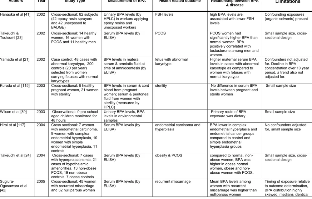
At this time, only a few epidemiological studies have been conducted to investigate the relationship between health related endpoints and BPA exposure (Table 3). Several human studies have focused on identifying sources or levels of BPA exposure. It is clear that additional epidemiological studies are needed to establish relationships between BPA exposure and health outcomes, especially considering the extensive literature that now exist for adverse effects on animals following exposure to low doses of BPA.
Sources and estimates of BPA exposure
Two studies have been conducted to estimate BPA exposure levels in young children. The first involved just 9 children and was designed to examine their potential exposures at home and in daycare [39]. BPA was detected in indoor and outdoor air samples, floor dust and play area soil in both locations at similar levels. BPA was also detected in liquid and solid food at daycare and at home. Based on these environmental levels, the authors estimated that the average BPA exposure level for young children is 42.98 ng/kg per day. A second observational study performed by the same group of investigators examined BPA exposures in 257 preschool children [40]. This study verified that BPA could be found in more than 50% of indoor air, hand wipe, solid food and liquid food samples. This study’s results suggested that 99% of exposures of preschool children originated in the diet; the estimated exposure from dietary sources was 52-74 ng/kg per day, and estimated inhalation exposure was 0.24-0.41 ng/kg per day.
In another study of interest, BPA was measured in the urine of male workers who apply epoxy resins containing bisphenol A diglycidyl ether (BADGE) [41]. Urinary BPA levels were significantly higher in 42 men exposed occupationally than in 42 non-exposed workers.
BPA exposure and human health effects
As stated above, human studies of possible health effects of BPA exposure are extremely limited. BPA levels in blood have been associated with a variety of conditions in women including obesity, endometrial hyperplasia, recurrent miscarriages, abnormal karyotypes and polycystic ovarian syndrome. Two studies found that women with PCOS had higher serum levels of BPA than women without PCOS and that levels of BPA were positively correlated with circulating androgen levels [23,24]. A negative correlation between BPA and FSH was found among men in the study of epoxy resin workers described above [41] however, the epoxy resin workers were also exposed to organic solvents. Due to the cross-sectional design of these studies, it cannot be determined whether BPA increases androgen levels or if androgen levels affect metabolism of BPA. Three studies found higher BPA exposure for health-related outcomes that are
associated with chromosomal abnormalities. One study found higher maternal serum BPA among women carrying fetuses with an abnormal karyotype compared to women carrying fetuses with a normal karyotype [21]. Maternal age, an important potential confounder was not controlled in this study. In another epidemiology study, an association between serum BPA levels and recurrent miscarriage was reported [42]; mean BPA levels were more than three times as high in 45 women with a history of three or more consecutive first-trimester miscarriages compared to 32 nonparous women without fertility problems. Additionally, among 35 women that then became pregnant, there was some evidence of lower BPA among the women who subsequently had a successful pregnancy as compared to those that miscarried again. However, it is important to note that the distribution of exposure among the women with recurrent miscarriage was highly skewed with only a few women with high exposure levels and that the median exposure levels were identical in the two groups. Finally, sister chromatid exchange measured in peripheral lymphocytes was positively associated with urinary BPA levels in adults [43].
Although providing interesting preliminary data on potential health risks, these epidemiology studies have several limitations. Overall, the studies have small sample sizes, limited details on subject selection criteria, and they generally are cross-sectional designs that include limited control for potential confounders. These limitations in design contribute to the limited ability to make conclusions based on the epidemiology of potential health risks of BPA. Finally, due to their design, it was not possible to determine whether altered BPA metabolism is a secondary effect due to the dysfunctions and conditions examined in these studies.
3. Levels of BPA in the environment
Most studies have focused on the potential for BPA exposure from dietary sources. In fact, a significant number of studies have been dedicated to determining BPA levels in foods, especially foods stored in cans with epoxy resin linings. A few other potential sources of BPA exposure, namely drinking water, air and dust, have received far less attention. While several studies have examined BPA leaching from landfills,
additional studies are needed to examine these other potential sources and routes of exposure.
Most of the studies described below conclude with a statement about the low level of BPA leaching from a single studied source. Very few studies have estimated total BPA exposure from multiple sources. Using literature from contamination in the environment (water, air, soil) and food contamination (can surfaces, plastic containers), the daily human intake of BPA was estimated at less than 1 microgram/kg body weight/day [44]. Alternatively, the European Commission’s Scientific Committee on Food [45] estimated BPA exposure to be 0.48-1.6 micrograms/kg body weight/day from food sources, while Thomson et al. estimated that New Zealanders consume as much as 4.8 micrograms/day from dietary sources alone [46].
BPA from plastics, baby bottles & other consumer products
In 1993, Krishnan et al. found that autoclaving cell culture media in polycarbonate flasks led to the release of an unknown estrogenic substance [47]. Using NMR and mass spectrometry, it was determined that the flasks were leaching BPA. At that time, Krishnan and colleagues speculated that these results could impact other scientific experiments using media autoclaved in polycarbonate flasks.
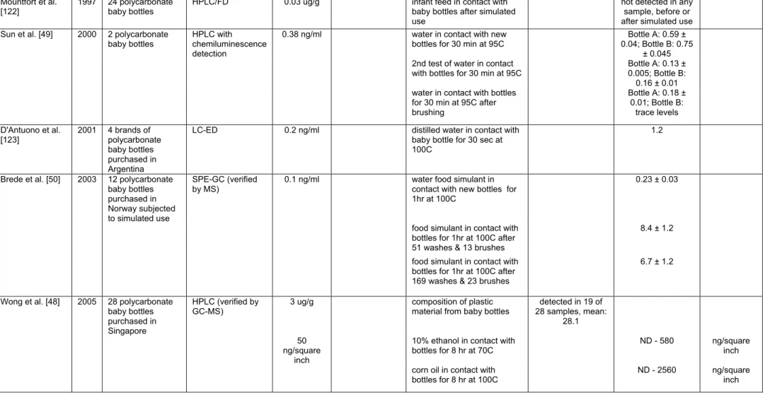
Subsequent studies have examined leaching from polycarbonate baby bottles using a variety of methods including HPLC, LC-ED, and GC-MS (Table 4A). BPA leaching has been observed from polycarbonate baby bottles manufactured in many different countries [48]. Different results have been obtained from various groups studying the effects of washing, boiling, and brushing on BPA leaching. Sun et al. found that BPA leached from polycarbonate bottles, but not glass bottles, on their first use [49]. However, during subsequent use, BPA concentrations were below the LOD. Alternatively, Brede et al. found that rounds in a dishwashing machine, boiling water and brushing led to significantly higher concentrations of BPA leaching into water [50,50]. Based on these measured levels of leaching, average dietary exposure to BPA was estimated for infants from birth through 3 months of age, the period when infants consume exclusively liquid foods [48]; these calculations estimated that newborns, because of their lower body weight, are exposed to the highest levels of BPA (24
micrograms/kg body weight/day). By 3 months of age, dietary exposure estimates drop to 15 micrograms/kg body weight/day.
Other polycarbonate containers (e.g., Tupperware) intended to be used as reusable food containers, have the potential to leach BPA. Many of these containers are marketed for use in the microwave, although heating may increase BPA leaching levels. Nerin et al. examined the composition of a microwavable polycarbonate plastic container [51]. BPA was found in the plastic at a concentration of 30 microgram/g plastic and the potential migration level was estimated at 6.5 microgram/g of food. However, this study only made leaching estimates, and its authors acknowledged that assessments of actual leakage from plastic products are still needed. In another study with potential implications for food safety, BPA levels in plastic stretch film used in food packaging were examined [52]. An examination of 5 polyvinyl chloride stretch films indicated measurable BPA content in 4 samples that ranged from 43 to 483 mg/kg film. The migration of BPA from these products was tested into water, acetic acid (3%) and olive oil. Three of 5 films showed leaching into water and acetic acid, while 4 of 5 leached BPA into olive oil, illustrating the potential for BPA contamination of consumer food products.
Chemical analysis has also been performed on some papers and cardboards used as food containers (Table 4A). BPA is often used as a developer in paper production, so its presence in food-contact papers is not unexpected. In an analysis of twenty different brands of kitchen paper towels (also called kitchen rolls), extracts from paper towels made with virgin paper contained no BPA, with the exception of one brand, with 0.12 mg/kg [53]. In contrast, paper towels made from recycled paper had BPA levels ranging from 0.55-24.1 mg/kg. In a second study examining 28 paper products in food-contact use, 67% of the twelve products made from recycled paper contained BPA at a range of 0.19-26 mg/kg [54]. Of the 16 products made from virgin paper, thirteen contained detectable levels of BPA, albeit at much lower concentrations (range: 0.034-0.36 mg/kg). A final study examined BPA levels in paper and cardboard containers used for take-out food [55]. Forty containers were collected in four European countries and the portion of the container in direct contact with food was analyzed. BPA
than in paper. Collectively, these studies indicate that a wide range of food-contact papers and cardboards serve as potential sources of BPA contamination in foods. However, no studies measured the actual contamination of food items in contact with these papers and cardboards. Additional studies to examine actual leaching rates are still needed.
Leaching of BPA from food cans & containers
Metallic food cans are protected from rusting and corrosion by the application of epoxy resins as inner coatings. Many of these resins are synthesized by the condensation of BPA with epichlorhydrin to create BADGE [2]. When incomplete polymerization occurs, residual BPA may leach from the epoxy resin and has the potential to contaminate stored foods.
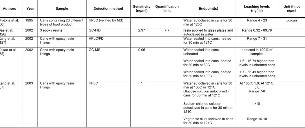
Several studies have documented conditions that support or enhance BPA migration from the coating of cans (Table 4B). These studies have obtained cans from manufacturers and performed carefully controlled studies on the influence of heating time, heating temperature, storage time, storage temperature, and other factors on the level of BPA migration. One of the earliest studies quantified BPA leaching at a range of 4-23 microgram of BPA per can [56]. Kang et al. conducted a comprehensive study and found that heating temperature had a significant effect on BPA migration, to a greater extent than heating time [57]. Vegetable oil and sodium chloride solutions were also found to significantly increase BPA leaching. Takao et al. also found an influence of temperature on the release of BPA from coated cans [58]. While low levels of BPA were detected in water stored in unheated cans, when cans were heated to 100 °C, a normal temperature for the preservation of canned foods, the BPA concentrations in the water increased 1.7-55.4 times (mean: 18.2x) the unheated concentration.
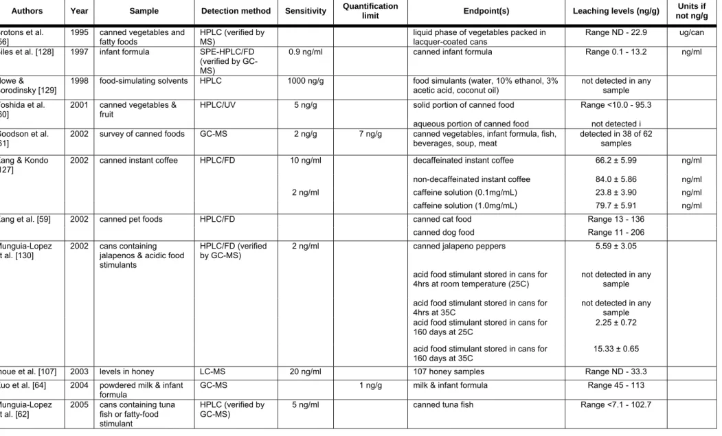
Many studies have also examined BPA levels leaching from epoxy resins lining cans to specific foods (Table 4C). BPA has been detected in canned pet foods [59], vegetables [56,60,61] and fish [61,62]. Others have found BPA contamination in infant formula [63,64]. Thomson et al. used information available from the literature to estimate total dietary estrogen exposures for New Zealand population subgroups [46]. The available literature led the authors to conclude that BPA accounts for approximately
34% of the estrogenic exposure in the New Zealand diet, with estimated intakes of 4.1-4.8 micrograms/day.
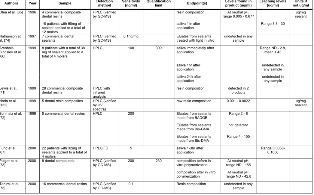
Leaching of BPA from dental products
Several resin-based monomers are used in dentistry as preventative sealants, adhesives and restorative materials. Since the 1960’s, BPA diglycidyl methacrylate has been used as a component of many dental restorative materials. These monomers are typically polymerized in situ to levels of double bond conversion that range from 60 to
80%. Small quantities of unreacted monomers have been shown to leach from polymerized dental materials (see Table 4D) and the potential exists for either residual BPA carried over from the manufacture of these monomers or from biological breakdown of the leached monomers to BPA in vivo.
In a study of 18 adults, Olea et al. applied approximately 50mg total of sealant to 12 molars [65]. Total saliva was collected continuously for one entire hour before and one entire hour after the application procedure. After the treatment, all samples were found to contain variable amounts of BPA, ranging from 3.3 to 30.0 micrograms/ml saliva. Subsequent studies, using different composite applications and saliva collection techniques, have added some controversy to this topic. Arenholt-Bindslev et al. applied 38mg of fissure sealant to 4 molars in 8 volunteers and found detectable levels of BPA in small saliva samples taken immediately after placement of the sealant [66]. However, no BPA was detected in samples collected at 1 hour or 24 hours after sealant application. Fung et al., however, detected BPA in some saliva samples of dental patients collected at 1 and 3 hours after the application of dental materials [67]. The number of detectable saliva samples decreased with sealant dose and the time after application. No BPA was detected in saliva samples collected at 1, 3 or 5 days after treatment, and BPA was not detected in any serum specimens collected at the same time as the saliva samples. Zafra et al. collected saliva samples from 8 patients undergoing dental procedures and found BPA in all specimens [68]. BPA levels ranged from 15.3 to 32.4 ng/ml. Sasaki et al. used an ELISA method to detect BPA in saliva samples from 21 patients treated with one of 9 commercially available dental resins
composite resins; however, gargling was found to remove measurable levels of BPA from subsequent saliva samples.
In a recent study, Joskow et al. examined BPA in urine and saliva of 14 adults treated with one of two different dental sealants [70]. Saliva samples were collected before, immediately after, and 1 hour after sealant application. Urine samples were collected before and at 1 and 24 hours after sealant placement. The total concentrations of BPA were measured by two different isotope dilution-MS-based techniques. Saliva levels were found to be highest immediately following treatment while the highest mean urinary levels were measured 1 hour following sealant application. These highest mean saliva and urine levels were 42.8 and 27.3 ng/ml, respectively, in patients treated with one dental sealant. Levels measured in the saliva and urine of patients treated with the second sealant were 0.54 and 7.26 ng/ml, respectively. These findings indicate that sealants produced by different manufacturers release markedly different amounts of BPA, and further research is needed to identify the sealants that leach the lowest amount of BPA for the shortest periods of time.
Finally, several additional studies have shown significant differences in either the composition or the leaching levels of dental sealants from different manufacturers [66,71,72,73,69,70] while other studies have been unable to detect BPA in either dental sealants or eluates [74,75,76]. Additionally, the storage of saliva samples can affect the detection of BPA [77]. Saliva samples were spiked with BPA, BPA dimethylacrylate (Bis-DMA), or triethylene glycol dimethacrylate (TEGDMA). The samples were stored at –20 °C or –70 °C, and then tested by HPLC and GC-MS (LOD: 1ng/ml). After storage at –20 °C, BPA levels were found to be higher than in the original samples, while Bis-DMA levels were decreased, indicating that this conjugate is unstable and may be deconjugated during storage. However, BPA Bis-DMA and TEGDMA were all stable in salivary samples stored at –70 °C. These results may affect the interpretation of other studies that used sealant products containing Bis-DMA and examined BPA in saliva following sample storage.
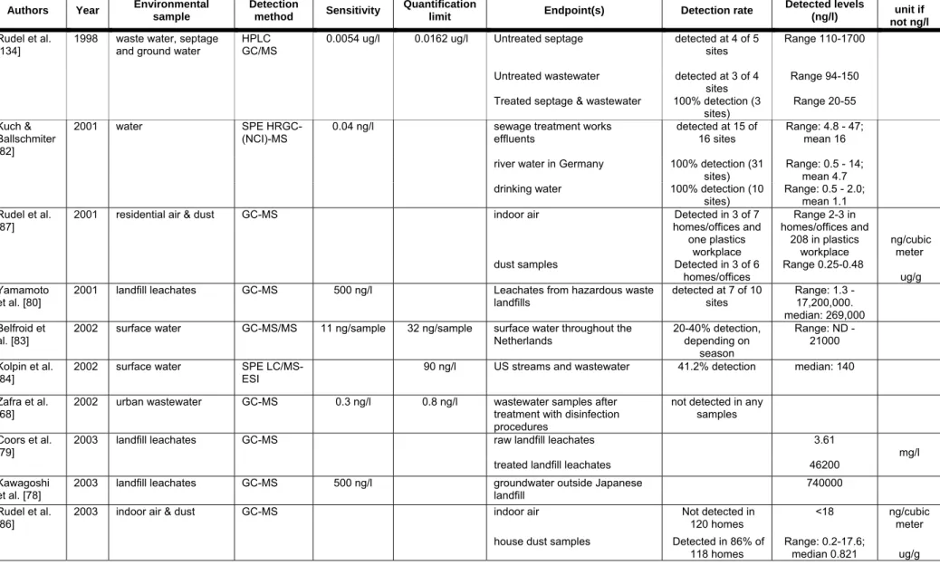
Sewage leachates and water
Several studies have demonstrated that BPA can be detected in landfill leachates (Table 5). Kawagoshi and colleagues used both chemical analysis (GC-MS) and a yeast two-hybrid system to analyze estrogenic compounds leaching into groundwater from a landfill located in Osaka North Port, Japan [78]. Several xenoestrogens and anti-estrogens were detected, but BPA was identified as the greatest contributor to the measured estrogenic activity, with a contribution ratio estimated at 84% and levels detected at 740 ng/ml. In a study of leachates from a landfill in West Germany, the BPA concentration measured from the raw leachate was 3.61 mg/L [79], in the upper range of levels detected in Japan [80]. While treatment of raw leachates using methods similar to those used to care for landfill waste throughout Europe removed 97% of the estrogenic activity, traces of BPA remained [79]. The authors from these studies suggest that BPA degradation from plastic waste buried in the landfill is the primary contributor to these high levels. These findings contrast with the view of plastic products as primarily posing a problem because of their resistance to degradation in contrast with biodegradable materials. The reality is that the leaching of chemicals such as BPA from plastics in landfills has the potential to contribute to contamination of the environment, particularly because such a large volume is produced annually and such a small proportion is recycled [81].
To assess the potential for BPA to reach drinking water, samples from sewage treatment works effluents, rivers, creeks and drinking water reservoirs were collected in Germany [82]. Using an extraction derivation reaction to convert contaminants into their pentaflouorobenzoylate esters followed by GC-MS, Kuch and Ballschmiter achieved an LOD of 20 picograms per liter for BPA. BPA was detected in all river samples in concentrations ranging from 500 pg/L to 16 ng/L; BPA levels in drinking water ranged from 300 pg/L to 2 ng/L. BPA was also detected in surface water in 96 samples collected from 38 different locations distributed equally throughout the Netherlands [83]. Twenty percent of samples collected showed detectable levels of BPA (LOD: 11 ng/L) and nine locations had levels over 100 ng/L. Another comprehensive study of wastewater contaminants found that BPA was detectable in 41.2% of 139 streams
sampled across 30 US states [84]. This study found a median level of detection of 0.14 micrograms per liter, and a maximum measure of 12 micrograms per liter.
Air & dust
Air and dust levels of BPA serve as another potential source for human BPA exposure (Table 5). Because of the large amounts of BPA produced annually, it is plausible that BPA enters air particles during production at plastics manufacturing plants. It has been speculated that the presence of BPA in other environmental samples (water, soil, etc.) could lead to its vaporization, despite its low vapor pressure, allowing it to be adsorbed into the core portion of airborne particles [85].
In a survey of 120 homes for the presence of endocrine disrupting chemicals, Rudel et al. found BPA present in 86% of house dust samples at concentrations ranging from 0.2-17.6 micrograms/gram [86]. Another study from the same group found BPA in 3 of 6 residential and office dust samples [87]. BPA was also detected in air samples, including a sample from a plastics workplace (208 ng/m3). An additional study measured BPA levels in urban ambient outdoor air particulates in Osaka, Japan [85]. BPA was detected in air samples with an average level of 0.51 ng/m3. This study also found mild seasonal variation in BPA levels, with increasing levels from autumn to winter and decreasing levels from winter to spring.
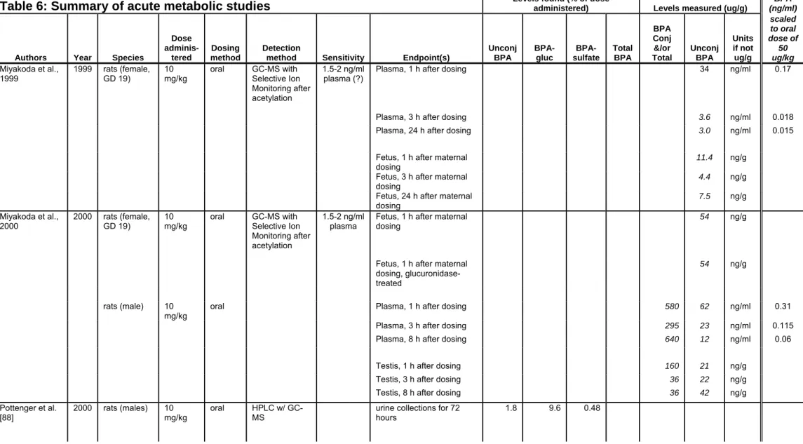
4. BPA metabolism in humans & animals
The metabolic elimination pathways for BPA need to be considered for human risk assessment. However, only a limited number of human studies have addressed these issues for several reasons, including ethical considerations and difficulties in identifying individuals that are completely unexposed to BPA from the environment [31,70]. In contrast, many studies have been dedicated to addressing the question of BPA metabolism in animal models, particularly rodents (Table 6). However, a major weakness to current metabolic studies is that, while current evidence indicates that humans are experiencing multiple exposures each day, virtually all of the current metabolic studies are based on kinetics following a single, usually high dose. A clear research need is pharmacokinetic studies that involve multiple exposures to BPA to
more accurately reflect typical human exposures as supported by the substantial literature of exposures from multiple sources that have been detailed in prior sections of this review. The conclusion reached by some investigators based on acute metabolic studies is that human exposure should essentially be non-existent [88,89]. However, these conclusions are contradicted by the extensive measurements of parent, unconjugated BPA in blood and tissues at ng/ml levels (see Table 1), which would be impossible according to these conclusions.
With regard to measurable background levels of BPA, there are many other estrogenic environmental contaminants as well as contaminants with other modes of activity that are present in most people examined [31,32]. In addition to BPA, humans are thus exposed to at least dozens of other chemicals that show estrogenic activity, and the likelihood of at least additive effects in humans by other estrogenic endocrine disrupting chemicals is currently not taken into account in regulating human exposure levels to these chemicals.
While oral, dietary exposure is currently considered a major route of human exposure to BPA, the wide range of sources of human exposure detailed in Tables 4 and 5 document the additional importance of exposures that avoid the first-pass hepatic metabolism following oral exposure. Specifically, animal studies involving subcutaneous exposure by injection and by osmotic pump are relevant to human exposures by dermal contact with air, dust and water. Intravenous and intraperitoneal exposure in animals are relevant to inhalation exposure to BPA carried by airborne dust, which has direct access to the systemic circulation. In addition, both of these routes are relevant to human exposures through intravenous medical tubing and exposure to implanted plastics used in surgery.
Another critical issue is that it is well known that the fetus and neonate show very limited first-pass metabolic capability for BPA and other endocrine disruptors [90], and the pharmacokinetics of BPA based on adult oral exposure can not be used to predict pharmacokinetics in the fetus, neonate or child; the maxim in pediatric medicine that “children are not little adults” is relevant to this issue. Given the ppb levels of parent BPA reported to be present in human blood and tissues, it cannot be assumed that
exposure. Accounting for all sources of BPA in human blood is an important research need.
Animal models of BPA pharmacokinetics and relation to circulating levels of free, unconjugated (aglycone), biologically active BPA
The routes by which adult animals are exposed to BPA affect the resulting circulating levels. Studies have used oral gavage, spiked water, intravenous and intraperitoneal injections, slow release capsules, and osmotic pumps, and results of many of these studies are detailed in Table 6, particularly in reference to levels of free, unconjugated BPA in circulation. As noted above, BPA may be absorbed by transdermal exposure by bathing in BPA-contaminated water, or by exposure via inhalation, and both routes avoid the first-pass conjugation that occurs with oral administration. Metabolism of BPA converts a majority of the parent compound to BPA glucuronide(s) and BPA sulfate(s), the levels of which are reported in many studies.
The estrogenic activity of the BPA conjugates has been reported as very low to none [91,88,92], and the active molecules are limited to unconjugated aglycones. The possibility that conjugates may be deconjugated locally in tissues to release biologically active BPA is an interesting hypothesis; to date there is no published information indicating that this is occurring. Because the parent unconjugated BPA is the only form shown to be biologically active and the published measures of human circulating BPA are solely of the unconjugated, bioactive form (Table 1), this review of the pharmacokinetics of BPA will focus particularly on circulating levels of the parent form in animal studies for comparison to the human circulating BPA levels.
A major portion of the animal literature on low-dose effects has used oral administration of low-dose BPA. This subset of the published animal response studies (reviewed in the In Vivo Panel Report) will be compared to the adult animal metabolic studies of oral exposure at higher doses. This allows for estimates of the circulating levels of parent, unconjugated BPA in animals that are showing adverse effects in low-dose in vivo studies, which has not been measured directly in any study of oral
pharmacokinetics. The estimate of the ranges of circulating levels of BPA that are active in low-dose animal studies will be compared to current measurements of circulating
levels of parent, unconjugated BPA that have been measured in human blood and tissues (Table 1), and to the concentrations of BPA that are active in human and animal cell culture studies in vitro (reviewed in the In Vitro Panel Report).
Direct rodent studies of metabolism of BPA administered orally in the low-dose range
A substantial proportion of the literature on low-dose effects has used oral exposure. Unfortunately, very few studies have measured BPA in the blood of animals treated with low doses of BPA (< 5 mg/kg bw), and none have measured after serial oral doses.
In the published study most relevant to low-dose developmental effects observed in rodents, tritiated BPA of high specific activity was orally administered to gestational day 17 pregnant mice at 25 μg/kg [17]. While unconjugated BPA was not measured after oral dosing in this study, the total radioactivity present in blood was measured at 0.027 ng BPA equivalents/g at 24 h after oral dosing, the only time point measured in the study [17]. Since unconjugated BPA is only a fraction of the total metabolites circulating after administration, the circulating level of unconjugated BPA in the study would be below the measured value of total BPA-derived radioactivity. In a second published study of oral low-dose pharmacokinetics [93], male rats were dosed orally with 500 μg BPA/kg body weight and free (unconjugated) BPA was calculated in blood at approximately 0.8, 0.3 and 0.03 ng/ml at 15 min, 6 h and 24 h after dosing, respectively (Table 6). Only total radioactivity (comprised mostly of conjugated BPA) in blood after oral dosing was reported in the same study at lower doses of BPA [93] or in other studies at low oral doses [94,95,17] including in pregnant animals. The median human level of unconjugated BPA (~2 ng/ml) was above the levels of unconjugated BPA in low-dose exposed rodents.
BPA is thought to bind to plasma proteins in rodents, monkeys and humans (reviewed in [89]). Because pharmacokinetics are altered by protein binding, the potential uptake of BPA into other tissues, including estrogen-target tissues, may be affected. This is a topic that requires additional study to properly address its implications for risk assessment purposes [96].
Processes used to estimate the range of circulating BPA in rodents in response to different doses of BPA, and comparison to median human exposure levels
While not available directly in any one study, existing published data can be used to estimate the circulating level of BPA in animals responding to low doses of BPA, and these estimated levels can be compared to current human circulating levels. This can be derived by addressing the following issues linking the oral low-dose exposure studies in animals, reports of the BPA pharmacokinetics in animals at different doses, and the reported human circulating levels of BPA. This process involves the following published conclusions: the importance of route of exposure (oral route selected), the form of BPA in circulating in blood (unconjugated, biologically active BPA), the reported proportionality of circulating level with dose across a wide range of doses, similar pharmacokinetics in nonpregnant and pregnant adults [97], only slight increases in circulating BPA following one exposure compared to multiple exposures [98], and rodent pharmacokinetics compared to pharmacokinetics in humans. These published conclusions link over 40 animal studies of adverse effects at oral doses below the reference dose for BPA, 11 studies of BPA pharmacokinetics following oral dosing, 9 reports of circulating BPA levels in pregnant and nonpregnant women, and 19 reports of effects BPA at or below 10 nM (2.3 ng/ml) on human and animal cell function in mechanistic studies in vitro (see In Vitro panel report).
As indicated above, the USEPA reference dose for BPA is currently 50 μg/kg/d. As detailed in the report from the In Vivo panel, there are over 40 studies reporting effects at or below this RfD. However, data are very limited regarding blood or tissue levels at or below the reference dose. To estimate these circulating levels for comparison to current human exposure (Table 1), the following steps were used to estimate the range of blood levels that would occur if a 50 µg/kg dose were administered to rodents: of the 21 acute metabolic studies (Table 6) in which BPA was administered to rodents, 17 contained data on blood levels of BPA and metabolites after oral administration, and of these 17 studies, 11 contained measurements of unconjugated BPA, which is the form measured in blood in human studies. Also, as indicated previously, only unconjugated BPA is biologically active. We thus used data from these 11 studies in this analysis to describe the pharmacokinetics of BPA after oral
administration to adult rodents (pregnant females, non-pregnant adult females and adult males).
There are several bases for the following analysis. Because all but one of the metabolism studies were performed at doses higher than 50 μg/kg, this raised the question of whether it is valid to use the high dose studies to estimate blood levels that would occur after administration of the RfD. For this analysis to be valid, it was necessary to determine whether there was proportionality of circulating level with administered dose. This is in fact supported by the conclusions of several studies [88,99,100,93,101] using an oral route of exposure, which is why only data from this route of exposure was used in this analysis.
We then used the data from all 11 studies at a number of different doses, and linearly scaled the reported results to a single administered dose of 50 μg/kg. For example, circulating levels reported after dosing at 500 μg/kg were divided by 10, while circulating levels after dosing at 10 mg/kg were divided by 200, in order to scale the reported data to 50 μg/kg. The results of this scaling are shown in the last column of Table 6. The complete set of 18 data sets from all 11 studies are graphed in Figure 1. The data are presented as a log-log plot, which allows data spanning a wide range to be displayed on a single graph. In addition the time-courses were approximately linear in the log-log plot. Even though there were differences in the values reported in these 11 studies with regard to measured unconjugated BPA in blood, in no case did any data point from these 11 studies reach the median human level of unconjugated BPA.
Subsets of the data shown in Figure 1 are presented in Figure 2 to address two issues. One is the validity of scaling circulating levels from different doses to one reference dose, specifically, the impact of the administered dose on the data obtained after the scaling procedure (Fig. 2 A, B and C); publications report proportionality with dose where encountered [88,99,100,93,101]. The second is variability due to the type of animal (pregnant female, non-pregnant adult female or adult male) used in the study (Fig. 2 E, F and G) to address pooling the small set of pregnant animal data with the larger set of nonpregnant animal data; the conclusion of at least one report is that the pharmacokinetics do not vary between nonpregnant and pregnant rodents [100].
Figure 2 Panel A shows the results from scaling data to 50 μg/kg across the extremes of the complete data set for administered dose: from 1 g/kg, 0.5 mg/kg and 25
μg/kg (represented as a single point), and the scaled profiles were quite similar, with all points close to the linear regression line (dark black line in the figure) of all data from all studies. Further, in Panel B, a plot of all the data for 100 mg/kg administered dose, and Panel C, the data for 10 mg/kg administered dose, there was again no trend that contradicted the assumption of proportionality based on this analysis. Taken together, the data in Panels A, B and C support proportionality of circulating unconjugated BPA based on administration of high doses down to the RfD.
The second issue of animal type was important because many of the in vivo
animal studies involve administration of BPA to pregnant female rodents, and there are a number of biomonitoring studies that have addressed the blood levels of unconjugated BPA in pregnant and nonpregnant women. However, there are only a limited number of metabolism studies that involved pregnant rodents. The data in Panel D from studies with pregnant rodents were within the range of the data from non-pregnant females (Panel E) and adult males (Panel F). Thus, the scaling procedure did not appear to show a bias based on the type of animal used in the study. As indicated previously, this finding is consistent with the conclusion of Domoradzki et al. [100] that BPA metabolism does not differ significantly between pregnant and non-pregnant females.
The data in Figure 1 and 2 support scaling and combining metabolism data across a wide range of doses and species to estimate circulating levels of BPA in rodents when administered doses within the "low dose" range that cause adverse effects. Specifically, from the combined data in Figure 1, at 1 hr after oral BPA administration, the blood levels of unconjugated BPA ranged from 0.003 - 0.3 ng/ml. At 24 h the values ranged from 0.002 - 0.06 ng/ml (Table 6). Peak levels of BPA achieved in the first 30 min after oral administration ranged from 0.01 to 1.14 ng/ml. Median values across the studies were 0.11 ng/ml at 0-30 min, 0.047 ng/ml at 1 h, and 0.007 ng/ml at 24 h.
There are two main conclusions from these findings. The first is that many adverse effects that have been reported in animals at or below the RfD (See In Vivo
panel report) occur in animals at circulating levels of unconjugated BPA below median current human exposure levels (~1-3 ng/ml). Second, unless humans metabolize BPA much more slowly than animals, human exposure to BPA would have to exceed the reference dose of 50 µg/kg/day. In fact, it has been reported that the metabolism and clearance of BPA is more rapid in humans than in rodents [89], suggesting that human exposure to BPA is substantially higher than the RfD based on a comparison to blood levels achieved in rodents at all time points after BPA exposure scaled to the reference dose of 50 μg/kg/day. Given an assumption of equivalent pharmacokinetics in humans and rodents, at 1 hour after administration of 50 μg/kg, rodent blood levels are over 10-fold below median human blood levels, and to achieve these levels human would have to be exposed to a dose greater than 500 μg/kg. If human metabolism and clearance is more rapid than rodent clearance, which is concluded by studies which have addressed the issue [102,103,89], then the human exposure to achieve the current human circulating levels would have to be well above 500 μg/kg/day (well above 32 mg/day/adult considering a 65 kg human). This is consistent with the observation of Shin at al. [98] that in their pharmacokinetic models, an oral intake of 100 mg BPA/day would explain the mean human circulating level of 1.49 ng/ml reported by Takeuchi & Tsutsumi [23]. Therefore, these models indicate that i) humans are exposed to BPA at a much higher level than has been estimated from known exposure sources, and/or ii) Humans are exposed through multiple routes, making the metabolic response different from that observed in animal models, and/or iii) metabolism of BPA following chronic, low-dose exposure is not predicted by the acute high-dose studies used to generate the current pharmacokinetic models. Finally, while many responses have been observed in human and animal cells at and below concentrations of 1 nM (0.23 ng/ml) (see In Vitro Panel Report) median human blood levels of unconjugated BPA are clearly higher. It is thus completely plausible that at current human exposure levels, BPA is impacting cell and organ function in humans (see In Vitro and In Vivo panel reports).
Comparisons of human exposure levels & animal studies
Kurebayashi, 2005). These levels are thus lower than concentrations that have been measured in human blood (Table 1). Collectively, these data indicate that the levels being studied in animals which lead to biological effects are relevant to current human exposure levels; current human exposures are higher than the levels in animals responding to BPA. Because few comprehensive studies have focused on human metabolism of BPA, and differences in pharmacokinetics are suspected between species, additional research in this area is needed.
One additional area of research that has remained largely unexplored is the potential differences in BPA metabolism between different groups of people. Several animal studies have indicated strain differences in rats and mice with regard to BPA metabolism. While some human studies have examined polymorphisms for enzymes involved in BPA metabolism [104,43], studies using larger and more widespread populations are needed.
Animal models of BPA metabolism- digestion & excretion
Because BPA is suspected to enter the human body mainly through the oral route, several studies have examined the absorption and metabolism of BPA in the intestine and liver. One comprehensive study compares the metabolism and excretion of BPA in rats dosed with 0.10 mg radiolabelled BPA/kg body weight either by oral or intravenous (i.v.) exposure [95,95]. This relatively low dose was chosen because previous studies used oral doses of 100 mg/kg or more, levels thought to saturate the metabolic and excretory mechanisms responsible for the elimination of BPA from the body. With this lower dose, the i.v. and oral dosing led to a urinary excretion of 8.4 and 6.3% of the radioactivity, respectively, within 24 hours of treatment. Fecal excretion from the i.v. and oral dosing was 77.6 and 81.6% of the administered dose, respectively. Collectively, Kurebayashi and colleagues concluded that there are similar metabolic kinetics in these two modes of exposure, and that fecal excretion is the main route of BPA elimination in the rat.
Several studies have determined that the liver plays an essential role in metabolizing BPA in vivo in animal models. Glucuronidation is a metabolic pathway in
BPA-glucuronide has been shown by many to be the major BPA metabolite in animals and humans and has little or no estrogenic activity in several in vitro assays. Yokota et al.
identified and examined UGT2B1, a liver enzyme responsible for glucuronidation of BPA and other xenoestrogens [105]. Interestingly, a study of rat liver S9 fractions, containing both microsomal and cytosolic fractions, indicates that the liver may also produce a BPA metabolite with increased estrogenic activity [106]. However, the authors of this study acknowledge that this metabolic pathway is probably not significant under normal circumstances, and is likely only active when glucuronidation is efficient.
An additional study used segmented everted rat intestine to measure transport and conjugation of BPA in each portion of the intestine [107]. Addition of BPA to the mucosal side of the intestine led to absorption and transport to the serosal side; there were no significant differences in this transport among the five portions of the intestine. However, the appearance of BPA on the serosal side was accelerated by the treatment with a high dose (100 micromolar). This study also examined glucuronidation of BPA by each segment of the rat intestine. Following BPA administration, BPA-glucuronide was expelled into the mucosal side and transported to the serosal side of the intestine; the level increased with the incubation time. Interestingly, in the small intestine, the greatest amount of BPA-glucuronide was secreted into the mucosal side, but in the colon, secretion was greatest to the serosal side. The authors therefore suggested that while the proximal intestine may protect against the absorption of BPA in rats, the colon may be more susceptible to BPA transport. The authors also proposed the possibility that BPA-glucuronide secreted into the mucosal side of the proximal intestine could be deconjugated by glucuronidases produced by bacteria in the colon. This BPA would then be free and could be reabsorbed [108,107]. These authors also suggest that the effects of BPA may be enhanced by repeated, continuous exposure [108].
Animal models of BPA metabolism- transfer to the developing fetus
The metabolic changes associated with pregnancy could cause alterations in the metabolism and excretion of BPA from both pregnant animals and women. Takahashi and Oishi examined oral administration of 1 g BPA/kg to pregnant rats on day 18 of
microgram/g), reached a peak concentration at 20 minutes after dosing (14.7 microgram/g) and gradually decreased over a period of 10 hours. BPA was also detected in fetuses within 10 minutes of dosing (2.00 microgram/g); a maximum concentration was reached at 20 minutes (9.22 microgram/g) and levels gradually decreased with time. The concentration after 6 hours was 5% of the level detected at maximum. This study illustrated that absorption of BPA by both the pregnant mother and the fetus in this model was rapid and the placenta did not block BPA transmission. An additional study of mice and Japanese monkeys dosed with 100 mg/kg BPA during pregnancy showed that BPA could be detected in several fetal tissues, including serum, liver, brain, uterus and testes within 30 minutes (mice) and 1 hour (monkeys) of treatment [110].
Zalko et al. demonstrated in a mouse model that much lower doses (25 microgram/kg) of BPA were also able to cross the placental barrier [17]. Twenty-four hours after BPA administration, fetuses accounted for 4% of the administered radioactivity, with an average of 3.7 ng/g. The placenta maintained 0.55% of the administered BPA (3.14 ng/g) and the amniotic fluid contained 0.34% (4.85 ng/ml).
Human metabolism of BPA- acute exposure studies
Only a small number of studies have attempted to determine the pharmacokinetics of BPA metabolism in human subjects (Table 6). Volkel and colleagues administered 5 mg radioactive BPA/person (54-90 micrograms/kg body weight) and report that elimination of BPA was complete within 24 hours of dosing [102,102]. Maximal plasma concentrations were reached 80 minutes after dosing and rapidly declined for the next 6 hours. BPA was detected only in its glucuronidated form, and not as free BPA. The results of this study indicated that in the human, BPA was absorbed from the gastrointestinal tract quickly, conjugated with glucuronic acid in the liver, and BPA-glucuronide was rapidly filtered from the blood by the kidneys and excreted in urine. This metabolic pathway differed from that of the rat, where a large amount of BPA-glucuronide is transported into bile and enters the digestive system [105,105].
In another metabolic study, BPA was administered (25 micrograms/person) and then free BPA and BPA conjugates were measured in urine and blood by isotope dilution LC-MS; LODs were 1.14 ng/ml (BPA) and 10.1 ng/ml (BPA-gluc) [103]. In the three men examined, 85% of the applied BPA dose was recovered in urine after 5 hours, mostly as BPA-glucuronide. In the three women examined, 75% of BPA was recovered as BPA-glucuronide after the same period of time, indicating the potential for some gender differences in BPA absorption, metabolism and/or excretion, as suggested by other studies [33,31,31,33]. In two of six individuals, free BPA was detected in the urine at levels of approximately 1 ng/ml; free BPA was not detected in the urine of the other 4 individuals [103], although this study was limited by its small numbers of subjects and relatively poor sensitivity. The levels of BPA in blood samples following this acute exposure were not reported in this study.
Some authors have suggested that human microsomes may not be able to glucuronidate BPA as extensively as rat microsomes, making the metabolic kinetics different for the human compared to other mammals [111]. Alternatively, Pritchett et al. predict that when metabolic levels measured in isolated hepatocytes are extrapolated to the entire liver, the hepatic capacity for BPA glucuronidation is higher in humans than in mice or rats [112]. Additional studies are needed to rectify these theories. In the studies of Yoshihara and colleagues discussed above, rat liver extracts were found to produce a BPA metabolite with increased estrogenic activity [106]. Interestingly, this metabolite was also produced in vitro by mouse, monkey and human liver S9 fractions, suggesting
that some aspects of BPA metabolism may be conserved across mammalian species. Together, data from these studies and others are being used to generate models for BPA kinetics following intravenous and oral route exposures [89]. These models indicate that BPA metabolism may be different in rats and humans, including endpoints such as BPA clearance rates, intestinal glucuronidation, and excretion rates. Additional studies are needed to validate these models or produce new ones. However, as already noted, these models are based on acute, single exposure kinetics instead of the chronic exposures that are most relevant to humans exposed environmentally.
Dozens of studies have been dedicated to monitoring levels of BPA in human tissues, blood, urine, and other fluids; extensive evidence exists to demonstrate that most humans are exposed to BPA. Unconjugated BPA has been measured repeatedly in human blood (serum and plasma), breast milk, amniotic fluid, and placental tissue in the low ng/ml or ng/g range using various analytical techniques. Additionally, BPA conjugates have been repeatedly found in the low ng/ml range in the urine of over 90% of individuals tested in several countries and continents. Of particular concern are the levels that have been detected in the blood of pregnant women, fetal blood, umbilical cords, placenta and amniotic fluid. Because the developing fetus is acutely sensitive to hormones and chemical exposures, the levels detected are a cause for concern.
It has been proposed that xenoestrogens such as BPA could play a role in reproductive cancers (testicular, prostate, breast, uterine, ovarian, etc.), fertility problems (low sperm count, decreased sperm quality), and other endocrine related endpoints. At this time, only a few small studies have explored the associations between BPA levels and human health issues. However, these limited data indicate that additional studies are warranted on human health and BPA exposure. Currently, there is limited evidence to suggest that BPA levels vary between men and women and/or with several endocrine-related syndromes and diseases, including polycystic ovarian syndrome and obesity, which are brought about in animals by exposure to low doses of BPA.
There is extensive evidence that many consumer products contain and release BPA. BPA content has been measured in food containers, epoxy resins, plastics, baby bottles, and dental sealants, and leaching rates have been measured from many of these products under normal conditions of use. BPA has been detected in a wide range of foods stored in cans with epoxy resins. Additionally, BPA has been measured in freshwater, seawater, landfill leachates, air, and dust particles. Collectively, these studies indicate that exposure to BPA is widespread, from many different sources in the environment. There are several studies that have generated estimates of current exposure from leaching levels of consumer products. These studies have estimated that human exposure ranges from under 1 microgram/kg/day to almost 5 micrograms/kg/day (0.325 mg/day/adult). However, pharmacokinetic modeling data suggest that oral
intakes up to 100 mg/day/adult would be required to explain the reported human circulating levels. Additional studies and mathematical models of potential exposures are needed, particularly because many sources of BPA exposure have been identified.
The consistent finding that BPA is detected in almost all individuals in developed nations implies that humans are exposed to BPA continuously. Because of the rapid metabolic clearance of BPA, and the measurable levels of BPA that have been detected in human blood and urine, Welshons and colleagues have identified two potential issues: 1) BPA intake may be actually much higher than has been suggested, and/or 2) long-term, daily intake leads to bioaccumulation of BPA, leading to steady-state levels that are not represented by any of the current models for BPA metabolism based on single, acute administration (Welshons, 2006).
The levels of BPA measured in human serum, urine and other tissues are within the range shown to cause effects in laboratory animals, and impact cell function in mechanistic studies in cell culture. Therefore, it is plausible and even likely that these levels are biologically active in humans, with obvious potential to cause disease or dysfunction. This review has highlighted several areas of research that must be addressed to answer additional questions that have been posed.
Conclusions and Levels of Confidence for Different Outcomes
A. Based on available evidence, we are confident of the following:
BPA levels in human tissues and fluid
Human studies have shown that most children, as well as adult men and women, including pregnant women, have measurable levels of BPA in body fluids and tissues sampled. Unconjugated BPA has been measured repeatedly in human blood (serum and plasma) with a central measure of the distribution in the 0.3 to 4.4 ng/ml range (1 to 19.4 nM), and in breast milk, amniotic fluid, and placental tissue in the low ng/ml or ng/g range. The measurements of BPA in maternal serum, fetal serum, umbilical cord blood, amniotic fluid and placenta indicate that the developing human fetus may be exposed to BPA in the 1 to 3 ng/ml range (4 to 13 nM). The ng/ml levels in human serum are
is at or below 0.5 ng/ml. Studies using mass spectrometry detection methods are considered highly reliable, while there is considerably less confidence in studies employing ELISA.
Conjugates of BPA in urine are measured in the low ng/ml range, and are repeatedly found in over 90% of individuals tested (8 of 13 cited publications), including a study of a reference adult population.
Sources of BPA in the environment
There is extensive evidence that many consumer products contain and release BPA. There is also extensive evidence that many of these products leach BPA under normal conditions of use. BPA has been detected in baby bottles, epoxy resins, and other consumer plastics. BPA has also been detected in a wide range of foods stored in cans with epoxy resins. There is very good evidence to indicate that BPA can be detected in environmental samples, including air, dust and water. Evidence for this is supported by studies of landfill leachates which indicate substantial release of BPA from landfills.
BPA metabolism in humans & animals
There is extensive evidence for the kinetics of BPA metabolism in rodent models following acute exposures to relatively high doses. Acute studies in both animals and humans indicate rapid metabolism and clearance. BPA can be detected in the blood shortly after treatment, and in collected urine and feces. However, acute studies do not reflect the situation in humans, where exposure is more likely chronic and low-level. Therefore, additional studies of chronic, low-level exposure to BPA are needed in both animal models and human subjects.
B. Based on the available evidence, we consider the following to be likely but requiring confirmation: