Magalhães, P., Schanstra, J. P., Carrick, E., Mischak, H. and Zürbig, P.
(2016) Urinary biomarkers for renal tract malformations. Expert Review of
Proteomics, 13(12), pp. 1121-1129. (doi:10.1080/14789450.2016.1254555).
This is the author’s final accepted version.
There may be differences between this version and the published version.
You are advised to consult the publisher’s version if you wish to cite from
it.
http://eprints.gla.ac.uk/183853/
Deposited on: 11 April 2019
Enlighten – Research publications by members of the University of Glasgow
Full Terms & Conditions of access and use can be found at
http://www.tandfonline.com/action/journalInformation?journalCode=ieru20
Download by: [Petra Zürbig] Date: 02 November 2016, At: 01:54
Expert Review of Proteomics
ISSN: 1478-9450 (Print) 1744-8387 (Online) Journal homepage: http://www.tandfonline.com/loi/ieru20
Urinary biomarkers for renal tract malformations
Pedro Magalhães, Joost P. Schanstra, Emma Carrick, Harald Mischak & Petra
Zürbig
To cite this article: Pedro Magalhães, Joost P. Schanstra, Emma Carrick, Harald Mischak & Petra Zürbig (2016): Urinary biomarkers for renal tract malformations, Expert Review of Proteomics, DOI: 10.1080/14789450.2016.1254555
To link to this article: http://dx.doi.org/10.1080/14789450.2016.1254555
Accepted author version posted online: 28 Oct 2016.
Submit your article to this journal
Article views: 3
View related articles
Publisher:
Taylor & Francis
Journal:
Expert Review of Proteomics
DOI:
10.1080/14789450.2016.1254555
Review
Title: Urinary biomarkers for renal tract malformations
Authors: Pedro Magalhães1,2, Joost P. Schanstra3,4, Emma Carrick5, Harald Mischak1,5 and
Petra Zürbig1
Author affiliations
1Mosaiques Diagnostics GmbH, Hannover, Germany 2Hannover Medical School, Hannover, Germany
3Institut National de la Santé et de la Recherche Médicale (INSERM), U1048, Institute of
Cardiovascular and Metabolic Disease, Toulouse, France
4Université Toulouse III Paul-Sabatier, Toulouse, France
5Institute of Cardiovascular and Medical Sciences, University of Glasgow, UK
*Corresponding author
Petra Zürbig
Mosaiques diagnostics GmbH, Rotenburger Str. 20, 30659 Hannover, Germany Phone: +49 511 554744 15
Email: zuerbig@mosaiques-diagnostics.com
Abstract
Introduction: Renal tract malformations (RTM) are congenital anomalies of the kidneys and urinary tract, which are the major cause of end stage renal disease in children. Using
immunoassay-based approaches (ELISA, western blot), individual urinary proteins including transforming growth factor β, tumor necrosis factor and monocyte attractant proteins 1 were found to be associated to RTM. However, only mass spectrometry (MS) based methods leading to the identification of panels of protein-based markers composed of fragments of the extracellular matrix allowed the prediction of progression of RTM and its complications.
Areas covered: In this review, we summarized relevant studies identified in “Pubmed” using the keywords “urinary biomarkers” and “proteomics” and “renal tract malformations” or “hydronephrosis” or “ureteropelvic junction obstruction” “posterior urethral valves or “vesicoureteral reflux”. These publications represent studies on potential protein-based biomarkers, either individually or combined in panels, of RTM in human and animal models.
Expert commentary: Successful use in the clinic of these protein-based biomarkers will need to involve larger scale studies to reach sufficient power. Improved performance will potentially come from combining immunoassay- and MS-based markers.
Keywords: proteomics; children; urine; biomarkers; renal tract malformation
1.Introduction
Renal tract malformations (RTM) are congenital anomalies of the kidneys and/or lower urinary tract. Severe RTM is life-threatening and only a limited number of RTM can be successfully (surgically) corrected, e.g. ureteropelvic junction obstruction (UPJO) and to a lesser extent posterior urethral valves (PUV). Severe, not surgically correctable, RTM will
lead in many cases to end stage renal disease (ESRD) (1;2). Hence, key issues in the clinical management of RTM, which is often detected in utero, are the prediction of disease progression and management of its complications.
Hippocrates linked health problems to diagnostic changes in the urine two millennia ago. Presently, measurement of urinary components such as proteins to test for the presence of kidney disease, using dipsticks and standard laboratory tests, is routine in diagnostics. However, these tests lack sensitivity and consequently detect kidney disease often at a late stage since there has to be significant kidney damage before frank proteinuria occurs. Therefore, identification of earlier and prognostic markers using, for example proteomics techniques, would be desirable. Indeed the use of proteome analysis for the discovery of clinically relevant proteins, known as “clinical proteomics”, is perceived to be the most relevant discipline to foster the translation of basic discoveries into clinical applications for the benefit of the patient (3). Clinical proteomics needs to adhere to strict guidelines to generate clinically useful biomarkers of disease. Therefore, Mischak et al. asserted that successful clinical proteome studies should in general use stringent statistical approaches for biomarker definition and that results should always be confirmed in independent test sets (4). In addition, they described a brief set of practical and feasible recommendations for
investigators to properly identify and qualify proteomic (or any other) biomarkers, which could also be used as reporting requirements. Such recommendations should help put proteomic biomarker discovery on solid ground.
2. Urine as a source of biomarkers of RTM
Easily accessible biological samples, such as plasma, serum or urine are valuable sources of biomarkers. Particularly urine which can be collected in large quantities and in non-invasive fashion and is less complex than for example plasma (5;6). Under physiological conditions, 70% of urinary proteins are produced by kidney and urinary tract and hence urine is a potential source of biomarkers for RTM. However, urine also allows to obtain information related to various other organs because of the glomerular filtration of blood (7). The urinary
proteome is relatively stable, because it is stored for hours in the bladder at 37°C and any proteolysis has been completed at time of voiding (8). Therefore, the urinary proteome does not change significantly and urine can be stored for 6 h at room temperature or 3 days at 4ºC as well as for several years at -20°C (9). In addition to full length proteins, urine also contains many naturally occurring low molecular weight proteins (peptides), which can be directly analysed by mass spectrometry, without additional sample processing, such as tryptic digestion and the depletion of abundant blood-derived proteins (10;11). Variability in the protein/peptides concentration is the main disadvantage of urine. These variations are caused by the differences of the protein quantity during the day, circadian rhythms, exercise, diet and metabolic or catabolic processes (10;12). However, this can be corrected by using normalization methods (13).
3. Proteome analysis-based approaches for the identification and validation of biomarkers
The main goal of clinical proteomics is the identification of biomarkers, providing information to improve and personalize medicine (14). Since, by definition, the proteome is complex, its analysis needs in general two steps: firstly, fractionation of the proteins into smaller less complex subsets followed by, secondly, analysis of the protein abundance and protein identity by mass spectrometry (MS) of those fractions. Fractionation is/has been often performed by chromatographic techniques including two-dimensional gel electrophoresis (2-DE), liquid chromatography (LC) and capillary electrophoresis (CE). The protein fractions are then analysed off- or on-line by MS. 2-DE, cumbersome and low-throughput, has now mostly been abandoned. The advantages and disadvantages of these proteomics techniques have been extensively described in several reviews (10;15;16) and will not be discussed here. Enzyme-linked immunosorbent assays (ELISA) are frequently cited for validation and clinical application of protein-based markers of disease. However, antibodies have the limitation that they only recognize a particular epitope and therefore one antibody cannot differentiate between isoforms (17;18) or highly similar peptides (19-21) that are often disease specific.
Another drawback of the use of antibodies is antibody cross-reactivity and protein/protein interactions that may modify the quantification of the results (22;23). Furthermore, although ELISAs supports multiplexing this is limited to a maximum number of proteins which often does not cover the several 10-100 of proteins of which biomarker panels are currently
composed of (24-26). Finally, although MS-based techniques need an initial large investment for the MS-equipment, ELISAs are relatively expensive in use and in the long run will cost more than MS-based techniques (27;28).
Selected reaction monitoring (SRM) and multiple reaction monitoring (MRM) are
selective/targeted MS-based techniques for protein quantification and identification of small proteins/peptides. High specificity, sensitivity, fast analysis, and the quantification of targeted proteins/peptides are some advantages of these proteome analysis based methods that can be employed in biomarker discovery, verification and validation (29-31). These techniques might become the references for clinical analysis of sets of proteome-based markers although large scale studies proving the feasibility still need to be undertaken. To our knowledge MRM has not yet been used in the context of RTMs. Finally, in recent years several studies have also demonstrated the importance of CE-MS in validation of
biomarkers, due to low cost and high-throughput, as well as high reproducibility, allowing this proteomic approach to be essential for clinical application.
4. Renal tract malformations
RTM are considered as the primary cause of chronic renal failure in children (30-50%). RTM can be unilateral or bilateral involving the upper or low urinary tract or only the kidney and combinations thereof, as depicted in figure 1 (30;31). For example ureteropelvic junction obstruction (UPJO) is often unilateral and represents an obstruction at the intersection of the kidney pelvis and the ureter, blocking normal urine flow and inducing in severe cases a hydronephrotic kidney (32). Posterior urethral valves (PUV) normally disappear during
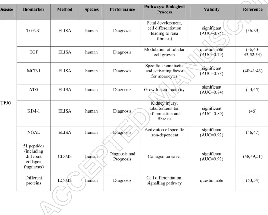
development, but in PUV remain and block urine outflow from the bladder leading to bilateral obstruction of urinary pathways with associated (often severe) lesions in the kidney (33;34). Vesicoureteral reflux (VUR) is characterized by a backward flow of the urine from the bladder towards the kidneys often due to urinary tract infection or urinary tract abnormalities. Severe VUR can lead to renal scarring, arterial hypertension and impaired renal function (35). RTM display a wide spectrum of pre- and postnatal outcomes ranging from death in utero to normal postnatal function. Most of the diagnostic methods of RTM are based on pre- and post-natal imaging, either ultrasound or radiological tests. However, imaging even if in some cases combined with fetal urine/amniotic fluid biochemistry, does often not help to provide the prediction of RTM progression and or its complications (31). Hence a number of studies have looked at the value of proteome analysis in RTM. Table 1 summarizes potential human urinary proteome analysis-based biomarkers of RTM that will be described in detail in the next sections.
4.1. UPJO
UPJO is the most common cause of upper tract obstruction and can be detected by
ultrasound before birth (20;32). Currently UPJO is treated surgically in the easily detectable severe cases. However for the other patients with mild forms of obstruction close follow-up during the first years of life is required which is necessitating imaging and frequent hospital visits to determine the progression of the disease. ELISA and MS-based urinary proteome studies have been performed with the aim to identify urinary biomarkers of UPJO. Several groups used sandwich ELISA kits to discover and identify potential biomarkers of UPJO based on predefined knowledge obtained in animal models of obstruction or processes repeatedly observed in kidney disease progression (e.g. fibrosis, inflammation).
Taha et al. were the first to study urinary concentrations of transforming growth factor β1 (TGF-β1) in UPJO. Using a quantitative sandwich ELISA kit and a cohort of 35 children with UPJO and 30 healthy children, they observed that the expression of TGF-β1 was significantly increased in urine of children with UPJO (36). Interestingly, urinary TGF-β1 decreased after
pyeloplasty (corrective surgery to remove the UPJO). These results were confirmed by Gawłowska-Marciniak et al., who used ELISA for their investigation of urine from the bladder and renal pelvis of 45 patients undergoing pyeloplasty and 25 controls (37) and by Sager et al. who used bladder urine from 19 UPJO patients and 19 matched controls (38). In another, more recent study, based on a cohort of 25 healthy controls and 25 children with UPJO, the authors also detected increased TGF- β1 levels in urine of patients with UPJO and a
significant correlation of TGF-β1 with hydronephrosis grade (p=0.0001) (39).
Studies of association between urinary EGF concentrations and UPJO were less successful because several studies described contradictory results. In 2000, ELISA was used in a study examining 24 patients with UPJO and 15 healthy children. Grandaliano et al. (40)established that EGF levels were significantly lower in UPJO patients when compared with healthy
controls. These results, employing ELISA, were confirmed by Bartoli et al. (41) using a cohort of 76 UPJO patients and 30 healthy children. Li et al. (42) analyzed the urinary EGF values in 33 healthy subjects and 12 patients with UPJO (surgical group) and they also observed, using ELISA, a significant decrease in abundance in patients group, during the first six months of life. The data presented also supported the hypothesis that urinary EGF changes over time are inversely correlated with Society of Fetal Urology (SFU) hydronephrosis grade. However, Taha et al. did not find significant differences in urinary EGF concentrations
between controls and surgical groups as well as preoperative and postoperative values one year after surgery (36). Furthermore, using a cohort of 28 UPJO children and 13 controls and a bead-based multiplex sandwich immunoassay, EGF levels according to Madsen et al. (43) were significantly increased in UPJO patients (submitted to pyeloplasty) compared to
controls. Furthermore, a 3 months and 1 year follow-up showed that these values normalized with similar values between patients and controls. Based on these contradictory results, it seems that EGF is not a valid biomarker for UPJO, because EGF levels were decreased in some studies (40-42), increased in another (43) or without changes (36).
The same studies also investigated specific urinary cytokines, such as monocyte
patients subjected to pyelopasty (preoperative and postoperative) and healthy controls, Grandaliano et al. (40) demonstrated that preoperative values of MCP-1were significantly increased. These findings were verified by Bartoli et al. (41) and Madsen et al. (43). They reported increased concentrations of this cytokine in urine of preoperative UPJO patients and a decrease in postoperative UPJO patients (similar to healthy).
Several, more anecdotal, studies focusing on the association of urinary angiotensinogen (AGT), kidney injury molecule-1 (KIM-1) and neutrophil gelatinase-associated lipocalin (NGAL) to UPJO were recently published: Taranta-Janusz et al. compared 31 children with severe hydronephrosis (who needed surgery); a non-surgical group, 20 patients with mild hydronephrosis, and 19 healthy children to study the urinary AGT concentration with the use of a commercially available ELISA (44). They observed that increased levels of AGT were directly correlated with UPJO children. AGT was also found in a later study (45), but the researchers turned no special attention to this protein.
A study involving 20 children with severe hydronephrosis, two groups of healthy individuals, 20 patients with mild non-obstructive hydronephrosis and 25 healthy children, demonstrated using immunoenzymatic ELISA commercial kits an increase in urinary NGAL and KIM-1 and established a correlation with worsening obstruction (46). The results for NGAL were
confirmed in another study, presenting an increase in urine from patients with obstructed kidneys at the time of surgery. This increase was followed by a decrease and stabilization to the same level as that of the controls (47).
Although these individual proteins were associated, with different degrees of confidence, to the severity and grade of UPJO, none of the studies evaluated their predictive power which would be the main use of urinary markers in UPJO.
MS-based proteomics approaches have been performed to predict the progression of UPJO, and the results were validated in several studies. The urinary proteome of 13 healthy
newborns, 19 UPJO patients with mild obstruction (non-operated individuals (no-OP)) and 19 UPJO patients with severe obstruction (operated individuals (OP)) was analysed using
CE-MS by Decramer et al. (51). They established a classifier based on 51 UPJO-specific urinary peptide biomarkers to differentiate resolution of UPJO or the requirement of pyeloplasty. Validation of these 51 biomarkers combined in a support vector machine (SVM) panel in an independent group of OP and no-OP patients (n=16) resulted in 94% sensitivity and 80% specificity. In a blinded cohort of 36 UPJO patients with 9 months of follow-up this panel predicted with 95% accuracy whether an infant with UPJO needed surgery. This biomarker panel was further validated in 19 children where the classifier displayed 83% sensitivity and 92% specificity. However in UPJO patients >1 year of age, this classifier lost sensitivity (66%) and specificity (20%) (52). This is probably due to the change of the urinary proteome after the age of 1-2 years (53). It also points to the fact that the context of use of disease classifiers will often not be beyond the population in which it was defined.
Bandin et al. used CE-MS-based urinary proteome analysis to determine whether early surgically corrected UPJO would normalize the urinary proteome after long-term follow-up compared to conservatively followed UPJO patients (54). Studying 42 patients with UPJO 5 year after either pyelopasty or spontaneous resolution of the obstruction, they observed that the urinary proteome was very similar in patients with early surgical correction of UPJO and age matched controls. However the urinary proteomes of UPJO patients leading to
spontaneous resolution or late surgical intervention did not normalize and displayed a significantly different pattern (54). This suggests ongoing renal or ureteral remodeling in the conservatively followed patients that is not visible clinically.
LC-MS/MS coupled to bioinformatics analysis was used in a number of studies to evaluate the urinary proteome of infants with UPJO. According to these studies, the most prominent proteins found were related to pathways involving inflammation, oxidative stress, fibrosis and renal disease (48;55-57). Mesrobian et al. (56;57) have identified the proteome differences between normal infants and infants with UPJO. In the first study, using 21 healthy infants and 25 infants with grade IV unilateral ureteropelvic junction obstruction, they identified 31
proteins changing at different time-points (56). In the second study, differences in individual urine samples were assessed in 21 healthy infants and 25 infants with grade IV unilateral
UPJO followed for 5 years. The urinary proteome from patients with UPJO was different from the age-matched controls, based on the activation of processes of inflammation, apoptosis, tubular injury and fibrosis, and oxidative stress (57).
A proteomic study of urine samples (n=5/group) from newborns with UPJO at different stages and controls was performed. Urine protein profiles of these patients were obtained by label free quantitative nanoLC-MS/MS and 970 urinary proteins were identified (52). In a recent case-control study (8 controls and 8 subjects with unilateral obstruction), Froehlich et al. (45) reported correlation between identified and quantified proteins associated with this type of RTM. They identified 1113 proteins, but only 76 were significantly different between the two groups. In particular biological processes such as inflammation and renal disease pathways showed significant variations, targeted oxidative stress proteins were also presented over expressed.
Based on these studies, potential regulated proteins in RTM such as arginase 1, glutathione S-transferase Mu 1 (GSTM1) and heat shock 70 kDa protein 1A (HSPA1A) were further studied and validated. Using Western Blot and MRM analysis, a decrease in arginase 1 in UPJO was validated in independent samples. In addition, in a mouse model of obstructive nephropathy, arginase 2 and total arginase activities were found to be increased (52).
GSTM1 and HSPA1A were also validated by Western Blot and confirmed that these proteins were elevated in urine of UPJO patients, supporting the idea that alterations in the
processing of reactive oxygen species (ROS) are related to UPJO.
These urinary proteome analysis studies were also an opportunity to re-evaluate the unclear association of urinary EGF to UPJO obstruction observed using ELISA. However, again contradicting results were obtained. Lacroix et al. (52) observed that urinary EGF decreased in UPJO using a combination of targeted (MRM) and non-targeted MS/MS analyses (using an independent cohort, n=10/group), and also demonstrated that EGF was among the top differentially excreted proteins. In contrast, Froehlich et al. (45) did not report EGF among the differentially expressed urinary proteins in their study. Hence EGF appears, again, not to be a reliable marker of UPJO. This might be due to the fact that urinary EGF is also modified in
other renal diseases including acute kidney injury, diabetic nephropathy, chronic kidney disease, IgA nephropathy, Lupus nephritis etc. (55) or even in different types of cancer (56), which demonstrates that EGF is not exclusively specific to UPJO.
4.2. PUV
Posterior urethral valves (PUV), the prototypic bilateral RTM (37), is an abnormal congenital obstructing membrane that is located within the posterior male urethra; this valve is the most common cause of bladder outlet obstruction in male children (36). The valve is believed to result from abnormal embryologic development of the fetal posterior urethra. The classic categorization of posterior urethral valves into types I, II, and III was developed by Young et al. in 1919 (60) and has undergone modification over time based on clinical observation and a better understanding of the embryologic events that lead to normal urethral development. This anomaly is not related with genetic disorders; however it was reported with familial inheritance (61). In PUV the major aim is the timing of surgical intervention (pre- or post-natal) and the prediction of the progression stage of chronic kidney disease (CKD) (34). CE-MS has been applied in a cohort of 28 patients with PUV with the aim to identify fetal urinary biomarkers that allow prediction of post-natal renal function (62). In that study Klein et al. identified 26 specific fetal urinary peptides biomarkers that characterized early ESRD. Twelve of these 26 peptides were combined in a classifier called the “12PUV” classifier, which displayed a sensitivity of 86% and a specificity of 95% for the prediction of post-natal renal function in an independent validation cohort of 38 patients with PUV. The main markers associated to early ESRD in PUV were collagen fragments. One particularity of this study was the abundance of these collagens in disease. Collagens were increased in fetal urine of patients with PUV displaying early ESRD. This is the opposite to what is observed in
postnatal urine in patients with CKD (62) suggesting that instead of fibrosis in CKD, patients with PUV display increased extracellular matrix turnover due to disruption of nephrogenesis (visible as dysplasia or hypoplasia in fetopathology). One fetal urinary biomarker of early ESRD was identified as a fragment of the XLαs variant of the G-protein-α subunit (GNAS).
GNAS and its variants represent imprinted genes (specific maternally or paternally
transmitted active copies of genes due to methylation patterns) and are described to have major effects on growth in utero and after birth.
Trnka et al. studied the potential correlation between post-natal urinary protein levels and kidney function in patients with PUV (63). In total, 47 subjects were studied: 27 patients with PUV and 20 controls, performing immunoblotting technique as well as determination of glomerular filtration rate (GFR). They were able to demonstrate significant differences in the excretion of some proteins between the two groups. Aquaporin-2 was significantly
decreased. On the other hand, the urinary protein-to-creatinine, whole-urine TGF-β and L1 cell adhesion molecule (L1CAM) were considered the best potential biomarkers, because they were significantly increased in urine of PUV patients and presented better correlation with GFR.
Using post-natal urine of 30 patients with PUV, Mandelia et al. investigated a number of urinary proteins and also the effects of angiotensin-converting enzyme inhibitors (ACE-I) on renal recovery (64). The pre- and postoperative protein levels of TGF-β1, tumor necrosis factor-α (TNF-α), interleukin-6 (IL-6) and microalbuminuria were measured over a period of 1 year. TGF-β1, TNF-α, and microalbuminuria were increased in patients with PUV. These proteins can be potential biomarkers because they reflect potential activation of the renin– angiotensin system, as well as they can provide an early recognition of children with ongoing renal damage. The treatment with ACE-I indicated an improvement of the kidney physiology and decrease of urinary TGF-β1 and microalbuminuria, allowing the retarding of renal injury in PUV patients and preserve the renal function in PUV patients (64).
4.3. VUR
Vesicoureteral reflux (VUR) can be detected and diagnosed by voiding cystourethrography which is an invasive and uncomfortable method for the children (38). VUR is frequently genetically heterogeneous, and emerges from interruption of complex signaling pathways as well as cell differentiation, which may be influenced by environmental factors (65). Urine from
73 children was collected and analyzed by CE-MS. To identify potential biomarkers, 18 patients with primary VUR (grade IV or V) and 19 patients without VUR were used. Nine urinary peptides were found, which were differentially regulated and a VUR-classifier was generated based on these peptides. A subsequent blinded evaluation has been performed on 17 patients with VUR grade IV or V and 19 patients without VUR with 88% sensitivity and 79% specificity. Five of the nine urinary biomarkers were successfully sequenced. They included 3 collagen alpha-1 (I) chain fragments, a sodium/ potassium-transporting ATPase and a CD99 antigen fragment.
5. Urinary RTM biomarkers from animal models
The UPJO animal model is the most frequently used RTM animal model. This model was used to study potential protein-based urinary markers of the disease including interleukin-1β (IL-1β), interleukin-2 (IL-2), interleukin-6 (IL-6), interleukin 10 (IL-10)), TNF-α, and interferon-γ (IFN-interferon-γ), etc. that are known to act as intercellular mediators in the cellular and molecular events of in UPJO.
Madsen et al. performed a study using two different rat UPJO models (the basic rat model is depicted in figure 2) to analyze the urinary abundance of these different potential biomarkers with the use of a bead-based multiplex sandwich immunoassay (66). One complete acute obstruction model (complete unilateral ureteral obstruction (CUUO) for 48 hours) and one partial chronic obstruction model (partial unilateral ureteral obstruction (PUUO) for 10 weeks) was employed. In the CUUO model IL-1β, IL-6, TNF-α, and IL-10 showed significant
differences in comparison with controls: IL-1β and IL-10 were detected at significantly decreased levels, with IL-6 and TNF-α found at significantly higher concentrations in urine from the left obstructed kidney. In the PUUO model only IL-6 showed a significant increase in urinary abundance. Another study compared the protein profiles in a PUUO model in
neonatal rats (obstruction at 1 day of age, randomized into 4 groups: 1, 2 and 5 days of obstruction and sham surgery) using 2-D gel electrophoresis coupled to MS (2DE-MS) (67). Forty-three proteins with differential urinary abundance were identified, which were reported
to be involved in the regulation of the cytoskeleton and actin, glucose metabolism, cell apoptosis, mitochondrial energy metabolism, oxidative stress, and endoplasmic reticulum stress. Electron transfer flavoprotein subunit beta (ETFB) identified as downregulated in urine by 2DE-MS was validated by immunoblot analysis. ETFB mRNA levels was also decreased in renal tissue from PUUO rats.
Another 2DE-MS study using an adult CUUO (three time-points (12 h, 24 h and 72 h)) rat model was performed by Zhao et al. They identified 39 proteins with different urinary abundance between the sham operated group and the CUUO group (68). Cell apoptosis, energy metabolism, injuries of mitochondrion and oxidative stress were some of the biological processes associated with these proteins. In addition, based on immunoblot analysis and immunofluorescence staining and assessment of the mRNA levels in renal tissue, they confirmed changes of 3 proteins: peroxiredoxin-1 (PRDX-1) was increased, glutathione peroxidase 1 (GPX1) and glutathione S-transferase P1 (GSTP1) were decreased. This study allowed a better understanding between oxidative stress and obstructive nephropathy.
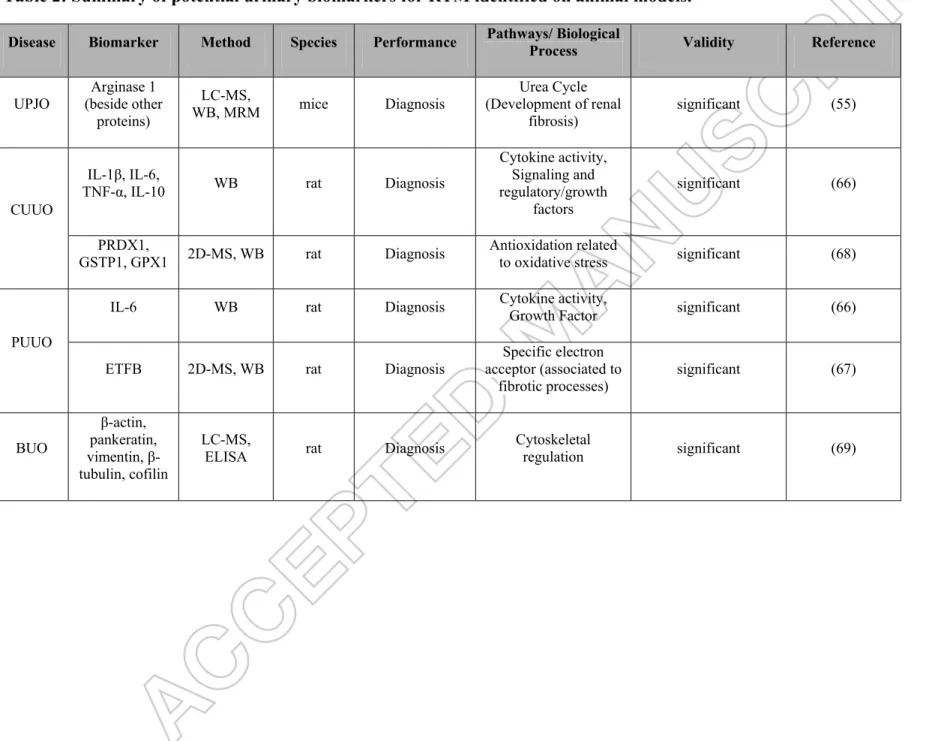
To study the acute urinary proteomic alterations induced by bilateral ureteral obstruction (BUO), MS-based proteomics was applied in rats that were subjected for three different time-points (2, 6, 24 h) to BUO (69). In this study 109 proteins associated with different biological processes involved in cytoskeleton and cytoskeletal regulation were identified. Western blots confirmed the selected results, demonstrating acute downregulation of proteins belonging to all three cytoskeletal components: the microfilament protein β-actin and the intermediate filament proteins pankeratin and vimentin, as well as β-tubulin, an important microtubular protein, were all downregulated. Furthermore, there was a significant upregulation of cofilin, an actin-binding protein. Table 2 summarizes potential urinary biomarkers of RTM identified in animal models.
In recent years a substantial number of studies have performed urinary proteome analysis in order to identify potential biomarkers of RTM. Only a few urinary biomarkers have been found to be unequivocally associated to UPJO. This is the case for urinary TGF-β1 that in all studies increased in UPJO, and decreased after relief of the obstruction. Studies on urinary EGF, although initially promising, turned out to be less clear and with time it appeared that EGF can be a urinary marker for many diseases thereby losing its specificity for UPJO. This also points to the important fact that to be marked as a “biomarker” validation should be performed in independent studies.
TGF-β1 and EGF were investigated as “isolated” markers of UPJO obstruction using ELISA and might perform better in a panel of markers. It has now been repeatedly observed that complex disease such as RTM cannot be faithfully described with single markers and panels of markers are more likely to “catch” the perturbed system, similarly to everyday medicine where a diagnosis is rarely based on a single observation. MS-based proteomics serves this goal. In this context it was observed, as described in detail above (sections 4.1-4.3), that panels of urinary peptides could predict the outcome in the different RTM, such as 51 UPJO-specific biomarkers (51), 12 related to PUV (62) and 9 concerning VUR (38).
7. Five-year view
In the coming years these initial successes obtained with single or multiple biomarkers still need to be confirmed to be applicable in the clinic. This involves larger scale studies (several hundreds of individuals/trial) the reach sufficient power. This might involve, in addition to the peptide panels discovered with MS-based tools, combination of markers (e.g. ELISA panels including TGF-β1, EGF, cytokines observed in animal models, etc.).
Finally, studies including other omics traits in urine (non-coding RNAs, metabolites, etc.) might allow adding additional markers to the panels and i) increase their specificity for the stratification of patients with RTM and ii) improve the understanding of the pathophysiology to allow optimised management of this frequent disease in children.
8. Key issues
• Urine is an important source of potential biomarkers, presenting several advantages such as the collection in large quantities, as a non-invasive method and as its stability. • Biomarker validation in an independent cohort is a crucial step for clinical proteomics, and it can be performed through mass spectrometry techniques (e.g. LC-MS, CE-MS) or immunoassay techniques (e.g. ELISA, WB).
• RTM are congenital anomalies of the kidneys and urinary tract, and are more common in infants, but can be also presented in childhood and adulthood.
• Development of non-invasive methods, such as urinary proteomics, can be useful to understand the progression of the different types of RTM, as well as to allow an early diagnosis, prognosis and a better clinical decision-making.
• Several studies in clinical proteomics have been performed, and a panel of biomarkers is more efficient than the use of a single biomarker.
Studies with high numbers of samples coupled to statistical analysis and an independent validation should be conducted to generate a classifier for RTM.
Funding
H. Mischak., J.P. Schanstra and P. Zürbig were supported in part by the European
Commission Seventh Framework Programme (FP7/2007-2013) under grant agreement no. 608332 (iMODECKD) and grant agreement no. 305608 (EURenOmics). Furthermore, P. Mischak received funding from the European Union's Horizon 2020 Research and Innovation Programme under the Marie Sklodowska-Curie grant agreement no. 642937
Declaration of Interest
H. Mischak is cofounder and a shareholder of mosaiques diagnostics GmbH. P.Magalhães and P. Zürbig are employees of mosaiques diagnostics GmbH. The authors have no other relevant affiliations or financial involvement with any organization or entity with a financial interest in or financial conflict with the subject matter or materials discussed in the
manuscript apart from those disclosed.
References
Reference annotations * Of interest
** Of considerable interest
(1) Benfield MR, McDonald RA, Bartosh S, et al. Changing trends in pediatric transplantation: 2001 Annual Report of the North American Pediatric Renal Transplant Cooperative Study. Pediatr Transplant 2003 Aug;7(4):321-35.
(2) Wuhl E, van Stralen KJ, Verrina E, et al. Timing and outcome of renal replacement therapy in patients with congenital malformations of the kidney and urinary tract. Clin J Am Soc Nephrol 2013 Jan;8(1):67-74.
(3) Celis JE, Moreira JM. Clinical proteomics. Mol Cell Proteomics 2008 Oct;7(10):1779. (4) Mischak H, Allmaier G, Apweiler R, et al. Recommendations for biomarker
identification and qualification in clinical proteomics. Sci Transl Med 2010 Aug 25;2(46):46ps42.
(5) Klein J, Buffin-Meyer B, Mullen W, et al. Clinical proteomics in obstetrics and neonatology. Expert Rev Proteomics 2014 Feb;11(1):75-89.
(6) Schaub S, Wilkins J, Weiler T, et al. Urine protein profiling with surface-enhanced laser-desorption/ionization time-of-flight mass spectrometry. Kidney Int 2004 Jan;65(1):323-32.
(7) Thongboonkerd V, Malasit P. Renal and urinary proteomics: current applications and challenges. Proteomics 2005 Mar;5(4):1033-42.
(8) Theodorescu D, Schiffer E, Bauer HW, et al. Discovery and validation of urinary biomarkers for prostate cancer. Proteomics Clin Appl 2008;2(4):556-70.
(9) Mischak H, Delles C, Klein J, et al. Urinary proteomics based on capillary electrophoresis-coupled mass spectrometry in kidney disease: discovery and validation of biomarkers, and clinical application. Adv Chronic Kidney Dis 2010 Nov;17(6):493-506.
(10) Decramer S, Gonzalez de PA, Breuil B, et al. Urine in clinical proteomics. Mol Cell Proteomics 2008 Oct;7(10):1850-62.
(11) Klein J, Bascands JL, Mischak H, et al. The role of urinary peptidomics in kidney disease research. Kidney Int 2016 Mar;89(3):539-45.
(12) Weissinger EM, Wittke S, Kaiser T, et al. Proteomic patterns established with capillary electrophoresis and mass spectrometry for diagnostic purposes. Kidney Int 2004 Jun;65(6):2426-34.
(13) Schiffer E, Mischak H, Novak J. High resolution proteome/peptidome analysis of body fluids by capillary electrophoresis coupled with MS. Proteomics 2006 Oct;6(20):5615-27.
(14) Mischak H, Delles C, Vlahou A, et al. Proteomic biomarkers in kidney disease: issues in development and implementation. Nat Rev Nephrol 2015 Apr;11(4):221-32.
(15) Fliser D, Novak J, Thongboonkerd V, et al. Advances in urinary proteome analysis and biomarker discovery. J Am Soc Nephrol 2007;18(4):1057-71.
(16) Kolch W, Mischak H, Pitt AR. The molecular make-up of a tumour: proteomics in cancer research. Clin Sci (Lond) 2005 May;108(5):369-83.
(17) Brennan DJ, O'Connor DP, Rexhepaj E, et al. Antibody-based proteomics: fast-tracking molecular diagnostics in oncology. Nat Rev Cancer 2010 Sep;10(9):605-17. (18) Kiernan UA. Quantitation of target proteins and post-translational modifications in
affinity-based proteomics approaches. Expert Rev Proteomics 2007 Jun;4(3):421-8. (19) Jankowski J, Schanstra JP, Mischak H. Body fluid peptide and protein signatures in
diabetic kidney diseases. Nephrol Dial Transplant 2015 Aug;30 Suppl 4:iv43-iv53. (20) Pejchinovski M, Hrnjez D, Ramirez-Torres A, et al. Capillary zone electrophoresis
on-line coupled to mass spectrometry: A perspective application for clinical proteomics. Proteomics Clin Appl 2015 Jun;9(5-6):453-68.
(21) Schanstra JP, Zurbig P, Alkhalaf A, et al. Diagnosis and prediction of CKD progression by assessment of urinary peptides. J Am Soc Nephrol 2015 Jan 14;26:1999-2010.
(22) Good DM, Thongboonkerd V, Novak J, et al. Body Fluid Proteomics for Biomarker Discovery: Lessons from the Past Hold the Key to Success in the Future. J Proteome Res 2007 Oct 31;6(12):4549-55.*This manuscript presents an overview about validation phase of urinary proteomics biomarkers.
(23) Rodriguez-Suarez E, Siwy J, Zurbig P, et al. Urine as a source for clinical proteome analysis: From discovery to clinical application. Biochim Biophys Acta 2013 Jul 2.
(24) Good DM, Zürbig P, Argiles A, et al. Naturally occurring human urinary peptides for use in diagnosis of chronic kidney disease. Mol Cell Proteomics 2010
Nov;9(11):2424-37.
(25) Kuznetsova T, Mischak H, Mullen W, et al. Urinary proteome analysis in hypertensive patients with left ventricular diastolic dysfunction. Eur Heart J 2012 Sep;33(18):2342-50.
(26) Zimmerli LU, Schiffer E, Zurbig P, et al. Urinary proteomic biomarkers in coronary artery disease. Mol Cell Proteomics 2008 Feb;7(2):290-8.
(27) Wang P, Whiteaker JR, Paulovich AG. The evolving role of mass spectrometry in cancer biomarker discovery. Cancer Biol Ther 2009 Jun;8(12):1083-94.
(28) Parker CE, Borchers CH. Mass spectrometry based biomarker discovery, verification, and validation--quality assurance and control of protein biomarker assays. Mol Oncol 2014 Jun;8(4):840-58.
(29) Lange V, Picotti P, Domon B, et al. Selected reaction monitoring for quantitative proteomics: a tutorial. Mol Syst Biol 2008;4:222.
(30) Mermelekas G, Vlahou A, Zoidakis J. SRM/MRM targeted proteomics as a tool for biomarker validation and absolute quantification in human urine. Expert Rev Mol Diagn 2015;15(11):1441-54.
(31) Shi T, Su D, Liu T, et al. Advancing the sensitivity of selected reaction monitoring-based targeted quantitative proteomics. Proteomics 2012 Apr;12(8):1074-92. (32) Albalat A, Mischak H, Mullen W. Clinical application of urinary
proteomics/peptidomics. Expert Rev Proteomics 2011 Oct;8(5):615-29. (33) Chevalier RL. Prognostic factors and biomarkers of congenital obstructive
different types of congenital obstructive nephropathy (e.g. UPJO, PUV), as well as, potential biomarkers of this disease based in imaging, serum, urine and amniotic fluid, in order to obtain an early diagnosis and to monitor the progression of the disease.
(34) Schanstra JP, Mischak H. Proteomic urinary biomarker approach in renal disease: from discovery to implementation. Pediatr Nephrol 2015 May;30(5):713-25.
(35) Caubet C, Lacroix C, Decramer S, et al. Advances in urinary proteome analysis and biomarker discovery in pediatric renal disease. Pediatr Nephrol 2010 Jan;25(1):27-35. (36) Manzoni C, Valentini AL. Posterior urethral valves. Rays 2002 Apr;27(2):131-4. (37) Roth KS, Koo HP, Spottswood SE, et al. Obstructive uropathy: an important cause of
chronic renal failure in children. Clin Pediatr (Phila) 2002 Jun;41(5):309-14. (38) Drube J, Schiffer E, Lau E, et al. Urinary proteome analysis to exclude severe
vesicoureteral reflux. Pediatrics 2012 Feb;129(2):e356-e363.
(39) Taha MA, Shokeir AA, Osman HG, et al. Pelvi-ureteric junction obstruction in children: the role of urinary transforming growth factor-beta and epidermal growth factor. BJU Int 2007 Apr;99(4):899-903.
(40) Gawlowska-Marciniak A, Niedzielski JK. Evaluation of TGF-beta1, CCL5/RANTES and sFas/Apo-1 urine concentration in children with ureteropelvic junction obstruction. Arch Med Sci 2013 Oct 31;9(5):888-94.
(41) Sager C, Lopez JC, Duran V, et al. Transforming growth factor-beta1 in congenital ureteropelvic junction obstruction: diagnosis and follow-up. Int Braz J Urol 2009 May;35(3):315-23.
(42) Merrikhi A, Bahraminia E. Association of urinary transforming growth factor-beta1 with the ureteropelvic junction obstruction. Adv Biomed Res 2014;3:123.
(43) Grandaliano G, Gesualdo L, Bartoli F, et al. MCP-1 and EGF renal expression and urine excretion in human congenital obstructive nephropathy. Kidney Int 2000 Jul;58(1):182-92.
(44) Bartoli F, Penza R, Aceto G, et al. Urinary epidermal growth factor, monocyte
chemotactic protein-1, and beta2-microglobulin in children with ureteropelvic junction obstruction. J Pediatr Surg 2011 Mar;46(3):530-6.
(45) Li Z, Zhao Z, Liu X, et al. Prediction of the outcome of antenatal hydronephrosis: significance of urinary EGF. Pediatr Nephrol 2012 Dec;27(12):2251-9.
(46) Madsen MG, Norregaard R, Palmfeldt J, et al. Epidermal growth factor and monocyte chemotactic peptide-1: potential biomarkers of urinary tract obstruction in children with hydronephrosis. J Pediatr Urol 2013 Dec;9(6 Pt A):838-45.
(47) Taranta-Janusz K, Wasilewska A, Debek W, et al. Urinary angiotensinogen as a novel marker of obstructive nephropathy in children. Acta Paediatr 2013
Sep;102(9):e429-e433.
(48) Froehlich JW, Kostel SA, Cho PS, et al. Urinary Proteomics Yield Pathological Insights for Ureteropelvic Junction Obstruction. Mol Cell Proteomics 2016 Aug;15(8):2607-15.*This study documented an association between identified proteins and type of RTM, as well as established a correlation with biological processes.
(49) Wasilewska A, Taranta-Janusz K, Debek W, et al. KIM-1 and NGAL: new markers of obstructive nephropathy. Pediatr Nephrol 2011 Apr;26(4):579-86.
(50) Madsen MG, Norregaard R, Palmfeldt J, et al. Urinary NGAL, cystatin C, beta2-microglobulin, and osteopontin significance in hydronephrotic children. Pediatr Nephrol 2012 Nov;27(11):2099-106.
(51) Decramer S, Wittke S, Mischak H, et al. Predicting the clinical outcome of congenital unilateral ureteropelvic junction obstruction in newborn by urinary proteome analysis. Nat Med 2006 Mar 19;12(4):398-400.**This clinical proteomic study allowed the identification and validation of a urinary biomarkers panel related with ureteropelvic junction obstruction (UPJO) disease, presenting high sensitivity and specificity. This study predicted significant outcomes in UPJO patients, especially in young patients. (52) Drube J, Zürbig P, Schiffer E, et al. Urinary proteome analysis identifies infants but
not older children requiring pyeloplasty. Pediatr Nephrol 2010 Sep;25(9):1673-8. (53) Zürbig P, Decramer S, Dakna M, et al. The human urinary proteome reveals high
similarity between kidney aging and chronic kidney disease. Proteomics 2009;9(8):2108-17.
(54) Bandin F, Siwy J, Breuil B, et al. Urinary Proteome Analysis at 5-Year Followup of Patients With Nonoperated Ureteropelvic Junction Obstruction Suggests Ongoing Kidney Remodeling. J Urol 2012 Jan 18;187(3):1006-11.
(55) Lacroix C, Caubet C, Gonzalez-de-Peredo A, et al. Label-free quantitative urinary proteomics identifies the arginase pathway as a new player in congenital obstructive nephropathy. Mol Cell Proteomics 2014 Dec;13(12):3421-34.
(56) Mesrobian HG, Mitchell ME, See WA, et al. Candidate urinary biomarker discovery in ureteropelvic junction obstruction: a proteomic approach. J Urol 2010
Aug;184(2):709-14.*Using LC-MS, these consecutive studies demonstrated significant different protein expression, based on biological processes, between normal infants and infants with UPJO.
(57) Mesrobian HG, Kryger JV, Groth TW, et al. Urinary proteome analysis in patients with stable SFU grade 4 ureteropelvic junction obstruction differs from normal. Urology 2013 Sep;82(3):745-10.*Using LC-MS, these consecutive studies demonstrated
significant different protein expression, based on biological processes, between normal infants and infants with UPJO.
(58) Klein J, Bascands JL, Buffin-Meyer B, et al. Epidermal growth factor and kidney disease: a long-lasting story. Kidney Int 2016 May;89(5):985-7.
(59) Honda K, Ono M, Shitashige M, et al. Proteomic approaches to the discovery of cancer biomarkers for early detection and personalized medicine. Jpn J Clin Oncol 2013 Feb;43(2):103-9.
(60) Young HH, Frontz WA, Baldwin JC. Congenital obstruction of the posterior urethra. J Urol 2002 Jan;167(1):265-7.
(61) Borzi PA, Beasley SW, Fowler R. Posterior urethral valves in non-twin siblings. Br J Urol 1992 Aug;70(2):201.
(62) Klein J, Lacroix C, Caubet C, et al. Fetal urinary peptides to predict postnatal outcome of renal disease in fetuses with posterior urethral valves (PUV). Sci Transl Med 2013 Aug 14;5(198):198ra106.**Excellent paper, based in a large case record using CE-MS, which pointed out the identification and validation of fetal urinary biomarkers in patients with posterior urethral valves (PUV). The classifier was able to correctly predict the postnatal renal function.
(63) Trnka P, Ivanova L, Hiatt MJ, et al. Urinary biomarkers in obstructive nephropathy. Clin J Am Soc Nephrol 2012 Oct;7(10):1567-75.
(64) Mandelia A, Bajpai M, Agarwala S, et al. The role of urinary TGF-beta(1), TNF-alpha, IL-6 and microalbuminuria for monitoring therapy in posterior urethral valves. Pediatr Nephrol 2013 Oct;28(10):1991-2001.
(65) Williams G, Fletcher JT, Alexander SI, et al. Vesicoureteral reflux. J Am Soc Nephrol 2008 May;19(5):847-62.
(66) Madsen MG. Urinary biomarkers in hydronephrosis. Dan Med J 2013 Feb;60(2):B4582.
(67) Zhao Q, Xue Y, Yang Y, et al. Screening and identification of the differentially expressed proteins in neonatal rat kidney after partial unilateral ureteral obstruction. Mol Med Rep 2016 Jul;14(1):681-8.
(68) Zhao Q, Yang Y, Wang CL, et al. Screening and identification of the differential proteins in kidney with complete unilateral ureteral obstruction. Int J Clin Exp Pathol 2015;8(3):2615-26.* Important paper which established potential correlation between oxidative stress and obstructive nephropathy, based on MS-approach for the
identification and immunoblot analysis and immunofluorescence staining for validation.
(69) Stodkilde L, Palmfeldt J, Nilsson L, et al. Proteomic identification of early changes in the renal cytoskeleton in obstructive uropathy. Am J Physiol Renal Physiol 2014 Jun 15;306(12):F1429-F1441.
Figure 1: Different types of urinary tract malformations in humans.
Normal System Ureteropelvic Junction Obstruction (UPJO)
Vesicoureteral reflux (VUR) Posterior urethral
Figure 2: Example for an induced ureteral obstruction in rats. Reproduced with permission from the Danish Medical Journal. Madsen
MG. Urinary biomarkers in hydronephrosis. Dan Med J 2013 Feb;60(2):B4582 (66).
Table 1: Summary of potential urinary biomarkers for RTM in studies concerning human samples.
Disease Biomarker Method Species Performance Pathways/ Biological Process Validity Reference
UPJO
TGF-β1 ELISA human Diagnosis
Fetal development, cell differentiation (leading to renal fibrosis) significant (AUC=0.75) (36-39)
EGF ELISA human Diagnosis Modulation of tubular cell growth (AUC=0.79) questionable 43;52;54)
(36;40-MCP-1 ELISA human Diagnosis
Specific chemotactic and activating factor
for monocytes
significant
(AUC=0.78) (40;41;43)
ATG ELISA human Diagnosis Growth factor activity (AUC=0.84) significant (44;45)
KIM-1 ELISA human Diagnosis
Kidney injury, tubulointerstitial inflammation and fibrosis significant (AUC=0.80) (46)
NGAL ELISA human Diagnosis Activation of specific iron-dependent (AUC=0.92) significant (46;47) 51 peptides
(including different collagen fragments)
CE-MS human Diagnosis and Prognosis Collagen turnover (AUC=0.92) significant (48;49;51)
Different
Arginase 1 (beside other proteins) LC-MS, WB, MRM human Diagnosis Urea Cycle (development of renal fibrosis) significant (52) GSTM1, HSPA1A (beside other 74 proteins)
LC-MS, WB human Diagnosis Stress response, transferase significant (45)
PUV 12 peptides (including different collagen and GNAS fragments)
CE-MS human Diagnosis and Prognosis Signal transductionCollagen turnover,
significant
(AUC=0.92) (58)
AQP2, TGFβ,
L1CAM 2D-MS, WB human Diagnosis Cell differentiation, transport
significant (AUCTGFβ=0.79) (AUCL1CAM=0.80)
(59) TGF-β1,
TNF-α ELISA human Diagnosis Cell differentiation significant (60)
VUR 9 peptides (including different collagen fragments, sodium/ potassium-transporting ATPase fragment, CD99 antigen fragment)
Table 2: Summary of potential urinary biomarkers for RTM identified on animal models.
Disease Biomarker Method Species Performance Pathways/ Biological Process Validity Reference
UPJO Arginase 1 (beside other proteins) LC-MS, WB, MRM mice Diagnosis Urea Cycle (Development of renal fibrosis) significant (55) CUUO IL-1β, IL-6,
TNF-α, IL-10 WB rat Diagnosis
Cytokine activity, Signaling and regulatory/growth factors significant (66) PRDX1,
GSTP1, GPX1 2D-MS, WB rat Diagnosis Antioxidation related to oxidative stress significant (68)
PUUO
IL-6 WB rat Diagnosis Cytokine activity, Growth Factor significant (66)
ETFB 2D-MS, WB rat Diagnosis acceptor (associated to Specific electron fibrotic processes) significant (67) BUO β-actin, pankeratin, vimentin, β -tubulin, cofilin LC-MS,