Contents lists available atScienceDirect
Behavioural Brain Research
j o u r n a l h o m e p a g e : w w w . e l s e v i e r . c o m / l o c a t e / b b r
Short communication
Na
+
,K
+
-ATPase activity impairment after experimental traumatic brain injury:
Relationship to spatial learning deficits and oxidative stress
Frederico Diniz Lima
a, Mauren Assis Souza
a, Ana Fl ´avia Furian
a,b, Leonardo Magno Rambo
c,
Leandro Rodrigo Ribeiro
c, Felipe Villa Martignoni
a, Maur´ıcio Scopel Hoffmann
a,
Michele Rechia Fighera
d, Luiz Fernando Freire Royes
c, Mauro Schneider Oliveira
a,b,∗,
Carlos Fernando de Mello
aaCentro de Ciˆencias da Sa´ude, Laboratorio de Psicofarmacologia e Neurotoxicidade, Departamento de Fisiologia, Universidade Federal de Santa Maria,
97105-900 Santa Maria, RS, Brazil
bPrograma de P´os-Graduac¸˜ao em Ciˆencias Biol´ogicas: Bioqu´ımica, Universidade Federal do Rio Grande do Sul, 90035-003 Porto Alegre, RS, Brazil
cCentro de Educac¸˜ao F´ısica e Desportos, Departamento de M´etodos e T´ecnicas Desportivas, Universidade Federal de Santa Maria, 97105-900 Santa Maria, RS, Brazil dCentro de Ciˆencias da Sa´ude, Departamento de Pediatria, Universidade Federal de Santa Maria, 97105-900 Santa Maria, RS, Brazil
a r t i c l e i n f o
Article history:
Received 26 March 2008
Received in revised form 13 May 2008 Accepted 15 May 2008
Available online 23 May 2008
Keywords: Na+,K+-ATPase Protein carbonylation TBARS Barnes maze FPI
a b s t r a c t
Traumatic brain injury (TBI) is a devastating disease that commonly causes persistent mental disturbances and cognitive deficits. Although studies indicate that oxidative stress and functional deficits occurring after TBI are interrelated events, the knowledge of the mechanisms underlying the development of such cog-nitive deficits has been limited. Thus, in the present study, we investigated the effect of fluid percussion brain injury (FPI) on a spatial learning task and levels of oxidative stress markers, namely, protein car-bonylation and thiobarbituric acid-reactive substances (TBARS) and Na+,K+-ATPase activity 1 or 3 months after FPI in rats. Statistical analysis revealed that FPI increased the scape latency and mean number of error in Barnes maze test 1 and 3 months after FPI. We also found that protein carbonylation and TBARS content increased in the parietal cortex 1 and 3 months after FPI. In addition, 3 months after FPI, protein carbonylation levels increased both in ipsilateral and contralateral cortices of FPI animals. Indeed, statis-tical analysis revealed a decrease in Na+,K+-ATPase activity in the cerebral cortex of 1 month FPI animals. Furthermore, the decrease in enzyme activity found 3 months was larger, when compared with 1 month after FPI. These results suggest that cognitive impairment following TBI may result, at least in part, from increase of two oxidative stress markers, protein carbonylation and TBARS that occurs concomitantly to a decrease in Na+,K+-ATPase activity.
© 2008 Elsevier B.V. All rights reserved.
Traumatic brain injury (TBI) is one of the major events in civil-ian life[3]and is a leading cause of disability and death[13]. The patients who survive present severe long-term neurological abnor-malities, including deficits in learning and memory[26]. Functional deficits result from both direct, immediate mechanical disruption of brain tissue (the primary injury) and indirect, delayed (sec-ondary) injury mechanisms. Secondary mechanisms are potentially amenable to post-injury therapeutic intervention because of their delayed onset and progression over hours to days and months after the initial trauma and include intracranial hemorrhage, brain swelling, raised intracranial pressure, and hypoxic/ischemic brain
∗Corresponding author at: Programa de P ´os-Graduac¸˜ao em Ciˆencias Biol ´ogicas: Bioqu´ımica, Universidade Federal do Rio Grande do Sul, 90035-003 Porto Alegre, RS, Brazil. Tel.: +55 55 3220 9378; fax: +55 55 3220 8241.
E-mail address:[email protected](M.S. Oliveira).
damage, among others[17]. The oxidative damage mediated by reactive oxygen (ROS) and nitrogen species is a significant com-ponent of the secondary injury cascade that accompanies TBI [24]. In fact, increased levels of markers of oxidative stress have been found in human cerebrospinal fluid after severe TBI[2,31] and also in animal models of TBI, such as in the fluid percus-sion and controlled cortical impact models of TBI [23,32]. It is also remarkable that pharmacological treatment with antioxidants can significantly ameliorate motor and cognitive outcomes after TBI. For instance, the antioxidant ␣-phenyl-N-tert-butyl nitrone markedly improve neurological reflexes and the performance in Morris water maze test as well as significantly reduced the loss of ipsilateral hemispheric tissue [15]. The results of these stud-ies seem to indicate that oxidative stress and functional deficits occurring after TBI are interrelated events and that the study of the mechanisms involved in this interface can open new avenues to better understand the TBI pathology[5]. Thus, since Na+,K+-ATPase 0166-4328/$ – see front matter © 2008 Elsevier B.V. All rights reserved.
is especially sensitive to oxidative stress[12,18]and is a key enzyme involved in maintenance of brain excitability and its inhibition induces spatial learning deficits[35], we investigated the role of this enzyme in spatial learning deficits induced by experimental TBI. For this purpose, spatial learning performance, levels of oxidative stress markers, namely, protein carbonylation and thiobarbituric acid-reactive substances (TBARS) and Na+,K+-ATPase activity were
measured 1 or 3 months after a single episode of fluid percussion in the rat.
Adult male Wistar rats (270–300 g), maintained under con-trolled light and environment (12:12 h light/dark cycle, 24±1◦C, 55% relative humidity), with standard laboratory chow (Guabi, Santa Maria, RS, Brazil) and waterad libitum. All reagents were purchase from Sigma (St Louis, MO, USA) except TBA, which was obtained from Merck (Darmstadt, Germany). All experimental pro-tocols designed aiming to keep the number of animals used to a minimum, as well as their suffering. Animal utilization reported in this study has been conducted in accordance with the policies of the National Institute of Health Guide for the Care and Use of Laboratory Animals (NIH Publications No. 80-23) revised in 1996, and with the approval of the Ethics Committee for Animal Research of the Federal University of Santa Maria (23081.018516/2006-57).
Fluid percussion brain injury (FPI) was carried out as described previously by D’Ambrosio et al.[6,7]. Animals were anesthetized with a single i.p. injection of Equithesin (6 ml/kg), a mix-ture containing sodium pentobarbital (58 mg/kg), chloral hydrate (60 mg/kg), magnesium sulfate (127.2 mg/kg), propylene glycol (42.8%), and absolute ethanol (11.6%) and placed in a rodent stereo-taxic apparatus. A burr hole of 3 mm in diameter was drilled on the right convexity parietal cortex, 2 mm posterior to the bregma and 3 mm lateral to the midline, taking care to keep the dura mater intact. A plastic injury cap was placed over the craniotomy with dental cement. When the dental cement hardened, the cap was filled with chloramphenicol and the animal was removed from the stereotaxic device and returned to its homecage. After 24 h, the ani-mals were anesthetized with halothane, and had the injury cap attached to the fluid percussion device and placed in a heat pad maintained at 37±0.2◦C.
Traumatic brain injury was induced by a fluid percussion device developed in our laboratory. A brief (10–15 ms) tran-sient pressure fluid pulse (3.53±0.17 atm) impact was applied against the exposed dura to model severe TBI. Pressure pulses were measured extracranially by a transducer (Fluid Con-trol Automac¸˜ao Hidr ´aulica, Belo Horizonte, MG, Brazil) and recorded on a storage oscilloscope (Gould Ltd., Essex, UK). Sham-operated animals underwent an identical procedure, with the exception of the FPI. Immediately after these procedures, the cap was removed and the orifice was covered with dental cement.
The tests for spatial learning assessment as well as neurochemi-cal determinations (oxidative stress parameters and Na+,K+-ATPase
activity) were performed at 1 or 3 months after the FPI. For this pur-pose, sham and FPI animals were randomly assigned in four groups, as follows:
•Experiment 1 (performed at 1 month after FPI): sham (n= 8) and FPI (n= 8–9).
•Experiment 2 (performed at 3 months after FPI): sham (n= 8) and FPI (n= 8–9).
To test the performance of the animals in a spatial learning paradigm, we choose the Barnes maze test, described by Barnes [1]and adapted by Oliveira et al.[22]. The apparatus was located in a 4 m×4 m test room where four visuospatial cues made of rigid
black paper (rectangle, circle, cross, triangle) were affixed to the walls but not directly over any one maze hole.
Briefly, on the first day of experiment, the rats were moved to the testing room and left undisturbed for 60 min. Following this habituation period, the rats were trained to find the escape hole; they were placed in the escape box for 1 min, then into a cylindrical opaque chamber (start box) in the center of the maze. With light on, the start box was removed and the rat allowed exploring freely and finding the escape box. A maximum latency of 180 s to find it was allowed. Each rat was given three trials per day, over four con-secutive days. In each trial we scored the time to reach the escape tunnel and the number of wrong holes visited. The arena, as well boxes, was wiped clean using distilled water both between each training session for a given rat and between each rat. Immediately after the Barnes maze test, the animals were transferred to the cen-tral area of a round open field (56 cm in diameter), which had its floor divided into 10 equal areas. The number of areas crossed was recorded for 20 min.
After the behavioral evaluation, the animals were killed by decapitation and had their brain exposed by the removal of the pari-etal bone. The ipsilateral cortex was rapidly dissected on an inverted ice-cold Petri dish. The contralateral cortex was subjected to the same procedure and served as internal control. Each tissue sam-ple was homogenized in cold 10 mM Tris–HCl buffer (pH 7.4) and then divided in aliquots for subsequent neurochemical analyses, as described below.
TBARS content was estimated in a medium containing 0.2 ml of brain homogenate, 0.1 ml of 8.1% SDS, 0.4 ml of acetic acid buffer (500 mM, pH 3.4), and 0.75 ml of 0.81% thiobarbituric acid (TBA). The mixture was finally made up to 2 ml with type I ultrapure water and heated at 95◦C for 90 min in a water bath using a glass ball as a condenser. After cooling to room temperature, absorbance was measured in the supernatant at 532 nm[21]. Total protein carbonyl content was determined by the method described by Yan et al.[34], adapted for brain tissue[27]. Assay of Na+,K+-ATPase activity was
performed according Wyse et al.[33]. Protein content was mea-sured colorimetrically by the method of Bradford[4], using bovine serum albumin (1 mg/ml) as standard. Statistical analysis was car-ried out by the Kruskal–Wallis test. Post hoc analysis was carcar-ried out, when appropriate, by the followed by Mann–Whitney test. Cor-relation analyses were carried out using the Pearson’s corCor-relation coefficient.
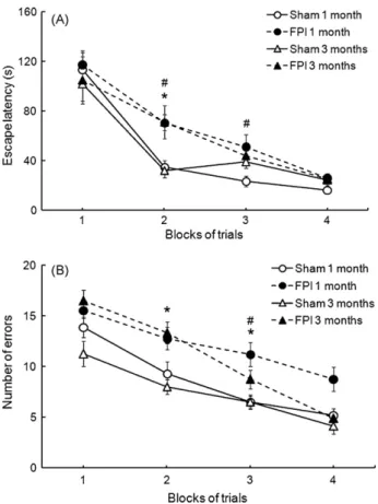
Fig. 1A and B shows the effect of FPI on the rat performance in the Barnes maze test. The first set of experiments was performed 1 month after FPI, and FPI animals showed an increased latency for escape at the second day of test, and number of errors at the second and third days of test when compared with sham animals, indicating a decreased performance of FPI animals in this spatial learning paradigm. There were no significant differences between sham and FPI animals in the latency for escape and number of errors in the first and in the fourth days of test. A decreased performance of FPI animals in the Barnes maze test was also found at 3 months after FPI, since FPI animals showed an increased latency for escape at the second and third days of test, and an increased number of errors at the third day of test, when compared with sham animals. We did not detect any motor deficit in the animals by analyzing the number of crossings in the open field, suggesting that a impaired performance in the Barnes maze is not related to motor disabilities. In order to evaluate the association between spatial learning deficits and levels of oxidative stress markers 1 or 3 months after FPI, we measured the levels of reactive species-induced damage to lipids (TBARS) and proteins (protein carbonylation). TBARS content increased in parietal cortex of FPI animals at 1 and 3 months, both in the ipsilateral and contralateral sides. Protein carbonylation lev-els are shown inFig. 2B. One month after FPI, protein carbonylation
Fig. 1.Effect of fluid percussion brain injury (TBI) on the (A) escape latency and (B) number of errors in the Barnes maze test. Data are the mean±S.E.M. forn= 8–9 animals in each group. *P< 0.05 compared with 1 month sham group.#P< 0.05 compared with 3 months sham group.
levels increased only in the ipsilateral cortex of FPI animals, in com-parison with sham animals. However, 3 months after FPI, protein carbonylation levels increased both in ipsilateral and contralateral cortices of FPI animals, when compared with sham animals. Such increase in protein carbonylation levels in FPI animals was larger at 3 months than at 1 month after FPI, indicating that protein car-bonylation increases over time after FPI, both in ipsilateral and contralateral cortices.
Since Na+,K+-ATPase is especially sensitive to oxidative stress,
we evaluated the effect of FPI on Na+,K+-ATPase activity (Fig. 2C).
Statistical analyses revealed a decrease in Na+,K+-ATPase activity
in the ipsilateral cerebral cortex of FPI animals both at 1 and 3 months after FPI, when compared with sham animals. Further-more, the decrease in enzyme activity 3 months after FPI was larger, when compared with decrease at 1 month after FPI, indicating that FPI-induced decrease in Na+,K+-ATPase activity is time-dependent.
Considering that a decrease in Na+, K+-ATPase activity plays
role in learning and memory deficits[35], we decided to inves-tigate whether Na+,K+-ATPase activity in the ipsilateral cerebral
cortex correlated with animal performance (escape latency) in the Barnes maze at the second day of test (in which the learn-ing deficits were more evident). Statistical analysis demonstrated that there is no correlation between these parameters, either in sham (Pearson’s coefficient = 0.267; P> 0.05 for n= 17) or FPI (Pearson’s coefficient = 0.089;P> 0.05 forn= 17) animals (data not shown).
In this study we showed that a single FPI episode in rat pari-etal cortex decreases Na+,K+-ATPase activity with a concomitant
increase in levels of oxidative stress markers, protein carbonyla-tion and TBARS. Moreover, animals showed an impaired ability to
Fig. 2. Effect of fluid percussion brain injury (TBI) on (A) TBARS content, (B) protein carbonylation levels and (C) Na+,K+-ATPase activity in rat parietal cortex. Data are the mean + S.E.M. forn= 8–9 animals in each group. *P< 0.05 compared with respective sham group.#P< 0.05 compared with 1 month after TBI group.
perform the Barnes maze, a spatial learning task. These alterations were found both at 1 or 3 months after FPI.
Chronic cognitive impairments such as deficits in attention, slow processing speed, or leaning and memory are frequently reported as a consequence of TBI in humans[26]and experimental animals [5]. Although the cognitive impairments are among the most debil-itating outcomes in patients, yet, to date, no treatment can prevent or counteract these effects in TBI patients[14,20,30]. Moreover, the mechanisms underlying the development of such cognitive deficits remain elusive. While neuronal death has been considered to be a major factor, the pervasive memory, cognitive and motor function deficits suffered by many mild TBI patients do not always corre-late with cell loss. In this context, it has been demonstrated that pharmacological treatment with neurotrophin-4/5 significantly protects against hippocampal cell death in mice, but not improves animal performance in three hippocampal-dependent behavioral tests[25]suggesting that cognitive impairment following TBI may result, at least in part, from alterations in surviving neurons[5].
In this study we showed that FPI induces a decrease in Na+,K+
-ATPase activity. These results are in full agreement with the view that FPI-induced neurochemical alteration, such Na+,K+-ATPase
activity inhibition, might be a factor contributing to a decreased rat performance in the Barnes maze task. Accordingly, the intrac-erebroventricular injection of ouabain, a selective inhibitor of Na+,K+-ATPase impairs rat performance in the Morris water maze,
another spatial learning and memory test[35]. Moreover, trans-genic mice lacking the␣subunit of Na+,K+-ATPase have severe
learning and memory deficits, as determined using the Morris water maze [19]. Altogether, these data may suggest that the presently reported deficits in spatial learning and decrease in Na+,K+-ATPase activity after TBI are interrelated events, and one
might suggest that decreased Na+,K+-ATPase activity may
con-tribute to the currently observed deficits in spatial learning after TBI. However, one may consider the fact that Na+,K+-ATPase
activ-ity in the ipsilateral cerebral cortex did not correlate with animal performance in the Barnes maze at the second day of test, sug-gesting that other factors may also contribute to impaired spatial learning performance after TBI.
The mechanisms underlying the presently observed decrease in Na+,K+-ATPase activity may include oxidative modification, since in
the present study we showed that increase in protein carbonylation and TBARS occurs concomitantly to a decrease in Na+,K+-ATPase
activity. In concordance with this view, it has been demonstrated that oxidative damage, mediated by reactive oxygen and nitrogen species, including the superoxide ion, hydrogen peroxide, hydroxyl radical, nitric oxide, and peroxynitrite, is well recognized as a signif-icant component of the secondary injury cascade that accompanies TBI[24]. Furthermore, while free-radical-induced damage has been proposed to be involved in traumatic cell injury and cell death, free radical scavengers, such catalase and glutathione peroxidase (GSH-Px) are associated with partial amelioration of traumatic injury [11,16,29].
Nevertheless, whether the presently reported increase in protein carbonylation and TBARS levels reflects a decrease in antioxidant defenses, an increase in reactive species formation or both remains to be determined, but it is interesting to point out that the increase in these markers of oxidative stress occur in par-allel with a decrease in Na+,K+-ATPase activity. It has long been
known that the Na+,K+-ATPase activity is particularly sensitive to
reactive species, which alter plasma membrane lipid composition [12]and the redox state of regulatory sulfhydryl groups[18]as well as other amino acid residues[28]. In addition, oxidative and nitrosative stress induce protein carbonylation[9], which is related to increased susceptibility to proteolysis and marked impairment of protein functionality[8]. Moreover, Dobrota et al.[10]suggested
that the most important consequence of oxidative modification is that Na+,K+-ATPase molecules have lost the ability to interact with
one another, decreasing oligomeric structure formation and leading to subsequent hydrolysis rate suppression.
In summary, the present study we showed that a single episode of FPI induces impairment in animal performance in a spatial learn-ing and memory paradigm, an increase of the oxidative stress markers and a decrease in Na+,K+-ATPase activity. Furthermore,
these findings add a new insight into the neurochemical mech-anism involved in the TBI-induced spatial learning impairment. Therefore, while the exact mechanism underlying of secondary injury after TBI is poorly understand, we think that these data may help in the understanding of TBI pathology.
Acknowledgements
Research supported by CNPq (grants 505527/2004-9 and 301552/2007-0) and CAPES. Mauro Schneider Oliveira is the recip-ient of CAPES fellowship. Carlos Fernando de Mello and Ana Fl ´avia Furian are the recipients of CNPq fellowships.
References
[1] Barnes CA. Memory deficits associated with senescence: a neurophysiological and behavioral study in the rat. J Comp Physiol Psychol 1979;93:74–104. [2] Bayir H, Kagan VE, Tyurina YY, Tyurin V, Ruppel RA, Adelson PD, et al.
Assessment of antioxidant reserves and oxidative stress in cerebrospinal fluid after severe traumatic brain injury in infants and children. Pediatr Res 2002;51:571–8.
[3] Beghi E. Overview of studies to prevent posttraumatic epilepsy. Epilepsia 2003;44(Suppl 10):21–6.
[4] Bradford MM. A rapid and sensitive method for the quantitation of micro-gram quantities of protein utilizing the principle of protein–dye binding. Anal Biochem 1976;72:248–54.
[5] Cohen AS, Pfister BJ, Schwarzbach E, Grady MS, Goforth PB, Satin LS. Injury-induced alterations in CNS electrophysiology. Prog Brain Res 2007;161:143–69. [6] D’Ambrosio R, Fairbanks JP, Fender JS, Born DE, Doyle DL, Miller JW. Post-traumatic epilepsy following fluid percussion injury in the rat. Brain 2004;127:304–14.
[7] D’Ambrosio R, Maris DO, Grady MS, Winn HR, Janigro D. Impaired K(+) homeostasis and altered electrophysiological properties of post-traumatic hip-pocampal glia. J Neurosci 1999;19:8152–62.
[8] Dalle-Donne I, Rossi R, Giustarini D, Milzani A, Colombo R. Protein carbonyl groups as biomarkers of oxidative stress. Clin Chim Acta 2003;329:23–38. [9] Dean RT, Fu S, Stocker R, Davies MJ. Biochemistry and pathology of
radical-mediated protein oxidation. Biochem J 1997;324(Pt 1):1–18.
[10] Dobrota D, Matejovicova M, Kurella EG, Boldyrev AA. Na/K-ATPase under oxida-tive stress: molecular mechanisms of injury. Cell Mol Neurobiol 1999;19: 141–9.
[11] Ikeda Y, Brelsford KL, Ikeda K, Long DM. Oxygen-free radicals in traumatic brain oedema. Neurol Res 1989;11:213–6.
[12] Jamme I, Petit E, Divoux D, Gerbi A, Maixent JM, Nouvelot A. Modulation of mouse cerebral Na+,K(+)-ATPase activity by oxygen free radicals. Neuroreport 1995;7:333–7.
[13] Langlois JA, Kegler SR, Butler JA, Gotsch KE, Johnson RL, Reichard AA, et al. Traumatic brain injury-related hospital discharges. Results from a 14-state surveillance system, 1997. MMWR Surveill Summ 2003;52:1–20.
[14] Maas AI. Neuroprotective agents in traumatic brain injury. Expert Opin Investig Drugs 2001;10:753–67.
[15] Marklund N, Clausen F, Lewen A, Hovda DA, Olsson Y, Hillered L. Alpha-phenyl-tert-N-butyl nitrone (PBN) improves functional and morphological outcome after cortical contusion injury in the rat. Acta Neurochir (Wien) 2001;143:73–81.
[16] Marmarou CR, Walker SA, Davis CL, Povlishock JT. Quantitative analysis of the relationship between intra-axonal neurofilament compaction and impaired axonal transport following diffuse traumatic brain injury. J Neurotrauma 2005;22:1066–80.
[17] Morales DM, Marklund N, Lebold D, Thompson HJ, Pitkanen A, Maxwell WL, et al. Experimental models of traumatic brain injury: do we really need to build a better mousetrap? Neuroscience 2005;136:971–89.
[18] Morel P, Tallineau C, Pontcharraud R, Piriou A, Huguet F. Effects of 4-hydroxynonenal, a lipid peroxidation product, on dopamine transport and Na+/K+ATPase in rat striatal synaptosomes. Neurochem Int 1998;33:531–40. [19] Moseley AE, Williams MT, Schaefer TL, Bohanan CS, Neumann JC, Behbehani
MM, et al. Deficiency in Na,K-ATPase alpha isoform genes alters spatial learning, motor activity, and anxiety in mice. J Neurosci 2007;27:616–26.
[20] Narayan RK, Michel ME, Ansell B, Baethmann A, Biegon A, Bracken MB, et al. Clinical trials in head injury. J Neurotrauma 2002;19:503–57.
[21] Ohkawa H, Ohishi N, Yagi K. Assay for lipid peroxides in animal tissues by thiobarbituric acid reaction. Anal Biochem 1979;95:351–8.
[22] Oliveira MS, Furian AF, Fighera MR, Fiorenza NG, Ferreira J, Rubin MA, et al. The involvement of the polyamines binding sites at the NMDA receptor in creatine-induced spatial learning enhancement. Behav Brain Res 2008;187: 200–4.
[23] Opii WO, Nukala VN, Sultana R, Pandya JD, Day KM, Merchant ML, et al. Proteomic identification of oxidized mitochondrial proteins following exper-imental traumatic brain injury. J Neurotrauma 2007;24:772–89.
[24] Potts MB, Koh SE, Whetstone WD, Walker BA, Yoneyama T, Claus CP, et al. Traumatic injury to the immature brain: inflammation, oxidative injury, and iron-mediated damage as potential therapeutic targets. NeuroRx 2006;3:143–53.
[25] Royo NC, LeBold D, Magge SN, Chen I, Hauspurg A, Cohen AS, et al. Neurotrophin-mediated neuroprotection of hippocampal neurons following traumatic brain injury is not associated with acute recovery of hippocampal function. Neuro-science 2007;148:359–70.
[26] Salmond CH, Sahakian BJ. Cognitive outcome in traumatic brain injury sur-vivors. Curr Opin Crit Care 2005;11:111–6.
[27] Oliveira MS, Furian AF, Royes LF, Fighera MR, Myskiw JC, Fiorenza NG, et al. Ascorbate modulates pentylenetetrazol-induced convulsions biphasically. Neuroscience 2004;128:721–8.
[28] Siems WG, Hapner SJ, van Kuijk FJ. 4-Hydroxynonenal inhibits Na(+)-K(+)-ATPase. Free Radic Biol Med 1996;20:215–23.
[29] Soustiel JF, Glenn TC, Shik V, Boscardin J, Mahamid E, Zaaroor M. Monitoring of cerebral blood flow and metabolism in traumatic brain injury. J Neurotrauma 2005;22:955–65.
[30] Tolias CM, Bullock MR. Critical appraisal of neuroprotection trials in head injury: what have we learned? NeuroRx 2004;1:71–9.
[31] Varma S, Janesko KL, Wisniewski SR, Bayir H, Adelson PD, Thomas NJ, et al. F2-isoprostane and neuron-specific enolase in cerebrospinal fluid after severe traumatic brain injury in infants and children. J Neurotrauma 2003;20:781–6. [32] Wu A, Ying Z, Gomez-Pinilla F. Dietary curcumin counteracts the outcome of traumatic brain injury on oxidative stress, synaptic plasticity, and cognition. Exp Neurol 2006;197:309–17.
[33] Wyse AT, Streck EL, Barros SV, Brusque AM, Zugno AI, Wajner M. Methyl-malonate administration decreases Na+,K+-ATPase activity in cerebral cortex of rats. Neuroreport 2000;11:2331–4.
[34] Yan LJ, Traber MG, Packer L. Spectrophotometric method for determination of carbonyls in oxidatively modified apolipoprotein B of human low-density lipoproteins. Anal Biochem 1995;228:349–51.
[35] Zhan H, Tada T, Nakazato F, Tanaka Y, Hongo K. Spatial learning tran-siently disturbed by intraventricular administration of ouabain. Neurol Res 2004;26:35–40.