American Dairy Science Association, 2006.
Technical Note:
Improved Method for Rapid DNA Extraction
of Mastitis Pathogens Directly from Milk
P. Cremonesi,*1B. Castiglioni,† G. Malferrari,‡ I. Biunno,§ C. Vimercati,# P. Moroni,# S. Morandi,储and M. Luzzana*
*Department of Biomedical Sciences and Technologies, University of Milan, Segrate, Milan, Italy †Institute of Agricultural Biology and Biotechnology-Italian National Research Council, Milan, Italy ‡Center for Bio-Molecular Interdisciplinary Studies and Industrial Applications (CISI), University of Milan, Segrate, Milan, Italy
§Institute of Biomedical Technologies-Italian National Research Council, Segrate, Milan, Italy #Department of Animal Pathology, Hygiene and Veterinary Public Health University of Milan, Italy
储Institute of Sciences of Food Production-Italian National Research Council, Milan, Italy
ABSTRACT
Efficient control against bovine mastitis requires sen-sitive, rapid, and specific tests to detect and identify the main bacteria that cause heavy losses to the dairy industry. Molecular detection of pathogenic microor-ganisms is based on DNA amplification of the target pathogen. Therefore, efficient extraction of DNA from pathogenic bacteria is a major step. In this study, we aimed to develop a specific, sensitive, and rapid method to extract DNA directly from the main gram-positive bacteria known to cause bovine mastitis ( Staphylococ-cus aureus,Streptococcus agalactiae,Streptococcus dys-galactiae, andStreptococcus uberis) found in milk sam-ples. The DNA extraction method is based on the lysing and nuclease-inactivating properties of the chaotropic agent, guanidinium thiocyanate, together with the nu-cleic acid-binding properties of the silica particles. An efficient protocol consisting of 6 basic steps (3 of which were done twice) was developed and applied directly to milk samples. Absence of PCR inhibitors and DNA quality were evaluated by PCR amplification of the spe-cies-specific DNA sequences of the target bacteria. The level of sensitivity achieved in our experiments is appli-cable to milk sample analysis without sample en-richment.
Key words: milk, DNA extraction, Staphylococcus
aureus, streptococci
INTRODUCTION
Mastitis (inflammation of the mammary gland) is a multifactorial disease and one of the most difficult pathologies to control. It can result from trauma or injury to the udder, chemical irritation, or from
infec-Received February 24, 2005. Accepted August 29, 2005.
1Corresponding author: [email protected]
tion caused by different bacterial species. Mastitis is the single most costly disease of dairy cattle resulting in the reduction of milk yield and quality. The estimated annual losses due to mastitis are about $184 per cow. These costs include reduced production, discarded milk, drug therapy, veterinarian costs, premature culling, and increased labor (National Mastitis Council, 1998). Microorganisms that cause mastitis are generally classified as either contagious or environmental based upon their primary reservoir and mode of transmission.
Staphylococcus aureusandStreptococcus agalactiaeare contagious pathogens and are commonly transmitted among cows by contact with infected milk. These patho-gens are of particular importance because they cause mainly subclinical forms of IMI that are often difficult to detect by the herdsman. Primary environmental pathogens include 2 types of bacteria: coliform species (e.g.,Escherichia coli,Klebsiella) and species of strepto-cocci other thanStrep. agalactiae. These bacteria arise from the environment in which the cow lives, entering into the udder between milkings, when teats are ex-posed to mud, manure, and dirty bedding materials.
Current identification methods are based on microbi-ological culture of milk and biochemical tests on the isolated bacteria, according to the National Mastitis Council guidelines (1998). At present, species identifi-cation by standard methods is labor intensive and takes at least 2 to 3 d to yield a positive result.
Due to limitations of culture methods, approaches using PCR have been developed to identify mastitis pathogens. Development of PCR-based methods pro-vides a promising option for the rapid identification of bacteria. Species-specific DNA sequences such as the highly conserved rRNA genes or the 16S-23S rRNA intergenic spacer of the ribosomal RNA operon can be used for the identification of bacterial species in hours, rather than days. Moreover, the sensitivity of PCR-based assays tends to be superior to bacterial cultures (Forsman et al., 1997; Phuektes et al., 2003) allowing
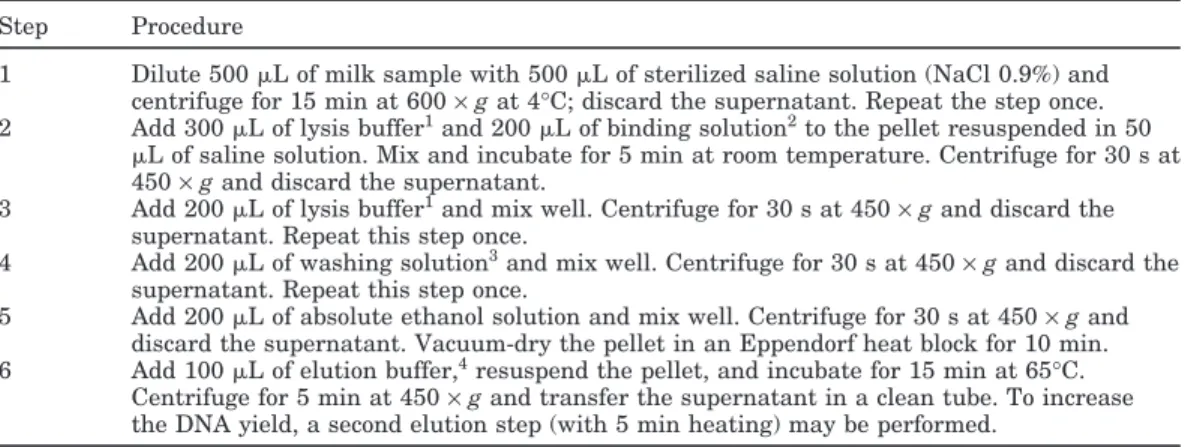
Table 1.Genomic DNA extraction protocol Step Procedure
1 Dilute 500L of milk sample with 500L of sterilized saline solution (NaCl 0.9%) and centrifuge for 15 min at 600×gat 4°C; discard the supernatant. Repeat the step once. 2 Add 300L of lysis buffer1and 200L of binding solution2to the pellet resuspended in 50
L of saline solution. Mix and incubate for 5 min at room temperature. Centrifuge for 30 s at 450×gand discard the supernatant.
3 Add 200L of lysis buffer1and mix well. Centrifuge for 30 s at 450×gand discard the supernatant. Repeat this step once.
4 Add 200L of washing solution3and mix well. Centrifuge for 30 s at 450×gand discard the
supernatant. Repeat this step once.
5 Add 200L of absolute ethanol solution and mix well. Centrifuge for 30 s at 450×gand discard the supernatant. Vacuum-dry the pellet in an Eppendorf heat block for 10 min. 6 Add 100L of elution buffer,4resuspend the pellet, and incubate for 15 min at 65°C.
Centrifuge for 5 min at 450×gand transfer the supernatant in a clean tube. To increase the DNA yield, a second elution step (with 5 min heating) may be performed.
1Lysis buffer: 3Mguanidine thiocyanate, 20 mMEDTA, 10 mMTris-HCl (pH 6.8), 40 mg/mL Triton
X-100, 10 mg/mLDL-dithiothreitol.
2Binding solution: 40 mg/mL silica (Sigma Aldrich, Milan, Italy) suspended in lysis buffer. 3Washing solution: 25% absolute ethanol, 25% isopropanol, 100 mMNaCl, 10 mMTris-HCl, pH 8. 4Elution buffer: 10 mMTris-HCl, pH 8.0, 1 mMEDTA.
the detection of small numbers of microorganisms. These factors can be extremely important when rapid and accurate identification of pathogenic bacteria is re-quired.
Different PCR-based methods have been developed for specific and sensitive detection of mastitis patho-gens in milk (Forsman et al., 1997; Phuektes et al., 2001; Riffon et al., 2001; Meiri-Bendek et al., 2002; Phuektes et al., 2003; Cremonesi et al., 2005). Direct isolation of high-quality DNA from the target bacteria found in milk, however, is often problematic and may require overnight selective-enrichment procedures (Phuektes et al., 2001; Meiri-Bendek et al., 2002; Ramesh et al., 2002). First, these difficulties are due to small concentrations of the pathogenic DNA present in a typical sample. Second, various factors affect DNA recovery, including the degree of cellular lysis, binding of DNA to particulate material, and degradation or shearing of DNA. Furthermore, in the case of gram-positive bacteria such as Staph. aureus and strepto-cocci, an optimal sample processing method should effi-ciently lyse resistant bacterial cell walls without dam-aging target DNA. In addition, many current methods typically require multiple steps or specialized equip-ment, rendering them impractical for use with large sample numbers (Boom et al., 1990). Finally, direct detection of pathogenic bacteria in food samples (Ramesh et al., 2002) is hampered by the presence of PCR-inhibitory substances frequently associated with the food matrix itself (Rossen et al., 1992). Particularly in milk, components such as Ca2+, proteinase, fats, and milk proteins may block DNA and shield it from access by polymerase (Wilson, 1997). Consequently, the devel-opment of a sample preparation strategy that can
effec-tively sequester high-quality DNA of the pathogenic bacteria from food samples before PCR amplification is needed.
In this paper, we describe a method for rapid DNA extraction directly from bovine and caprine raw milk to obtain material for PCR detection of gram-positive bacteria such asStaph. aureus,Strep. agalactiae,Strep. uberis, and Strep. dysgalactiae. This method is based on the ability of silica resin to bind DNA in the presence of high concentrations of guanidine thiocyanate as de-scribed previously (Malferrari et al., 2002), which guar-antees excellent disruption of bacterial cells.
MATERIALS AND METHODS DNA Extraction
Our DNA extraction procedure is described in Table 1. For preliminary experiments, sterile saline solution and sterile bovine milk were inoculated with <10 to 107cfu/mL of theStaph. aureusATCC 23235 reference strain andStrep. agalactiae,Strep. uberis, andStrep. dysgalactiaeisolated from bovine mastitis samples. The strains had been grown in brain-heart infusion broth (Scharlau, Barcelona, Spain) at 37°C for 24 h, identified by biochemical tests and the API Staph System (BioMe´r-ieux, Rome, Italy), and then stored at−70°C in a nutri-ent broth enriched with 15% glycerol. Genomic DNA was then isolated from both sterile saline solution and sterile bovine milk inoculated using the procedure de-scribed in Table 1, starting from step 2 when sterile saline solution was used.
To evaluate and optimize our extraction procedure, 30 bovine and caprine milk samples containing high
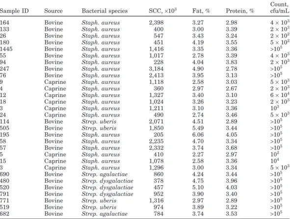
Table 2.Bovine and caprine milk samples processed and their characteristics
Count, Sample ID Source Bacterial species SCC,×103 Fat, % Protein, % cfu/mL
164 Bovine Staph. aureus 2,398 3.27 2.98 4×10³
133 Bovine Staph. aureus 400 3.00 3.39 2×10³
26 Bovine Staph. aureus 547 3.43 3.24 2×10²
180 Bovine Staph. aureus 451 4.19 3.55 5×10²
1445 Bovine Staph. aureus 1,416 3.35 3.36 >105
55 Bovine Staph. aureus 1,017 2.78 3.39 4×10³
94 Bovine Staph. aureus 228 4.04 3.83 2×10³
247 Bovine Staph. aureus 3,184 4.90 2.78 >105
76 Bovine Staph. aureus 2,413 3.95 3.13 >105
9 Caprine Staph. aureus 1,118 2.58 3.03 5×10³
4 Caprine Staph. aureus 360 2.97 2.67 2×10³
12 Caprine Staph. aureus 1,327 3.40 3.10 6×10⁴
18 Caprine Staph. aureus 1,024 3.26 3.23 2×10³
3 Caprine Staph. aureus 1,211 3.10 3.36 103
24 Caprine Staph. aureus 490 2.74 3.46 5×10³
114 Bovine Strep. uberis 2,071 4.51 2.89 >105
505 Bovine Strep. uberis 1,850 5.49 3.44 >105
195 Bovine Staph. aureus 205 6.06 4.05 >105
58 Bovine Staph. aureus 2,235 4.70 3.34 >105
57 Bovine Staph. aureus 2,332 3.74 3.68 >105
5 Caprine Staph. aureus 410 2.27 2.97 102
15 Caprine Staph. aureus 1,078 2.58 3.36 104
3 Caprine Staph. aureus 1,296 3.00 3.34 5×10³
690 Bovine Strep. agalactiae 860 4.24 3.44 >105
480 Bovine Strep. dysgalactiae 378 4.75 3.96 >105
520 Bovine Strep. dysgalactiae 457 5.10 4.03 >105
791 Bovine Strep. dysgalactiae 952 3.90 3.40 >105
771 Bovine Strep. uberis 1,316 2.97 2.89 >105
519 Bovine Strep. uberis 974 3.89 3.22 >105
682 Bovine Strep. agalactiae 784 3.74 3.53 >105
SCC were processed for bacterial DNA extraction and identification (Table 2).
ForStaph. aureus, total sample counts were obtained by following the FIL-IDF standard procedure (no. 145A; 1997). Briefly, 10-fold serial dilutions were plated onto Baird Parker rabbit plasma fibrinogen agar plates, which were then incubated at 37°C for 24 and 48 h, and observed for characteristic colony morphology. Streptococcal strains were grown at 37°C on blood agar plates (agar base supplemented with 5% defibrinated sheep blood; Oxoid, Milan, Italy) for 24 to 48 h.
Genomic DNA was isolated from milk samples follow-ing the procedure described in Table 1, startfollow-ing from step 1. The extraction protocol required approximately 90 min to process the samples from sample receipt to DNA rehydration.
At the same time, DNA extractions were carried out starting with 200L each of the 2 bovine milk samples described in Table 2 and using 2 commercial kits, the Puregene DNA Isolation kit for gram-positive bacteria (Gentra Systems, Minneapolis, MN) and the Wizard Genomic DNA Purification kit (Promega Italia, Milan, Italy) according to the manufacturer’s instructions or with minor modifications (Ercolini et al., 2004). The quantity and quality of DNA samples were measured
using a NanoDrop ND-1000 spectrophotometer (Nano-Drop Technologies, Wilmington, DE).
PCR Reactions
All PCR reactions were carried out in a GeneAmp PCR System 2700 (Applied Biosystems, Foster City, CA) in 0.2-mL tubes containing 12.5 L of 2× PCR Master Mix (Fermentas, M-Medical SRL, Milan, Italy), 0.1L of each of the primers, 5L of extracted DNA, and sterile water in a total reaction volume of 25L.
ForStaph. aureusdetection, a pre-PCR step was run at 94°C for 5 min followed by 30 PCR cycles under the following conditions: denaturation at 94°C for 1 min, annealing at 56°C for 1 min, and extension at 72°C for 1 min. After the final cycle, the preparation was kept at 72°C for 10 min to complete the reaction. TheStaph. aureusspecific primers for the 23S rRNA gene are de-scribed in Cremonesi et al. (2005): 23S-F 5′AGC TGT GGA TTG TCC TTT GG 3′; 23S-R 5′ TCG CTC GCT CAC CTT AGA AT 3′.
Streptococci primers (i.e., primers forStrep. agalac-tiae, Strep. uberis, and Strep. dysgalactiae) and PCR annealing temperatures were derived from Riffon et al. (2001). According to this protocol, a pre-PCR step at
94°C for 2 min was run followed by 35 PCR cycles under the following conditions: denaturation at 94°C for 45 s, annealing for 1 min at 60°C for Strep. agalactiae, at 59°C forStrep. uberisand at 57°C forStrep. dysgalac-tiae, respectively, and extension at 72°C for 2 min. After the final cycle, the preparation was kept at 72°C for 10 min to complete the reaction.
The β-casein primers and PCR conditions were de-rived from Klotz and Einspainer (2001). Briefly, a pre-PCR step at 94°C for 4 min was run followed by 30 PCR cycles under the following conditions: denaturation at 94°C for 50 s, annealing at 60°C for 50 s, and extension at 72°C for 50 s. After the final cycle, the preparation was kept at 72°C for 7 min to complete the reaction.
Ten microliters of each of the PCR-amplified products were analyzed by electrophoresis on 2% agarose gel stained with ethidium bromide (0.05g/L; Sigma Ald-rich, Milan, Italy). After an electrophoresis run-time of 30 min, the gels were photographed under UV light using the BioProfile system (Mitsubishi, Tokyo, Japan). Molecular size markers (100-bp and 1-kb DNA ladder; Finnzymes, Espoo, Finland) were included in each agar-ose gel.
Sensitivity Tests
The sensitivity of our extraction method was exam-ined using sterilized bovine milk inoculated with dilu-tions of mixed cultures ofStaph. aureus(ATCC 23235) andStrep. agalactiaestrains, starting from 107cfu/mL. Parallel dilutions ofStaph. aureusandStrep. agalactiae
strains were made in sterile saline solution. Dilutions obtained were then plated on sheep blood agar. Num-bers of bacterial colonies were counted after 24 h of incubation, and ranged from<102to 107cfu/mL for both
Staph. aureus and Strep. agalactiae strains. Unbal-anced mixed cultures were tested at the following con-centrations: 102cfu/mL ofStaph. aureus with 102cfu/ mL of Strep. agalactiae, 104 cfu/mL of Staph. aureus with 102 cfu/mL of Strep. agalactiae, 104 cfu/mL of
Staph. aureuswith 103cfu/mL ofStrep. agalactiaeand 107cfu/mL ofStaph. aureus with 102cfu/mL ofStrep. agalactiae, respectively. The PCR reactions forStaph. aureusand Strep. agalactiae strains were carried out separately following the PCR conditions described above.
RESULTS AND DISCUSSION
As described in previous studies (Gutierrez et al., 1997; Romero and Lopez-Goni, 1999; Ramesh et al., 2002), a PCR-based assay can be extremely useful for analyzing pure microbial cultures. However, when ap-plied directly to food samples, its efficiency can be
mark-edly reduced by poor sample preparation, which might inadvertently introduce inhibitory substances preclud-ing DNA amplification. Fats, proteinases, and high con-centrations of Ca2+(Wilson, 1997) have been proposed as potential inhibitors of PCR. In our protocol, modifi-cations made to the original procedures (Malferrari et al., 2002) consisted of pretreatment of samples to elimi-nate PCR inhibitors such as milk fats and proteins. Furthermore, the robustness of bacterial cells with spe-cial reference to gram-positive bacteria (in our case
StaphylococcusandStreptococcusspecies) necessitated the use of enzymes such as lysozyme, lysostaphin, and lyticase to guarantee total DNA release from lysed cells (Riffon et al., 2001; Meiri-Bendek et al., 2002). In our study, efficient lysis of cells and removal of inhibitors were accomplished by increasing the concentration of guanidine thiocyanate and lysis buffer solution, both of which increased the disruption of bacterial cells, re-sulting in stronger and more reproducible amplifica-tion, avoiding the combination of enzymes and incuba-tion condiincuba-tions, and maintaining good characteristics of the method without time-consuming procedures.
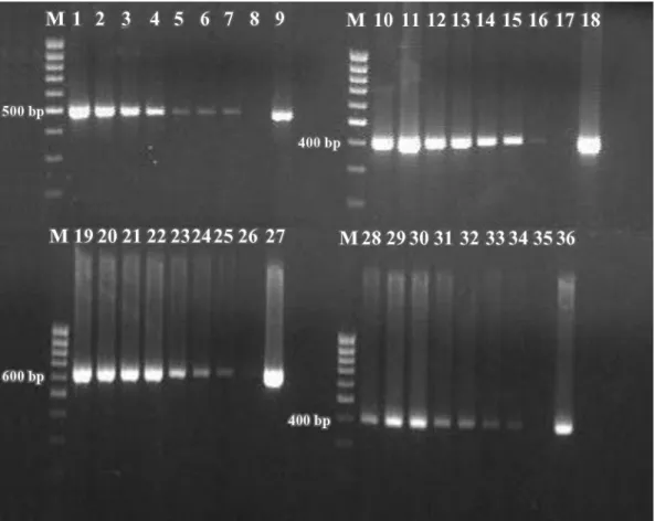
The specific PCR amplifications obtained from sterile bovine milk inoculated withStaph. aureus,Strep. aga-lactiae,Strep. dysgalactiae, and Strep. uberisranging from<10 to 107cfu/mL are shown in Figure 1. A quanti-tative decrease in the intensity of the amplicons re-flected a corresponding decrease in cell numbers. Am-plification of these species-specific DNA sequences is a necessary positive control to confirm the efficiency of DNA extraction as well as the quality of the DNA being amplified. This is an important step for studies in which detection of pathogenic DNA is carried out using PCR amplification. Furthermore, all samples obtained from sterile bovine milk inoculated with Staph. aureus,
Strep. agalactiae,Strep. dysgalactiae, andStrep. uberis
were successfully amplified, also confirming the ab-sence of potential inhibitory factors.
Sensitivity of the extraction procedure was found to achieve a detection of 10 cfu/mL for all species, both in milk and sterilized saline solution. The detection level of our method eliminates the need for bacterial enrich-ment culturing of Staphylococcus and Streptococcus
species.
Presence of coexisting bacteria in a milk sample can attenuate the specific detection of the target bacterial species (Ramesh et al., 2002). The sensitivity of the DNA extraction procedure for identifying a specific bac-terial pathogen in the presence of a coexisting microbe is shown in Figure 2.Staphylococcus aureusandStrep. agalactiaein concentrations ranging from<102to 107 cfu/mL could be detected simultaneously, even when they coexisted in milk at concentrations as low as 102 cfu/mL.
Figure 1.Sensitivity of PCR assay in detecting DNA from milk samples artificially inoculated withStaphylococcus aureus(ATCC 23235, reference strain),Streptococcus agalactiae,Streptococcus dysgalactiae, andStreptococcus uberistarget genes. The amounts of the bacteria used were from 2×10⁷to 2×10 cfu/mL forStaph. aureus(lanes 1 to 7, 499 bp), from 5×10⁷to 5×10 cfu/mL forStrep. agalactiae(lanes
10 to 16, 405 bp), from 7×10⁷to 7×10 cfu/mL forStrep. uberis(lanes 19 to 25, 624 bp), and from 2×10⁷to 2×10 cfu/mL forStrep. dysgalactiae(lanes 28 to 34, 401 bp). Positive controls forStaph. aureus(lane 9),Strep. agalactiae(lane 18),Strep. uberis(lane 27), and
Strep. dysgalactiae(lane 36) and negative controls (uninoculated milk, lanes 8, 17, 26, and 35) were included; M = 100-bp DNA ladder (Finnzymes, Espoo, Finland).
To validate our extraction assay, 30 bovine and cap-rine milk samples with high SCC were processed for bacterial DNA extraction and identification (Table 2). All samples extracted were successfully amplified using target bacteria-specific primers. Identification of patho-gens obtained by PCR assays was in full agreement with that obtained by microbiological methods (data not shown).
Furthermore, to evaluate the relative effectiveness of our method in recovering bacterial DNA from milk samples, we compared our procedure to 3 existing proto-cols. Two of the existing protocols are reported in the literature for direct DNA extraction of gram-positive bacteria from raw milk and are based on the use of a commercial kit used according to the manufacturer’s instructions (Furet et al., 2004) or with minor modifica-tions (Ercolini et al., 2004). The third DNA extraction was performed using a commercial kit to extract DNA from gram-positive bacteria in culture. Two bovine milk
samples containing>105cfu/mL ofStaph. aureuswere tested to compare these methods for DNA recovery and their compatibility with PCR detection.
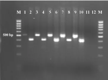
The quality and the quantity of DNA extracted are shown in Table 3. In addition, all DNA samples were analyzed by PCR using primers to amplify theβ-casein gene andStaph. aureus23S rRNA gene (Figure 3). As shown in Table 3, our method yielded higher concentra-tions of DNA than did the other methods. Reasons for decreased DNA recoveries reported for other methods are unknown, but might include lost DNA template through degradation or difficulty in disrupting bacterial cell walls. Furthermore, PCR analysis of samples ex-tracted using the diverse procedures provided good de-tection levels of the β-casein gene with the exception of the Puregene DNA Isolation kit; different detection levels of the target bacterial gene are probably due to differences in bacterial DNA extraction efficiency.
Figure 2.Sensitivity test using PCR for simultaneous detection using unbalanced mixes ofStaphylococcus aureus23235 ATCC refer-ence strain andStreptococcus agalactiaein milk samples. Lanes 1 and 2 = mix withStaph. aureus102cfu/mL andStrep. agalactiae102
cfu/mL, respectively; lanes 3 and 4 = mix withStaph. aureus104cfu/
mL andStrep. agalactiae102cfu/mL, respectively; lanes 5 and 6 =
mix withStaph. aureus104cfu/mL andStrep. agalactiae103cfu/mL,
respectively; lanes 7 and 8 = mix withStaph. aureus107cfu/mL and Strep. agalactiae102cfu/mL, respectively; lane 9 = positive control
for Staph. aureus; lane 10 = positive control forStrep. agalactiae; lanes 11 and 12 = milk without bacterial contamination; M = 100-bp DNA ladder (Finnzymes, Espoo, Finland).
Although sensitivity of a DNA extraction method is important, many additional factors must be considered, including time required, cost per test, and the need for specific reagents. In addition, a protocol that does not include specialized equipment or knowledge supports the routine isolation of DNA from a large series of sam-ples (Boom et al., 1990). Processing time varied among procedures. Sample processing by our method required approximately 90 min; processing time by the other methods varied from 130 to 140 min (Ercolini et al.,
Table 3.Efficiency of 4 different DNA extraction methods tested on 2 samples
DNA DNA
concentration, purity,
Sample DNA extraction procedure ng/L A260/A280
57 Ercolini et al., 2004 10.2 1.76
57 Wizard Genomic DNA Purification kit1 8.3 2.16
57 Our procedure 22.9 1.73
57 II Our procedure with second elution step2 22.2 1.88
57 Puregene DNA Isolation kit3 12.0 1.76
58 Ercolini et al., 2004 18.4 1.69
58 Wizard Genomic DNA Purification kit 10.5 2.03
58 Our procedure 23.1 1.79
58 II Our procedure with second elution step 14.7 1.92
58 Puregene DNA Isolation kit 8.1 1.93
1Promega Italia, Milan, Italy.
2A second elution step performed by our DNA extraction procedure (as described in Table 1). 3Gentra Systems, Minneapolis, MN.
Figure 3.Effect of DNA extraction using 4 different procedures on the PCR detection of Staph. aureusand bovine genomic DNA using primers forStaph. aureus23S rRNA gene (lanes 1 to 7, 499 bp) and bovineβ-casein gene (lanes 8 to 14, 453 bp). Lanes 1 and 8 = DNA extracted according to Ercolini et al. (2004); lanes 2 and 9 = DNA extracted by Wizard Genomic DNA Isolation kit (Promega); lanes 3 and 10 = DNA extracted following our procedure; lanes 4 and 11 = DNA extracted following our procedure, with a second elution step; lanes 5 and 12 = DNA extracted by Puregene DNA isolation kit (Gentra Systems). Lanes 6 and 13 = positive controls; lanes 7 and 14 = negative controls (no DNA); M = 100-bp DNA ladder (Finnzymes, Espoo, Finland).
2004), and up to 3 h for the Puregene DNA Isolation kit. In total, the method described herein requires a total time of less than 6 h (for DNA extraction, PCR amplification, and gel band visualization).
In addition to decreased processing time, our proce-dure reduced the number of manipulations needed to obtain pure DNA, improving the ease of sample han-dling, and minimizing the risk of cross-contamination. The present procedure for DNA preparation is rapid, simple, and reproducible, providing a more efficient pro-tocol applicable directly to milk samples, which remains unaffected by matrix-derived factors, potential inhibi-tors, and the presence of coexisting bacteria. Therefore, the DNA extraction method developed in the present study generates PCR-compatible templates without need for enrichment of the samples.
The procedure described herein could be automated using a liquid handling system to allow for high-throughput screening. Indeed, good preliminary results were obtained when the procedure was implemented on the Multiprobe II HT EX (Perkin Elmer) liquid han-dling system (data not shown). As described in Malfer-rari et al. (2004), the technology provided significant improvements in terms of efficiency, quality, and cost reduction.
ACKNOWLEDGMENTS
Part of this work was funded by the Fondazione Cari-plo (contract no. 2003.1824/10.8441) and by Italian FIRST 2004 (to Paolo Moroni).
REFERENCES
Boom, R., C. J. A. Sol, M. M. M. Salimans, C. L. Jansen, P. M. E. Wertheim-van Dillen, and J. Noordaa. 1990. Rapid and simple method for purification of nucleic acids. J. Clin. Microbiol. 28:495–503.
Cremonesi, P., M. Luzzana, M. Brasca, S. Morandi, R. Lodi, C. Vimer-cati, D. Agnellini, G. Caramenti, P. Moroni, and B. Castiglioni. 2005. Development of a multiplex PCR assay for the identification ofStaphylococcus aureus enterotoxigenic strains isolated from milk and dairy products. Mol. Cell. Probes 19:299–305. Ercolini, D., G. Blaiotta, V. Fusco, and S. Coppola. 2004. PCR-based
detection of enterotoxigenicStaphylococcus aureusin the early stages of raw milk cheese making. J. Appl. Microbiol. 96:1090– 1096.
Forsman, P., A. Tilsala-Timisjarvi, and T. Alatossava. 1997. Identifi-cation of staphylococcal and streptococcal causes of bovine masti-tis using 16S-23S rRNA spacer regions. Microbiology 143:3491–3500.
Furet, J. P., P. Que´ne´e, and P. Tailliez. 2004. Molecular quantification of lactic acid bacteria in fermented milk products using real-time quantitative PCR. Int. J. Food Microbiol. 97:197–207.
Gutierrez, R., T. Garcia, I. Gonzalez, B. Sanz, P. E. Hernandez, and R. Martin. 1997. A quantitative PCR-ELISA for the rapid
enumer-ation of bacteria in refrigerated raw milk. J. Appl. Microbiol. 83:518–523.
Klotz, A., and R. Espainer. 2001. Development of a DNA-based screen-ing method to detect cow milk in ewe, goat and buffalo milk and dairy products using PCR-LCR-EIA technique. Milchwis-senschaft 56:67–70.
Malferrari, G., E. Monferini, P. DeBlasio, G. Diaferia, G. Saltini, E. Del Vecchio, L. Rossi-Bernardi, and I. Biunno. 2002. High-quality genomic DNA from human whole blood and mononuclear cells. Biotechniques 33:1228–1230.
Malferrari, G., E. Monferini, S. Michelini, M. C. Proverbio, L. Biagi-otti, G. Saltini, F. Gervasi, P. DeBlasio, and I. Biunno. 2004. Automated system for nucleic acid extraction, purification and analysis. Nucleic Acids Purif. 6:52–54.
Meiri-Bendek, I., E. Lipkin, A. Friedmann, G. Leitner, A. Saran, S. Friedman, and Y. Kashi. 2002. A PCR-based method for the detection of Streptococcus agalactiae in milk. J. Dairy Sci. 85:1717–1723.
National Mastitis Council. Current Concepts of Bovine Mastitis.1998. 4th ed. Natl. Mastitis Council, Madison, WI.
Phuektes, P., G. F. Browning, G. Anderson, and P. Mansell. 2003. Multiplex polymerase chain reaction as a mastitis screening test forStaphylococcus aureus,Streptococcus agalactiae, Streptococ-cus dysgalactiaeandStreptococcus uberisin bulk milk samples. J. Dairy Res. 70:149–155.
Phuektes, P., P. Mansell, and G. F. Browning. 2001. Multiplex poly-merase chain reaction assay for simultaneous detection of Staphy-lococcus aureusand streptococcal causes of bovine mastitis. J. Dairy Sci. 84:1140–1148.
Ramesh, A., B. P. Padmapriya, A. Chandrashekar, and M. C. Vara-daraj. 2002. Application of a convenient DNA extraction method and multiplex PCR for the direct detection of Staphylococcus aureus andYersinia enterocoliticain milk samples. Mol. Cell. Probes 16:307–314.
Riffon, R., K. Sayasith, H. Khalil, P. Dubreuil, M. Drolet, and J. Lagace`. 2001. Development of a rapid and sensitive test for identi-fication of major pathogens in bovine mastitis by PCR. J. Clin. Microbiol. 39:2584–2589.
Romero, C., and I. Lopez-Goni. 1999. Improved method for purifica-tion of bacterial DNA from bovine milk for detecpurifica-tion ofBrucella
spp. by PCR. Appl. Environ. Microbiol. 65:3735–3737.
Rossen, L., P. Norskov, K. Holmstrom, and O. F. Rasmussen. 1992. Inhibition of PCR by components of food samples, microbial diag-nostic assays and DNA-extraction solutions. Int. J. Food Micro-biol. 17:37–45.
Wilson, I. G. 1997. Inhibition and facilitation of nucleic acid amplifi-cation. Appl. Environ. Microbiol. 63:3741–3751.