3DCRT for posterior fossa: Sparing of surrounding
organs at risk
Azza Helal
a,*, Mohamed Farouk Mostafa
b, Rasha Elsaka
b, Shady Fadel
ba
Medical Physics Unit, Diagnostic Imaging Department, Faculty of Medicine, Alexandria University, Egypt bClinical Oncology Department, Faculty of Medicine, Alexandria University, Egypt
Received 11 March 2013; accepted 16 May 2013 Available online 25 June 2013
KEYWORDS
Medulloblastoma;
Post fossa boost irradiation; 3DCRT;
Cochlea sparing
Abstract Introduction: Radiotherapy of the posterior fossa is routinely delivered using 3DCRT parallel-opposed lateral fields. However high incidence of sensorineural hearing loss, hypotha-lamic–pituitary dysfunction, thyroid and gonadal dysfunction during radiotherapy makes the need for treatment plan which provides adequate coverage of the target volume while sparing of the cochlea and other surrounding organs at risk (OARs) at same time inevitable.
Aim of the work: To compare the coverage of posterior fossa and the dose to surrounding OARs including non-posterior fossa brain, pituitary, cochlea, eyes, optic nerves, optic chiasm, cervical spinal cord, thyroid gland, pharynx, parotid glands and mandible using three different 3DCRT plans.
Methods: Ten patients underwent CT simulation for treatment planning of posterior fossa boost. The CT data were transferred to Precise Elekta treatment planning system where posterior fossa, non-posterior fossa brain, pituitary, cochlea, parotid glands, cervical spinal cord, thyroid gland, pharynx, mandible, eyes, lenses, optic nerves and optic chiasm were all contoured.
For each patient, three plans were carried out; two parallel opposed open lateral photon fields, a pair of wedged posterior oblique fields, and a pair of wedged posterior oblique fields and an open vertex field.
Abbreviations: 3DCRT, three dimensional onformal radiotherapy; OARs, organs at risk; DVPs, dose volume histogram parameters; CNS, central nervous system; COG, children’s oncology group; ACOD, alexandria clinical oncology department; CT, computed tomography; CSI, craniospinal irradiation; PTV, planning target volume; MLCs, multi-leave collimators; DVH, dose volume histo-grams; Rt.C, right cochlea; Lt. C, left cochlea; Non P.F, non posterior fossa brain.
* Corresponding author. Present address: Diagnostic Imaging Department, Faculty of Medicine, Alexandria University, Egypt.
E-mail addresses: [email protected], [email protected]
(A. Helal),[email protected](R. Elsaka),shadyfadel@hotmail. com(S. Fadel).
Peer review under responsibility of Alexandria University Faculty of Medicine.
H O S T E D BY
Alexandria University Faculty of Medicine
Alexandria Journal of Medicine
http://www.elsevier.com/locate/ajme
http://dx.doi.org/10.1016/j.ajme.2013.05.003
and OARs were compared and analyzed statistically using excel sheet 2003 and SPSS spreadsheet (SPSS base 18).
Results: Posterior fossa dose coverage and its homogeneity were adequate and comparable for the three plans. A part from high mean dose received by cochlea, plan 1 shows the best sparing for other OARs.
Conclusion: 3DCRT using parallel opposed fields is recommended for posterior fossa irradiation boost as it minimizes the exit dose to all structures other than the cochlea, however its mean dose was within the tolerance.
ª2013 Alexandria University Faculty of Medicine. Production and hosting by Elsevier B.V. All rights reserved.
1. Introduction
Primary central nervous system (CNS) tumors are the most common solid tumors in children. The posterior fossa is the site of about half of these tumors, including medulloblastomas, cerebellar and brainstem astrocytomas, and fourth ventricular ependymomas.1 In Egypt medulloblastoma is a common tumor, and constitutes about 19% of primary intracranial neoplasms.2
Most children with brain tumors (including medulloblas-toma) will require radiotherapy during the course of their management. Radiotherapy of posterior fossa tumors is tech-nically challenging, and radiation-induced side effects have been a primary concern in those patients. The concern has been the greatest for patients with average-risk medulloblas-toma, for whom long-term disease control is expected, and the side effects of therapy have more significant lasting impact.3
Radiotherapy of the posterior fossa is routinely delivered using 3DCRT parallel-opposed lateral fields. However it is technically challenging because of radiation-induced side effects for surrounding OARs. Although the exit doses may be in the surrounding normal brain and other critical struc-tures; the entrance dose from a vertex field may also contribute to additional dose to the normal supra tentorium.3
Although five-year free and overall survival rates were 81 and 86% respectively in one of the COG trials, ototoxicity was reported in approximately 25% of the patients in this trial.4,5
Ototoxicity is the most important adverse effect of treat-ment as radiation beams cover the inner ear. It impairs the child’s cognitive function and quality of life.6 Although the tolerance of the cochlea to radiation is not known, the inci-dence of Ototoxicity was low for a cochlear mean dose of 30 Gy or less and increased at doses greater than 40– 45 Gy.7 Ototoxicity has been found to be more significant when radiotherapy and cisplatin chemotherapy are used in combination.8,9 Hearing loss occurred after doses greater than 50–60 Gy.10
There has been some concern in the pediatric radiation oncology community regarding this method of delivering the posterior fossa boost. In two-dimensional radiotherapy, opposed lateral fields are used to treat the posterior fossa com-partment; exit doses of each field are confined mainly to the posterior fossa. The high incidence of sensorineural hearing loss during radiotherapy treatment planning of post fossa makes the need for treatment plan which provides adequate
coverage of the target volume while sparing the cochlea at same time inevitable.1
2. Aim of the work
This study aimed to compare the coverage of posterior fossa and the dose to surrounding OARs including non-posterior fossa brain, pituitary, cochlea, eyes, optic nerves, optic chiasm, cervical spinal cord, thyroid gland, pharynx, parotid glands and mandible for three different 3DCRT plans in a way to cre-ate an optimum plan that shows adequcre-ate target coverage and sparing of these OARs.
3. Methods
Ten children diagnosed as having medulloblastoma included in the present study were referred to Alexandria Clinical Oncol-ogy Department (ACOD), Alexandria Faculty of Medicine from January 2011 to March 2012 for craniospinal irradiation and post fossa irradiation boost. A computed tomography (CT) simulation was performed at a slice thickness of 3 mm from the top of the head to the mid-pelvis. Patients were immobilized using individual thermoplastic head masks with shoulder fixation which extended to the base of the skull to ensure reproducible positioning.
The CT data were transferred to Precise Elekta treatment planning system. After all patients had received craniospinal irradiation (CSI) with two lateral parallel opposed fields to the head and a matched posterior spine field, all required struc-tures necessary for posterior fossa irradiation were contoured. They include posterior fossa and OARs. The PTV was con-structed by adding a margin of 7 mm around the posterior fossa. OARs including non-posterior fossa brain, pituitary, cochlea, parotid glands, cervical spinal cord, thyroid gland, pharynx, mandible, eyes, lenses, optic nerves and optic chiasm were all contoured.
For each patient three plans were created using 6 MV pho-ton beams; (a) two parallel opposed lateral open fields, (b) a pair of posterior oblique wedged fields, and (c) a pair of pos-terior oblique wedged fields and an open vertex field (Fig. 1). The gantry and collimator angles of obliques and vertex beams were adjusted to spare most of OARs. Wedge angles used were 15, 25and 30. Couch angle was 90for vertex. The field size was defined by the beam’s eye view of the PTV and MLCs were used to shape the beams and to spare close by OARs. The dose prescribed was 18 Gy to posterior fossa in
10 fractions over 2 weeks (subsequent to a craniospinal treatment of 36 Gy).
For all plans, isodose distributions and DVHs were gener-ated. Plan evaluation and comparison depend on dose cover-age of PTV, its conformity and homogeneity and the sparing of OARs. The coverage of PTV was evaluated using D95%
and maximum dose. Dose homogeneity index within PTV was calculated for all patients by dividing the maximum dose of the PTV by its minimum. The DVPs which determine the tolerance and so the sparing of OARs were also compared.
4. Statistical analysis
For all patients, DVPs were recorded. The differences and % of reduction of the DVPs of PTV and different OARs between the plans were calculated and analyzed statistically using excel sheet 2003 and SPSS Wilcoxon signed Rank test (version 18).
5. Results
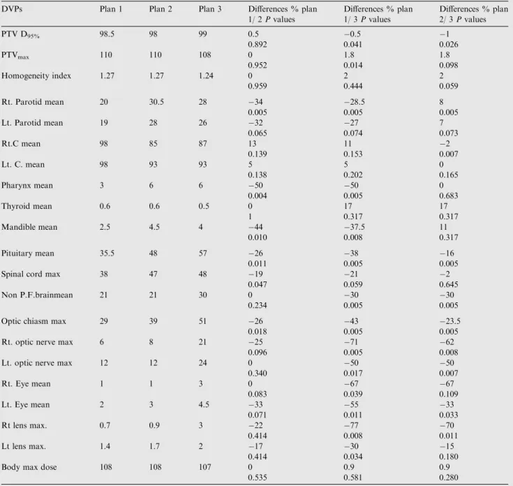
By reviewing the DVPs of the three treatment plans for all patients, the followings are the results as regards the dose dis-tribution of the PTV and OARs including non-posterior fossa brain, pituitary, cochlea, parotid glands, cervical spinal cord, thyroid gland, pharynx, mandible, eyes, lenses, optic nerves and optic chiasma (Table 1andFig. 2):Table 1gives the sta-tistical analysis and DVPs for PTV and OARs comparing the three plans.
5.1. PTV dosimetry
PTV coverage (D95%) is adequate and comparable for plans 1
and 2 and it is significantly better for plan 3. PTV max. dose is comparable for plans 1 and 2 and it is significantly lower for plan 3 compared to plan 1. Although the dose is more homo-geneous for plan 3 compared to plans 1 and 2, theses differ-ences are not significant. Body max dose is accepted and comparable for the three plans.
5.2. OAR dosimetry
Using two parallel opposed lateral open fields (plan 1) shows the least mean doses for parotids, pharynx and mandible fol-lowed by plan 3 and then plan 2. Plan 1 also shows the least
mean dose for pituitary and eyes and the least max. point dose to the spinal cord, optic chiasm, optic nerves and lenses fol-lowed by plan 2 and then plan 3.
Although Plan 2 shows the least mean dose for both cochlea followed by plan 3 then plan 1, Plan 1 & 2 show the least mean dose for non posterior fossa brain followed by plan 3. On the other hand, plan 3 shows the least mean dose for thy-roid followed by plan 1 & 2.Table 1shows the percentage of reduction in DVPs of different OARs between the three plans and whether these reductions are significant or not.
Although the three plans show adequate dose coverage and homogeneity within PTV, using two parallel opposed lateral open fields (plan 1) shows some advantages; the least mean dose for parotids, pharynx and mandible, pituitary, non post fossa brain and eyes and the least max. point dose to the spinal cord, optic chiasm, optic nerves and lenses. On the other hand it shows non significant greatest dose to both cochlea as they are entirely present within the parallel opposed fields. How-ever, the advantage of using a pair of posterior oblique wedged fields (plan 2) and a pair of posterior oblique wedged fields with vertex field (plan 3) is sparing of the cochlea.
6. Discussion
3DCRT for post. fossa boost allows delivery of the prescribed dose to posterior fossa while minimizing the dose to surround-ing OARs. This is important in children with medulloblastoma which is a common childhood malignancy with various critical structures such as the developing brain, bone, thyroid gland, pituitary gland, and cochlea may not be fully developed.3Also, as the majority of patients will survive (long-term survival in this disease is about 55–65%).11 Recent reports highlighted the importance of minimizing the dose to these OARs below their tolerance values during cranio spinal and/or posterior fossa boost irradiation, otherwise, late effects of radiation ther-apy such as somatic and carcinogenic effects may be observed during the follow-up period.3,11
The current study compared three techniques for irradia-tion of post. fossa; two parallel opposed open lateral photon fields, a pair of wedged posterior oblique fields, and a pair of wedged posterior oblique fields with an open vertex field. In the current study, although the average of the mean dose received by both cochlea was greater for plans 2 and 3 (98%,89%and90%) respectively compared with the figures
Figure 1 Field arrangements for the three techniques used in irradiating the posterior fossa: (A) parallel-opposed lateral fields; (B) two posterior oblique wedged fields; (C) two posterior oblique wedged fields with a vertex field. It also shows post fossa in red, both parotids in green, cochlea and pharynx in blue, spinal cord in violet.
achieved by Paulino et al.3(100%,
50%and42%) respectively, on the other hand, the average in the current study was still within tolerance (52 Gy = 16 Gy + 36 Gy; 16 Gy delivered by post fossa boost (90% X 18 Gy) added to 36 Gy from cra-niospinal irradiation).
In the current study the average of the mean dose to non-posterior fossa brain using plans 1 and 2 was comparable and lower than plan 3 which showed the highest average of the mean dose among the three plans (21%, 21% and30%) respectively. Although these figures were comparable to those achieved by Paulino et al.3(22%, 22% and39%) respectively, on the other hand, the mean dose of plan 3 in the current study was lower than what they achieved. In contrast to Paulino et al.3results, the average of the mean dose to the pituitary
gland using plans 2 and 3 was far higher in the current study (35.5%,48%and57%) respectively compared to in their study (36%,19%and21%)respectively.
The average of the mean dose to the mandible was the least for plan 1 followed by plan 3 and then plan 2 (2.5%, 4.5% and 4%) respectively this is in accordance to Paulino et al.3results (8%, 14% and 11%),but our figures were far lower.
The average of the mean dose to thyroid was lower in the current study (0.6%, 0.6%, and 0.5%) respectively compared to Paulino et al.3study (0%, 1% and 3%) respectively.
The average of the mean dose to both parotids was lower in plan 1 followed by plan 3 then plan 2 (19%, 29% and 27%) respectively this agrees with Paulino et al.3 (13%, 34% and 25%) respectively.
Table 1 Comparison of the average of DVPs of PTV and OARs irradiated by three different techniques for post fossa patients (all are in %).Pvalues for the differences between the plans are also shown.
DVPs Plan 1 Plan 2 Plan 3 Differences % plan 1/ 2Pvalues Differences % plan 1/ 3Pvalues Differences % plan 2/ 3Pvalues PTV D95% 98.5 98 99 0.5 0.5 1 0.892 0.041 0.026 PTVmax 110 110 108 0 1.8 1.8 0.952 0.014 0.098 Homogeneity index 1.27 1.27 1.24 0 2 2 0.959 0.444 0.059 Rt. Parotid mean 20 30.5 28 34 28.5 8 0.005 0.005 0.005 Lt. Parotid mean 19 28 26 32 27 7 0.065 0.074 0.073 Rt.C mean 98 85 87 13 11 2 0.139 0.153 0.007 Lt. C. mean 98 93 93 5 5 0 0.138 0.202 0.165 Pharynx mean 3 6 6 50 50 0 0.004 0.005 0.683 Thyroid mean 0.6 0.6 0.5 0 17 17 1 0.317 0.317 Mandible mean 2.5 4.5 4 44 37.5 11 0.010 0.008 0.317 Pituitary mean 35.5 48 57 26 38 16 0.011 0.005 0.005 Spinal cord max 38 47 48 19 21 2
0.047 0.059 0.645 Non P.F.brainmean 21 21 30 0 30 30
0.234 0.005 0.005 Optic chiasm max 29 39 51 26 43 23.5
0.018 0.005 0.005 Rt. optic nerve max 6 8 21 25 71 62
0.096 0.005 0.008 Lt. optic nerve max 12 12 24 0 50 50
0.340 0.017 0.007 Rt. Eye mean 1 1 3 0 67 67 0.083 0.039 0.109 Lt. Eye mean 2 3 4.5 33 55 33 0.071 0.011 0.033 Rt lens max. 0.7 0.9 3 22 77 70 0.414 0.008 0.011 Lt lens max. 1.4 1.7 2 17 30 15 0.414 0.034 0.180 Body max dose 108 108 107 0 0.9 0.9
In contrary to Paulino et al.3 study, the current study showed the average of the mean dose to the pharynx was far lower for the three plans (3%, 6% and 6%) respectively com-pared to the figures in their study (8%, 13% and 12%).
So the results of the current work agree with Paulino et al.3
in; plan 1 shows the lowest mean dose to mandible, parotids and pharynx, plans 1 and 2 show the lowest mean dose to non post fossa brain, and plans 2 and 3 achieve the lowest mean dose to cochlea. For both studies thyroid dose was very low for all plans. On the other hand the results are different in; the lowest dose to pituitary was in plan 1 in the current study and in plan 2 in their work.
In both studies; the main advantage of using posterior obl-iques and post oblobl-iques with vertex is minimizing the dose to both cochlea while the main disadvantage is higher dose to parotid glands, mandible and pharynx, and to non post fossa brain when a vertex field is used.
Fukunaga-Johnson et al.12compared the dose received by both cochlea, pituitary and mandible using plans 1 and 2. The cochlear dose for each patient was calculated as an aver-age between the right and the left cochlea due to symmetry of the problem so the dose distributions between the right and left cochlea were essentially equivalent. For plans 1 and 2, the average of the mean dose of both cochlea was (100%
post. fossa
Pituitary
A B C
Parotid
A B CNon post. fossa brain
A B C
Spinal cord
C A BCochlea
B C AFigure 2 Dose volume histograms for OARs. (A) parallel-opposed lateral fields; (B) two posterior oblique wedged fields; (C) two posterior oblique wedged fields with a vertex field. All are in %.
(98% and 89%) respectively in the current work. The average of the mean dose to the pituitary was far higher in their work (48% and 68%) respectively compared to (35.5% and 48%) respectively in the current work. On the other hand, the aver-age of mean dose of the mandible was far lower in their work (0.8% and 1.8%) respectively compared to (2.5% and 4.5%) respectively in the current work.
So for both studies; plan 1 shows the highest dose to cochlea, and the lowest dose to pituitary and mandible compared to plan 2. Moreover in the current study our figures for pituitary only were far lower than those achieved in their study.
In Breen et al.1study using opposing and oblique fields, the average of the mean dose to the cochlea delivered by both plans respectively was lower (97–100% and 65–84%) respec-tively compared to (98% and 89%) respecrespec-tively in our work. For none post fossa brain; the average of the mean dose was far higher (46–61% and 36–51%) respectively compared to (21% and 21%) in our work. For optic nerves; the max point dose was lower (2–2.8% and 4–13%) respectively compared to (6% and 12%) respectively in our work. For lenses; max. Point dose was higher (0.9–1.4% and 1.8–2.3%) respectively com-pared to (0.7–0.9% and 1.4–1.7%) respectively in our work. For pituitary; the average of the mean dose was higher (15– 38% and 56–60%) respectively compared to (35.5% and 48%) respectively in our work. For the spinal cord; the max point dose was higher (14–77% and 14–78%) respectively compared to (38% and 47%) respectively in our work). They attributed their results to; the use of posterior oblique fields can exclude the cochlea from the fields, thereby reducing their dose, however, as the exit dose will be in structures anterior and close by to the posterior fossa so their doses are increased compared to opposed lateral fields. Comparing both studies, except for the mean dose of cochlea and max. point dose of optic nerve which were higher in the current study, our figures are far lower than those achieved in their study.
3DCRT open opposed lateral fields showed the lowest dose to all critical structure except both cochlea which received the target dose because of their proximity to the target volume. However as the average of the mean dose to cochlea (53.6Gy) was still within tolerance (17.6Gy delivered by poster-ior fossa boost (98% X 18Gy) added to 36Gy from craniospi-nal irradiation) so we recommend to use it for posterior fossa boost irradiation.
7. Conclusion
3DCRT using parallel opposed fields is recommended for pos-terior fossa irradiation boost as it minimizes the exit dose to all structures other than the cochlea, however its mean dose was within the tolerance.
None declared.
References
1. Breen SL, Kehagioglou P, Usher U, Plowman PN. A comparison of conventional, conformal and intensity-modulated coplanar radiotherapy plans for posterior fossa treatment.British J Radiol 2004;77:768–74.
2. El-Gaidi MA. Descriptive Epidemiology of Pediatric Intracranial Neoplasms in Egypt.Pediatr Neurosurg2011;47:385–95. 3. Paulino AC, Ashwatha Narayana M, Najeeb Mohideen,
Andsam Jeswani, C. Posterior Fossa Boost In Medulloblastoma An Analysis Of Dose To Surrounding Structures Using 3Dimensional Conformal Radiotherapy.Int J Radiation Oncology Biol Phys2000;46(2):281–6.
4. Packer RJ, Gajjar A, Vezina G, et al. Phase III study of craniospinal radiation therapy followed by adjuvant chemother-apy for newly diagnosed average-risk medulloblastoma. J Clin Oncol2006;24:4202–8.
5. Merchant TE, Wang MH, Haida T, et al. Medulloblastoma. Long-term results for patients treated with definitive radiation therapy during the computed tomography era.Int J Radiat Oncol Biol Phys1996;36:29–35.
6. Fukunaga-Johnson N, Lee JH, Sandler HM, et al. Patterns of failure following treatment for medulloblastoma: is it necessary to treat the entire posterior fossa? Int J Radiat Oncol Biol Phys 1998;42:143–6.
7. Hua C, Bass JK, Khan R, et al. Hearing loss after radiotherapy for pediatric brain tumors: effect of cochlear dose.Int J Radiat Oncol Biol Phys2008;72:892–9.
8. Kortmann RD, Kuhl J, Timmermann B, et al. Postoperative neoadjuvant chemotherapy before radiotherapy as compared to immediate radiotherapy followed by maintenance chemotherapy in the treatment of medulloblastoma in childhood: results of the German prospective randomized trial HIT ’91.Int J Radiat Oncol Biol Phys2000;46:269–79.
9. Paulino AC, Lobo M, Teh BS, et al. Ototoxicity after intensity-modulated radiation therapy and cisplatin-based chemotherapy in children with medulloblastoma. Int J Radiat Oncol Biol Phys 2010;78:1445–50.
10. Huang E, Teh BS, Strother DR, et al. Intensity-modulated radiation therapy for pediatric medulloblastoma: early report on the reduction of ototoxicity. Int J Radiat Oncol Biol Phys 2002;52:599–605.
11. Kun LE. Malignant brain tumors including medulloblastoma, embryonal neuroectodermal tumors, and tumors of the pineal region, with a special discussion of the management of brain tumors in children of 3 years and younger. In: Cassady JR, editor. Radiation Therapy in Pediatric Oncology. first ed. Berlin, Heidel-berg: Springer-Verlag; 1994. p. 197–213.
12. Fukunaga-Johnson N, Sandler HM, Marsh R, et al. The use of 3D conformal radiotherapy (3D CRT) to spare the cochlea in patients with medulloblastoma. Int J Radiat Oncol Biol Phys 1998;41:77–82.