International Journal of Innovation Scientific Research and Review Vol. 02, Issue, 12, pp.599-603, December, 2020
Available online at http://www.journalijisr.com
Research Article
ISSN: 2582-6131
EVALUATION OF LIVER LEISONS ON BIPHASIC COMPUTED TOMOGRAPHY (CT)
SCAN- AN OBSERVATIONAL STUDY
*
Nazeeha Waseem
1, Kainat Saleem
2, Umair Waqas
3, Aroosh Akhtar
4, Saleha Liaqat
5
1 Medical Imaging Doctor, The University of Lahore, Gujrat 2, 4,5 Medical Imaging Technologists, Islamabad Diagnostic Centre
3 Lecturer, The University of Lahore, Gujrat
Received 14th September 2020; Accepted 10th November 2020; Published online 30th December 2020 ABSTRACT
Purpose: The purpose of this study was to evaluate liver lesions on dual phase computed tomography scan (CT scan) especially in the HCV patients having cirrhotic liver. One of the significant aspects is to improve the diagnostic facility for hepatocelluar carcinoma (HCC), besides its prevention. Early stage diagnosis is helpful in asymptomatic patients where liver function remains preserved. It also provides better opportunities for tumor treatment via medications and radio therapy. Radio imaging is an imperative tool to diagnose HCC in early stages. HCC shows typical vascular enhancement on arterial phase in CT scan and MRI, and washout in venous phase. This review summarizes the current knowledge about imaging techniques for early diagnosis and staging of HCC. Primary hepatocellular carcinoma is among 10 most common tumors, and is one of the most common primary liver malignancies in the world. Method and material: 130 patients were evaluated on ultrasound to have liver defect. 106 were confirmed to possess liver lesions that were evaluated on biphasic CT scan. After maintaining the IV line, injection was injected through pressure injector and acquisition was started. At 15-20 sec arterial phase was taken and at 50-60 second portal venous phase was taken. Result: Out of 106 patients, 64.71% possessed hepatocecullar carcinoma along with HCV and cirrhotic liver. The patients diagnosed with HCC were 23.53%. The percentage of patients diagnosed with HCC together with HCV was same as patients having HCC and cirrhotic liver i.e., 5.88%. Conclusion: Biphasic computer tomography is a great diagnostic tool for the detection of liver lesions where lesion conspicuity is dependent on vascularity of the lesion.
Keywords:
Dual phase computer tomography, HCC, HCV, cirrhotic liver, liver lesion.
INTRODUCTION
The most frequent disease of liver is focal liver lesions. Primarily liver lesions are of two types i.e. benign and malignant. Benign lesions are mostly cystic lesions, focal nodular, hyperplasia infections, hemangioma, hepatic adenoma, billary hamartoma, regenerative nodule, and focal fatty liver. However, malignant lesions are generally metastatic, hepatocellular carcinoma cholangio carcinoma, lymphoma and sarcoma (Algarni, Alshuhri, Alonazi, Mourad, and Bramhall, 2016). The liver infections may be viral, bacterial, parasitic and amebic. Hemangiomas are most common vascular liver lesions (Lv, Lin, Li, Li, and Chen, 2011). These lesions may result due to the dilation of existing blood vessels (Marrero, Ahn, and Reddy, 2014). The hepatic cyst may be abscess, cyst adenoma, metastatic cyst and hydatid cyst (Singh, Winick, and Tabbara, 1997). A simple cyst is regular thin walled, unilocular and generally asymptomatic, while a large cyst may be slightly symtomatic (Bailey, 1995). Presence of thick separation or masses inside the cyst may enhance its vascularity then it may be termed as abscess or neoplastic cyst. On the other hand if abscess is infectious due to Echinococcus then it is
known as hydatid cyst (Singh et al., 1997). Cell necrosis or blood
circulation alternation may derive regenerative nodules formation
(Hanna et al., 2008). Hepatic adenoma is rarely found benign tumor
usually formed in females consuming hormonal medicines or oral
contraceptives (Assy et al., 2009; Marrero et al., 2014). Focal
nodular hyperplasia is a solid and hyper vascular lesion (Murakami and Tsurusaki, 2014. Cholangio carcinomas may be intra hepatic or
extra hepatic cholangio carcinomas (Marrero et al., 2014). These are
hyper vascular malignant lesions (Murakami and Tsurusaki, 2014). Liver is the most common site where the mates can move from gastrointestinal tract, pancreas, breast and lungs.
*Corresponding Author: Nazeeha Waseem,
Medical Imaging Doctor, The University of Lahore, Gujrat
These mates can also be calcified (Assy et al., 2009). Computer
tomography is clinically very important in the diagnostics. It has high accuracy in diagnosis and staging of liver lesions. The development of rapid scanning technique has played a vital role in accurate
diagnosis (Hollett et al., 1995). It has become the standard for
detection and characterization of the liver lesions (Baron et al., 1996;
Hollett et al., 1995; Hwang, Kim, Yoo, and Lee, 1997; Ohashi,
Hanafusa, and Yoshida, 1993). Increased enhancement of tumor compared with the surrounding liver parenchyma during hepatic arterial phase is the cornerstone for diagnosis of lesion at multi phasic MDCT (Khan and Hafeez, 2017). By using the images some important parameters are taken e.g. size of lesion, vascularity of lesion etc. It is important to use high injection rates and appropriate bolus timing. The liver tumors will be strongly enhanced during the arterial phase (beginning 15-20 sec after the start of a bolus injection) but of similar density to enhanced normal parenchyma during the portal venous phase (50-60 sec). Some tumors are most conspicuous during early-phase arterial scanning (25 sec after the start of bolus injection), others later, during the late arterial phase 35 sec after the start of bolus injection. After the initial triphasic-phase protocol, delayed images at 5 and 10 minutes are obtained (Chapman and Nakielny, 2001). On the basis of lesion enhancement, the lesions can be divided into two types 1) Hyper vascular lesion 2) Hypo vascular lesion. The hyper vascular lesions are those having high concentration of blood vessels due to which has high ability to grow. For example: HCC, focal nodular hyperplasia etc. The hypo vascular lesions are those having low concentration of blood vessels due to
whichhaslowabilitytogrow. For example: fattyliver,livercirrhosisetc.
MATERIALS AND METHODS
The study was conducted at Islamabad Diagnostic Center Faisalabad (October 2019 to January 2020). The data was collected through CT
films and reports by using convenient (non-probability) sampling technique. Sample size was 130 patients and patients of all age groups especially diagnosed with hepatitis C were included. Exclusion criteria included patients having serum creatinine level more than 1.5mg/dl, no liver mass on abdominal ultrasound and pregnant ladies. All the patients fulfilling the inclusion criteria were included in the study. Multi detector 16 slice helical CT scan machine (Toshiba) was utilized for taking images. All CT reports were diagnosed by the experienced radiologists. A multi slice computer tomography was used to scan the liver in multiple times in a very short duration using a single bolus injection of contrast medium. Patients having normal renal function tests i.e. urea and creatinine level were considered to be fit for the contrast media administration. Patients were asked to sign the informed consent form and were guided regarding lying down in supine position and restrict movements was addressed.
Table1. Lesions on CT-Scan (Assy et al.,
Lesion On CT
Haemangioma Peripheral puddles, fill in from periphery, enhancement on delayed scan Focal fatty liver Sharp interface and low density
Focal nodular hyperplasia
Homogeneous enhance strongly with hepatic arterial phase Iso dense with liver; Central low density scar
Hepatoadenoma Homogenous > Heterogeneous,Peripheral feeders filling in from periphery Hepatocellular
carcinoma
Hyper vascular, often irregular borders, Heterogeneous > Homogeneous
abnormal internal vessel, Hallmark is venous washout Cholangiocarcinoma Hypo dense lesion. Delayed enhancement
Metastasis Complete ring enhancement
The 18-20 gauge cannula was passed and IV line was fixed. Pressure injector was used to inject the 100ml of contrast media. Initially scan gram image was taken to draw the field of view after that bolus injection was injected and scan was started.
taken after 15-20 seconds of the start of bolus injection. Then at 50 60 seconds after injection portal venous phase was taken. Early and late arterial phase were also be taken. Early arterial phase was taken at 15 seconds and late arterial phase was taken at 35 seconds, since some lesions are more conspicuous during early or late arterial phase. The lesions were best visualized in contrast enhanced axial images of arterial phase of biphasic CT. Mostly axial images were preferred but sagittal and coronal images were also taken for accurate evaluation. All data was analyzed by using Microsoft excel 365. Mean and percentages were calculated for all the numerical or quantitative variable such as gender, frequency of hyper vascular lesion, hypo vascular lesion and cirrhotic liver with lesion. Graphs, tables, component and multiple bar charts and pie chart were used to express different frequencies.
RESULTS
Out of 130 patients, 24 patients showed normal liver on CT scan e.g., they had other problems like cholelithiasis, hence were excluded from the study. 106 patients represented with liver lesions. Out of these 130 patients, 67(52%) were females and 63(48%) were males.
Figure1. Gender distribution among the sample size
F
52%
M
48%
Total
International Journal of Innovation Scientific Research and Review
probability) sampling technique. Sample size was 130 patients and patients of all age groups especially diagnosed with hepatitis C were included. patients having serum creatinine level more than 1.5mg/dl, no liver mass on abdominal ultrasound and pregnant ladies. All the patients fulfilling the inclusion criteria were included in the study. Multi detector 16 slice helical CT scan machine was utilized for taking images. All CT reports were diagnosed by the experienced radiologists. A multi slice computer tomography was used to scan the liver in multiple times in a very short duration using a single bolus injection of contrast medium. ts having normal renal function tests i.e. urea and creatinine level were considered to be fit for the contrast media administration. Patients were asked to sign the informed consent form and were guided regarding lying down in supine position and restriction of
et al., 2009)
Peripheral puddles, fill in from periphery, enhancement on delayed scan
Homogeneous enhance strongly with hepatic arterial phase Iso dense with
Homogenous > Heterogeneous,Peripheral feeders filling in from periphery
abnormal internal vessel, Hallmark is venous washout
20 gauge cannula was passed and IV line was fixed. Pressure injector was used to inject the 100ml of contrast media. image was taken to draw the field of view after that bolus injection was injected and scan was started. Arterial phase was 20 seconds of the start of bolus injection. Then at 50-60 seconds after injection portal venous phase was taken. Early and late arterial phase were also be taken. Early arterial phase was taken e was taken at 35 seconds, since some lesions are more conspicuous during early or late arterial phase. The lesions were best visualized in contrast enhanced axial images of arterial phase of biphasic CT. Mostly axial images were coronal images were also taken for accurate evaluation. All data was analyzed by using Microsoft excel 365. Mean and percentages were calculated for all the numerical or quantitative variable such as gender, frequency of hyper vascular ar lesion and cirrhotic liver with lesion. Graphs, tables, component and multiple bar charts and pie chart were used to
Out of 130 patients, 24 patients showed normal liver on CT scan e.g., ike cholelithiasis, hence were excluded from the study. 106 patients represented with liver lesions. Out of these 130 patients, 67(52%) were females and 63(48%) were males.
Gender distribution among the sample size
For hyper vascular lesion like for HCC, the lesions in the HCC showed arterial enhancement and also venous enhancement or washout phase. This was also noticed for focal nodular hyperplasia and metastatic patients. Out of 106 patients, 75patients presented hyper vascular lesions i.e., showed both phases of biphasic CT scan in 100% cases. In the hemangiomas there was complete enhancement in both phases and no washout phase.
The graph shows the enhancement in both phases.
Figure 2. Arterial and venous comparison for Hyper vascular
Figure3. Arterial to Venous Enhancement Percentage for Hemangioma This study concluded that all hyper vascular lesions have arterial enhancements as well as venous enhancements on biphasic computer tomography. Finally the result for hyper
100% in arterial phase. Out of 106 patients, 31 patients presented hypo vascular lesions and showed no enhancement in both phases of CT scan. In this result 29% represented with hypo vascular lesions and 71% with hyper vascular lesions.
enhancement pattern but basically they are showed enhancement or no enhancement on the bases of their vascularization.
Figure 4. Ratio of patient on the bases of 2 types of lesions
F M 100.00%, 100.00%, 100% 100.00% 0.00% 20.00% 40.00% 60.00% 80.00% 100.00% 120.00%
Research and Review
like for HCC, the lesions in the HCC showed arterial enhancement and also venous enhancement or washout phase. This was also noticed for focal nodular hyperplasia and metastatic patients. Out of 106 patients, 75patients presented ., showed both phases of biphasic CT scan In the hemangiomas there was complete enhancement in both phases and no washout phase.
The graph shows the enhancement in both phases.
Arterial and venous comparison for Hyper vascular Lesion
Arterial to Venous Enhancement Percentage for Hemangioma This study concluded that all hyper vascular lesions have arterial enhancements as well as venous enhancements on biphasic Finally the result for hyper vascular lesion was 100% in arterial phase. Out of 106 patients, 31 patients presented hypo vascular lesions and showed no enhancement in both phases of In this result 29% represented with hypo vascular lesions and 71% with hyper vascular lesions. Although all have different enhancement pattern but basically they are showed enhancement or no enhancement on the bases of their vascularization.
Ratio of patient on the bases of 2 types of lesions
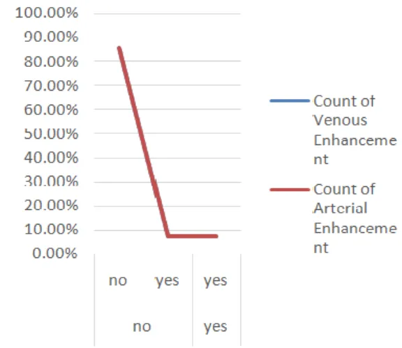
100.00%, 100% yes washout 100.00% yes yes yes Count of Arterial Enhancement Count of Venous Enhancement 600
Figure 5. Arterial to Venous Enhancement for Hypo vascular Lesion Conclusively, lesions of two types were presented and both types showed two different percentage of enhancement on arterial and venous phase.
Table 2. Lesions Enhancement
Lesions Arterial Enhancement Venous Enhancement
Hyper vascular 100% 100%
Hypo vascular 0.0% 0.0%
In these 71% hyper vascular patients, they possessed hepatocecullar carcinoma with HCV along with cirrhotic liver were 64.71%. The patients having only HCC were 23.53%. The patients having HCV and HCC are 5.88%. The patients having HCC with cirrhotic liver are 5.88%.
Figure 6. Relation between HCC, HCV and cirrhotic liver
DISCUSSION
The vascularity of a liver lesion is still a point of
work on animals showed that lesion in the liver acquires its blood
supply from hepatic artery. (Breedis et al., lien et al.) However recent
work evaluated that portal vein also contributes in supplying the blood. It may be through collaterals (Akeraman et al.,Lin
akeraman et al., Matsui et al., Kan et al.,). According to Breedis
adenocarcinoma is circulated by 80% hepatic artery supply and 20% portal vein supply. However, prominent blood supply of liver lesion is hepatic artery. As the size of tumor increases, the vascularity of lesion depends upon the type of lesion. The main two types of lesions are hyper vasular and hypo vascular. The hyper vascular malignant liver lesions are best seen on the arterial phase of
tomography. Hyper vascular lesions show full enhancement on arterial phase (Hollett et al.,) (Figure 7). This study correlated with these results as 100% enhancement of hyper vascular lesion was observed.
International Journal of Innovation Scientific Research and Review
Arterial to Venous Enhancement for Hypo vascular Lesion Conclusively, lesions of two types were presented and both types showed two different percentage of enhancement on arterial and
Lesions Enhancement
Venous Enhancement Percentage of patients
71% 29%
In these 71% hyper vascular patients, they possessed hepatocecullar carcinoma with HCV along with cirrhotic liver were 64.71%. The having only HCC were 23.53%. The patients having HCV and HCC are 5.88%. The patients having HCC with cirrhotic liver are
Relation between HCC, HCV and cirrhotic liver
The vascularity of a liver lesion is still a point of conflict. The early work on animals showed that lesion in the liver acquires its blood lien et al.) However recent work evaluated that portal vein also contributes in supplying the
terals (Akeraman et al.,Lin et al., N.B
Kan et al.,). According to Breedis et al.,
adenocarcinoma is circulated by 80% hepatic artery supply and 20% portal vein supply. However, prominent blood supply of liver lesion is hepatic artery. As the size of tumor increases, the vascularity of lesion depends upon the type of lesion. The main two types of lesions The hyper vascular malignant liver lesions are best seen on the arterial phase of computer tomography. Hyper vascular lesions show full enhancement on arterial phase (Hollett et al.,) (Figure 7). This study correlated with these results as 100% enhancement of hyper vascular lesion was
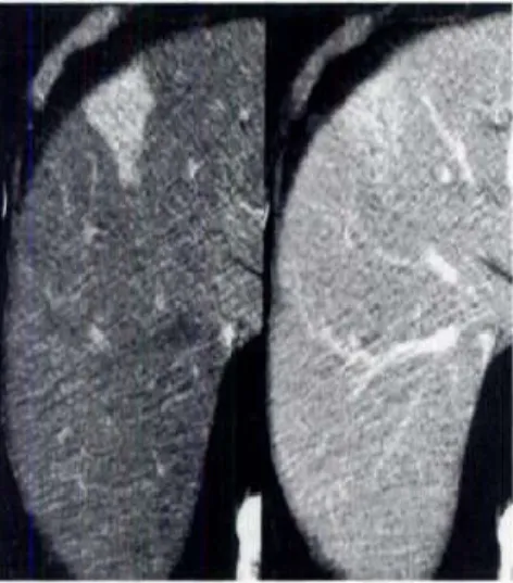
Figure 7. The Hyper vascular Lesion on
This figure represents that hyper vascular lesion show enhancement on the arterial phase because of the involvement of hepatic artery. Biphasic computer tomography is also convenient for hypo vascular lesion like adenomas and infections. Mostly hypo vascular lesions are best seen on the portal venous phase. There relative attenuation difference is checked between lesion and normal parenchyma of liver. (Figure 8)
Figure 8. Hypo vascular Lesion on Portal Venous
The study shows 7.14% enhancement on portal venous even though biphasic computer tomography is best for hyper vascular lesion but in some cases it was also seen the lesion become iso intense with liver after the contrast administration, especially in the cases w dose of contrast injection is not enough. Then it is more convenient to see lesion on unenhanced scan (Bressler
scan is not suitable for detection of small lesions because it becomes difficult to differentiate between small lesions and hyper tense vessels. It relates to the low sensitivity of the unenhanced CT for detection of liver lesion. Therefore, it can be stated that arterial phase is more suitable to detect the small lesions (Heiken
phase maximizes the possibility for detection of small lesions and hyper vascular lesions. Previously, biphasic computer tomography was used to evaluate only hepatocellular carcinoma with a very slow rate of contrast infusion and the delayed scan was taken at the 6 minutes after the contrast administered (Ohashi
arterial phase was required then it became compulsory that the scanner should be capable enough to capture images of the entire liver in a very short time such as 6 second after the infusion of contrast material arterial phase is taken and then patient is allowed to breath for few seconds like 10-20 second, at 50
venous phase is taken (Heiken et al
is set to maximize the arterial enhancement.
the lesion detection in all types of lesions as well as the venous phase. But alone venous phase is n
hyper vascular lesions are more enhanced on biphasic CT as compared to hypo vascular lesions. But there can also be chances of false detection of lesion like in Hollett
Research and Review
Hyper vascular Lesion on the Arterial Phase of CT Scan This figure represents that hyper vascular lesion show enhancement on the arterial phase because of the involvement of hepatic artery. Biphasic computer tomography is also convenient for hypo vascular infections. Mostly hypo vascular lesions are best seen on the portal venous phase. There relative attenuation difference is checked between lesion and normal parenchyma of liver.
Hypo vascular Lesion on Portal Venous Phase udy shows 7.14% enhancement on portal venous even though biphasic computer tomography is best for hyper vascular lesion but in some cases it was also seen the lesion become iso intense with liver after the contrast administration, especially in the cases when the dose of contrast injection is not enough. Then it is more convenient to see lesion on unenhanced scan (Bressler et al.). The unenhanced scan is not suitable for detection of small lesions because it becomes difficult to differentiate between small lesions and hyper tense vessels. It relates to the low sensitivity of the unenhanced CT for ore, it can be stated that arterial phase is more suitable to detect the small lesions (Heiken et al.). Arterial phase maximizes the possibility for detection of small lesions and Previously, biphasic computer tomography evaluate only hepatocellular carcinoma with a very slow rate of contrast infusion and the delayed scan was taken at the 6 minutes after the contrast administered (Ohashi et al.,). But when the arterial phase was required then it became compulsory that the scanner should be capable enough to capture images of the entire liver in a very short time such as 6 second after the infusion of contrast material arterial phase is taken and then patient is allowed to 20 second, at 50-60 second portal et al.,). An appropriate rate of injection is set to maximize the arterial enhancement. Arterial phase improve the lesion detection in all types of lesions as well as the venous phase. But alone venous phase is not reliable. As the result show that hyper vascular lesions are more enhanced on biphasic CT as compared to hypo vascular lesions. But there can also be chances of false detection of lesion like in Hollett et al.
Figure 9. A False Positive Liver Lesion Shown on the CT Arterial Phase in a female of 60 Years and Its Correction on MRI
CONCLUSION
Biphasic computer tomography is best diagnostic technique for the detection of liver lesion. Although some lesion does not show any enhancement on the arterial or venous or on both phases but biphasic study increases the conspicuous of lesion for the radiologist. And it becomes easier to identify and differentiate the lesions through contrast enhanced images.
REFERENCES
Ackerman, N. B. (1986). Experimental studies on the role of the portal circulation in hepatic tumour vascularity. Cancer, 58(8), 1653-1657.
Ackerman, N. B. The blood supply ofexperimental liver metastases. IV. Changes in vascularity with increasing tumor growth. Surgeiyl9l4, 75, 589-596.
Algarni, A. A., Alshuhri, A. H., Alonazi, M. M., Mourad, M. M., and Bramhall, S. R. (2016). Focal liver lesions found incidentally. World J Hepatol, 8(9), 446-451. doi: 10.4254/wjh.v8.i9.446 Assy, N., Nasser, G., Djibre, A., Beniashvili, Z., Elias, S., and Zidan,
J. (2009). Characteristics of common solid liver lesions and recommendations for diagnostic workup. World journal of gastroenterology: WJG, 15(26), 3217.
Bailey, H., Love, R. J. M. N., Mann, C. V., and Russell, R. C. G. (1992). Bailey and Love's short practice of surgery. London: Chapman and Hall Medical.
Baron, R. L., Oliver 3rd, J., Dodd 3rd, G., Nalesnik, M., Holbert, B. L., and Carr, B. (1996). Hepatocellular carcinoma: evaluation with biphasic, contrast-enhanced, helical CT. Radiology, 199(2), 505-511.
Bonaldi, V. M., Bret, P. M., Reinhold, C., and Atri, M. (1995). Helical CT of the liver: value of an early hepatic arterial phase. Radiology, 197(2), 357-363.
Breedis, C., and Young, G. (1954). The blood supply of neoplasms in the liver. The American journal of pathology, 30(5), 969.
Bressler, E. L., Alpern, M. B., Glazer, G. T., Francis, I. R., and Ensminger, W. D. (1987). Hypervascular hepatic metastases: CT evaluation. Radiology, 162(1), 49-51.
Chapman, S., and Nakielny, R. (2001). A guide to radiological procedures. Edinburgh [u.a.: Saunders.
Choi, B. I., Han, J. K., Cho, J. M., Choi, D. S., Han, M. C., Lee, H. S., and Kim, C. Y. (1995). Characterization of focal hepatic tumours
value of two‐phase scanning with spiral computed tomography.
Cancer, 76(12), 2434-2442.
Hanna, R. F., Aguirre, D. A., Kased, N., Emery, S. C., Peterson, M. R., and Sirlin, C. B. (2008). Cirrhosis-associated hepatocellular nodules: correlation of histopathologic and MR imaging features. Radiographics, 28(3), 747-769.
Heiken, J. P., Brink, J. A., McClennan, B. L., Sagel, S. S., Forman, H. P., and DiCroce, J. (1993). Dynamic contrast-enhanced CT of the liver: comparison of contrast medium injection rates and uniphasic and biphasic injection protocols. Radiology, 187(2), 327-331
Hiatt, J. R., Gabbay, J., and Busuttil, R. W. (1994). Surgical anatomy of the hepatic arteries in 1000 cases. Annals of surgery, 220(1), 50.
Hollett, M., Jeffrey Jr, R., Nino-Murcia, M., Jorgensen, M., and Harris, D. (1995). Dual-phase helical CT of the liver: value of arterial phase scans in the detection of small (< or= 1.5 cm) malignant hepatic neoplasms. AJR. American journal of roentgenology, 164(4), 879-884.
Hwang, G. J., Kim, M.-J., Yoo, H. S., and Lee, J. T. (1997). Nodular hepatocellular carcinomas: detection with arterial-, portal-, and delayed-phase images at spiral CT. Radiology, 202(2), 383-388. Ichikawa, T., Kitamura, T., Nakajima, H., Sou, H., Tsukamoto, T.,
Ikenaga, S., and Araki, T. (2002). Hypervascular hepatocellular carcinoma: can double arterial phase imaging with multidetector CT improve tumour depiction in the cirrhotic liver? American Journal of Roentgenology, 179(3), 751-758.
Kan, Z. U. X. I. N. G., Ivancev, K., Lunderquist, A., McCuskey, P. A., Wright, K. C., Wallace, S., and McCuskey, R. S. (1993). In vivo microscopy of hepatic tumours in animal models: a dynamic investigation of blood supply to hepatic metastases. Radiology, 187(3), 621-626.
Kanematsu, M., Oliver 3rd, J. H., Carr, B., and Baron, R. L. (1997). Hepatocellular carcinoma: the role of helical biphasic contrast-enhanced CT versus CT during arterial portography. Radiology, 205(1), 75-80.
Khan, A. A., and Hafeez, M. (2017). THE ROLE OF COMPUTED TOMOGRAPHY IN THE DIAGNOSIS OF HEPATOCELLULAR CARCINOMA IN VIRAL HEPATITIS B AND C PATIENTS. PJR, 27(3).
Kim, J. E., Kim, H. O., Bae, K., Cho, J. M., Choi, H. C., and Choi, D. S. (2017). Differentiation of small intrahepatic mass-forming cholangiocarcinoma from small liver abscess by dual source dual-energy CT quantitative parameters. European Journal of Radiology, 92, 145-152.
Liau, K. H., Blumgart, L. H., and DeMatteo, R. P. (2004). Segment-oriented approach to liver resection. Surg Clin North Am, 84(2), 543-561. doi: 10.1016/j.suc.2003.12.003
Lien WM, Ackerman NB. The blood supply of experimental liver metastases II. A microcirculatory study of the normal and tumor vessels of the liver with the use of perfused silicone rubber. Surgery 1970;68:334-340
Lien, W. M. (1970). The blood supply of experimental liver metastasis. Surgery, 68, 334-340.
Lin, G., Lunderquist, A., Hägerstrand, I., and Boijsen, E. (1984). Postmortem examination of the blood supply and vascular pattern of small liver metastases in man. Surgery, 96(3), 517-526. Lv, P., Lin, X. Z., Li, J., Li, W., and Chen, K. (2011). Differentiation of
small hepatic hemangioma from small hepatocellular carcinoma: recently introduced spectral CT method. Radiology, 259(3), 720-729. doi: 10.1148/radiol.11101425
Marrero, J. A., Ahn, J., and Reddy, K. R. (2014). ACG clinical guideline: the diagnosis and management of focal liver lesions. The American journal of gastroenterology, 109(9), 1328-1347. Monzawa S1, Ichikawa T, Nakajima H, Kitanaka Y, Omata K, Araki
T.Dynamic CT fordetecting small hepatocellular carcinoma:
usefulness of delayed phase imaging. AJR Am J Roentgenol 2007 Jan;188(1):147-53
Murakami, T., and Tsurusaki, M. (2014). Hypervascular benign and malignant liver tumors that require differentiation from hepatocellular carcinoma: key points of imaging diagnosis. Liver Cancer, 3(2), 85.
Netter, F. H. (2006). Atlas of human anatomy. Philadelphia, PA: Saunders/Elsevier.
Ohashi, I., Hanafusa, K., and Yoshida, T. (1993). Small hepatocellular carcinomas: two-phase dynamic incremental CT in detection and evaluation. Radiology, 189(3), 851-855.
Sibulesky, L. (2013). Normal liver anatomy. Clinical Liver Disease, 2(S1).
Singh, Y., Winick, A. B., and Tabbara, S. O. (1997). Multiloculated cystic liver lesions: radiologic-pathologic differential diagnosis. Radiographics, 17(1), 219-224.
Snell, R. S. (2012). Clinical anatomy by regions. Baltimore, MD: Lippincott Williams and Wilkins.
Soler, L., Delingette, H., Malandain, G., Montagnat, J., Ayache, N., Koehl, C., . . . Marescaux, J. (2001). Fully automatic anatomical, pathological, and functional segmentation from CT scans for hepatic surgery. Computer Aided Surgery, 6(3), 131-142. doi: 10.1002/igs.1016
Takahashi, S., Murakami, T., Takamura, M., Kim, T., Hori, M., Narumi, Y., ... and Kudo, M. (2002). Multi–detector row helical CT angiography of hepatic vessels: Depiction with dual-arterial phase acquisition during single breath hold. Radiology, 222(1), 81-88. Vignaux, O., Legmann, P., Coste, J., Hoeffel, C., and Bonnin, A.
(1999). Cirrhotic liver enhancement on dual-phase helical CT: comparison with noncirrhotic livers in 146 patients. AJR. American journal of roentgenology, 173(5), 1193-1197.
Weg, N., Scheer, M. R., and Gabor, M. P. (1998). Liver lesions: improved detection with dual-detector-array CT and routine 2.5-mm thin collimation. Radiology, 209(2), 417-426.