Caval Sonography in Shock
A Noninvasive Method for Evaluating Intravascular
Volume in Critically Ill Patients
onography has traditionally been used to assess anatomic abnormalities. However, its value in evaluating physiologic characteristics has recently been recognized, particularly in the care of patients in shock. As the use of point-of-care sonogra-phy grows in critical care and emergency medicine, noninvasive assessment of intravascular volume status is increasingly being used to guide therapy of the critically ill.
Although intravenous fluid is often the initial treatment in hypotensive patients, aggressive volume resuscitation may be detri-mental in some patients and in certain types of shock. Accurate diag-nosis of shock state can be challenging because physical findings of hypovolemic, distributive, cardiogenic, and obstructive shock often overlap. Pulmonary artery and central venous pressure catheters, which provide physiologic data such as cardiac output and right atrial pressure, are time-consuming, invasive, and carry consider-able risks. Central venous pressure has long been used to guide fluid management; however, data suggest that in critically ill patients, central venous pressure may not correlate with the effective intravas-cular volume.1Furthermore, invasive hemodynamic monitoring has not been shown to benefit patients.2
Given the importance of determining intravascular volume in shock, a rapid bedside sonographic examination can be instrumen-tal in guiding medical management of critically ill patients. Multiple sonographic protocols now exist for the evaluation of shock, dysp-nea, and cardiac arrest.3,4This article will describe the use of sonog-raphy of the inferior vena cava (IVC) in the evaluation of patients in shock.
Physiology: IVC Parameters
The IVC is a compliant vessel that distends and collapses with pres-sure and volume changes. Although the absolute IVC size varies widely among healthy individuals and may not by itself be diagnos-tic, the maximal IVC diameter has been shown to be lower in patients with hypovolemia.5
A better indicator of intravascular volume is collapsibility of the IVC. As intrathoracic pressure decreases with inspiration, venous blood is pulled from the lower half of the body into the right atrium. This action causes a transient, but normal, decrease in the IVC diameter. With expiration, the IVC diameter increases and returns
Dina Seif, MD, MBA, RDMS, Thomas Mailhot, MD, RDMS, Phillips Perera, MD, RDMS, Diku Mandavia, MD
Received August 12, 2012, from the Department of Emergency Medicine, Los Angeles County and University of Southern California Medical Cen-ter, General Hospital, Los Angeles, California USA (D.S., T.M., D.M.); and Division of Emer-gency Medicine, Stanford University Medical Cen-ter, Stanford, California USA (P.P.). Revision requested August 31, 2012. Revised manuscript accepted for publication September 25, 2012.
Drs Seif and Perera are educational con-sultants for SonoSite, Inc (Bothell, WA), and Dr Mandavia is employed by SonoSite, Inc.
Address correspondence to Dina Seif, MD, MBA, RDMS, Department of Emergency Medi-cine, Los Angeles County and University of South-ern California Medical Center, 1200 N State St, 1011, Los Angeles, CA 90033 USA.
E-mail: [email protected] Abbreviations
IVC, inferior vena cava
S
Invited paper
Videos online at www.jultrasoundmed.org
The Sound Judgment Series consists of invited articles highlighting the clinical value of using ultrasound first in specific clinical diagnoses where ultrasound has shown comparative or superior value. The series is meant to serve as an educational tool for medical and sonography students and clinical practitioners and may help integrate ultrasound into clinical practice.
to its baseline. These changes are known as respirophasic variability. The IVC collapsibility index, also known as the caval index, is defined as the difference between the maxi-mal (expiratory) and minimaxi-mal (inspiratory) IVC diameters divided by the maximal diameter. The caval index is used in spontaneously breathing patients to estimate right atrial pressure.6,7In patients with minimal respirophasic collapse, having the patient inspire forcefully, or sniff, will differenti-ate between patients with poor inspiratory effort and those with elevated right atrial pressure. The sniff method may provide more accurate estimation of volume status; how-ever, measurements taken during normal respiration are reasonably accurate as well.8
Recent guidelines from the American Society of Echocardiography support the general use of IVC size and collapsibility in assessment of volume status.9Studies have suggested the use of specific parameters for maximal IVC diameter and caval index to predict volume status.6,8In one of these studies, using 2 cm as the cutoff for the maximal IVC diameter resulted in good sensitivity and specificity for predicting elevated right atrial pressure.8A caval index greater than 50% suggests a low volume state,6especially in combination with a small IVC diameter. Conversely, a low caval index with a large IVC diameter suggests a high vol-ume state.
Inferior vena cava size does not predict right atrial pressure in patients receiving mechanical ventilation.10 Mechanical ventilation reverses the hemodynamics of venous return during the respiratory cycle. During positive pressure inspiration, intrathoracic pressure is increased, impeding blood flow from the IVC to the right atrium. During expiration, intrathoracic pressure is lower, and venous return increases. In a patient with normal right atrial pressure, this cyclic venous return produces minimal variation of the IVC size during the respiratory cycle. When a patient is volume depleted, however, the right atrium and IVC become more compliant, and the IVC size increases with positive pressure inspiration. Assessment of the IVC has been used in mechanically ventilated patients to predict whether fluid expansion is expected to increase the stroke volume and cardiac output. The variation of the IVC in positive pressure ventilation, known as the IVC distensi-bility index, is the difference between the maximum and minimum IVC diameters divided by the minimum diam-eter. In contrast to IVC collapsibility, which indicates volume status, the distensibility index has been used to assess preload dependence and predict fluid responsive-ness such that the absence of respiratory variation suggests that volume expansion is unlikely to be effective.11,12Fluid responsiveness is an emerging and important concept in
critical care that seeks to avoid unnecessary fluid adminis-tration, which may expose the patient to risks of volume overload, when a fluid challenge is not expected to improve hemodynamics and organ perfusion.
Anatomy and Scanning Technique
A low-frequency phased array transducer (3.5–5 MHz) is used to evaluate the IVC, which lies in the retroperitoneum to the right of the aorta. It is differentiated by its thinner walls and respiratory flow variation. The IVC passes pos-terior to the liver and is joined by the hepatic veins before it enters the thoracic cavity and drains into the right atrium. There exists considerable variability in the literature regarding the location at which the IVC diameter should be measured. Most studies agree that the measurement should be distal to the junction with the right atrium and within 3 cm of that point.6,8,12–14Other studies measure the IVC at or near the junction with the hepatic veins.11,15– 20 A study comparing commonly measured locations found that respiratory variation of the IVC at the junction with the right atrium did not correlate with variation at sites distal to the hepatic veins.21Guidelines from the Ameri-can Society of Echocardiography recommend an assess-ment of the IVC just proximal to the hepatic veins, which lie approximately 0.5 to 3 cm from the right atrium.9
To image the IVC, the probe is placed in the subx-iphoid 4-chamber position with the probe marker oriented laterally to identify the right ventricle and right atrium. As the probe is progressively aimed toward the spine, the convergence of the IVC with the right atrium will be seen. The IVC should be followed inferiorly, specifically look-ing for the confluence of the hepatic veins with the IVC (Figure 1). The IVC can also be evaluated in the long-axis
plane. For this view, the probe is turned from a 4-chamber subxiphoid to a 2-chamber subxiphoid orientation, with the probe now in a longitudinal orientation (Figure 2). Although this view allows visualization of the IVC through-out the length of the hepatic segment, the true size of the IVC may be underestimated in the long axis due to a com-mon error known as the cylinder tangent effect. This effect occurs when the ultrasound beam travels through the ves-sel longitudinally in an off-centered plane. One way to avoid underestimating the size of the IVC is to angle the probe laterally and medially until the greatest dimension is identified.
The diameter of the IVC should be measured per-pendicular to the long axis of the IVC at end-expiration and end-inspiration. The finding of a small-diameter IVC with large inspiratory collapse (high caval index) correlates with low volume states. This phenomenon may be observed in hypovolemic and distributive shock states (Figures 3 and
4 and Videos 1 and 2). Conversely, a large IVC with min-imal collapse (low caval index) suggests a high volume state such as cardiogenic or obstructive shock (Figures 5 and 6 and Videos 3 and 4). Movement of the diaphragm, especially during forceful inspiration or sniffing, may dis-place the IVC relative to the probe, making it difficult to obtain comparative measurements at the same location. In the short axis, the probe may need to be angled inferiorly during inspiration to locate the site measured at expiration. In the long axis, displacement of the IVC may require angling inferiorly and/or laterally (to avoid tangential measurement). In either orientation, it is recommended to observe the changes of the IVC through several respira-tory cycles.
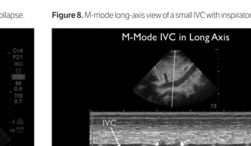
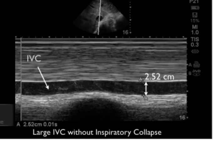
M-mode Doppler sonography of the IVC can be used to graphically document the absolute size and dynamic changes in the caliber of the vessel during the patient’s res-piratory cycle in both short and long axes (Figures 7–10). It should be noted, however, that M-mode sonography may introduce inaccurate measurements due to the dis-placement of the IVC relative to the probe during inspira-tion. Movement of the IVC out of the plane of the M-mode cursor may appear as vessel collapse on the M-mode trac-ing. It is therefore recommended that M-mode sonogra-phy be used after adequately visualizing IVC variability in the B-mode to avoid inaccurate estimation of vessel size and collapse.
Further studies are needed to define normal IVC parameters such as size, collapsibility, and distensibility (in mechanically ventilated patients). Until then, assessment of IVC collapsibility is useful in the critically ill patient whose caval index approaches the extremes. Additionally, caval sonography can be repeated during resuscitation to evaluate improvement of these parameters.
Figure 3. Short-axis view of a small IVC with inspiratory collapse. Figure 4.Long-axis view of a small IVC with inspiratory collapse.
Evidence
Incorporation of a goal-directed sonographic protocol including assessment of the IVC has been shown to improve the accuracy of physician diagnosis in patients with undif-ferentiated hypotension.22In a recent prospective study, point-of-care sonography evaluating cardiac contractility and IVC collapsibility in patients with suspected sepsis was shown to increase physician certainty and alter more than 50% of treatment plans.23Inadequate dilatation of the IVC after a fluid challenge was more sensitive than blood pres-sure for identification of hypovolemia in trauma patients.24 Another study in trauma patients showed the value of bed-side caval sonography in evaluation of fluid status and resus-citation of critically ill patients.25A study in acutely dyspneic patients presenting to the emergency department showed that IVC sonography rapidly identifies patients with con-gestive heart failure and volume overload.26
Rather than relying on a single measurement of the IVC, it may be more useful to follow changes in vessel size and collapsibility over time in response to an intervention. Studies have shown a decrease in the IVC diameter and increased collapsibility after blood loss15and fluid removal during hemodialysis.27In hypotensive emergency patients, volume resuscitation was associated with increases in the IVC diameter and less inspiratory collapsibility.14Just as a single blood pressure measurement is an incomplete rep-resentation of the hemodynamic status of a patient, sonog-raphy of the IVC should be repeated after interventions or changes in clinical parameters. Monitoring of the IVC diameter during resuscitation is an emerging area of research, and further studies are necessary to determine the exact parameters to interpret IVC size and collapsibil-ity.
Figure 6. Long-axis view of a large IVC without inspiratory collapse.
Figure 5. Short-axis view of a large IVC without inspiratory collapse.
Figure 8.M-mode long-axis view of a small IVC with inspiratory collapse.
Pitfalls
The IVC should be followed to the junction with the right atrium to avoid misidentification with the aorta. Because a single long-axis view may be inaccurate, it is recommended to assess the IVC in both short and long axes. Inferior vena cava determinations should be made at or near the conflu-ence with the hepatic veins. Measurements elsewhere may not reflect intravascular volume.
A dynamic evaluation of the degree of IVC collapse with inspiration may correlate better with the intravascu-lar volume than a single static measurement of the vessel size. Inferior vena cava size does not predict right atrial pressure in patients receiving mechanical ventilation. Care should be taken to maintain adequate visualization of the IVC throughout the respiratory cycle because the probe and IVC may be displaced by diaphragmatic and abdom-inal wall movements. Overestimation of intravascular
volume may occur in conditions that impede flow to the right heart, including valvular abnormalities, pulmonary hypertension, and heart failure.
Interpretation of caval physiology is hindered by con-ditions that restrict the physiologic variability of the IVC, such as liver cirrhosis and fibrosis,28masses causing exter-nal compression, and elevated intra-abdomiexter-nal pressure. Interpretation of the physiologic characteristics of the IVC should be done in context with the patient’s clinical sce-nario and adjunctive data.
Conclusions
Determination of shock state in critically ill patients is chal-lenging, but caval sonography may be a substitute for invasive hemodynamic monitoring. Assessment of the physiologic characteristics of the IVC provides a rapid distinction between low and high volume states and offers the clinician a rapid, noninvasive way to guide resuscita-tion in critically ill patients. In addiresuscita-tion to caval sonogra-phy, focused echocardiography and lung sonography have been suggested by an increasing number of resuscitation sonography protocols to further evaluate patients in shock.
References
1. Marik PE, Baram M, Vahid B. Does central venous pressure predict fluid responsiveness? A systematic review of the literature and the tale of seven mares. Chest2008; 134:172–178.
2. Shah M, Hasselblad V, Stevenson LW, et al. Impact of the pulmonary artery catheter in critically ill patients: meta-analysis of randomized clini-cal trials. JAMA2005; 294:1664–1670.
3. Perera P, Mailhot T, Riley D, Mandavia D. The RUSH exam: Rapid Ultrasound in SHock in the evaluation of the critically ill. Emerg Med Clin North Am2010; 28:29–56.
4. Perera P, Mailhot T, Riley D, Mandavia D. The RUSH exam: Rapid Ultrasound in SHock in the evaluation of the critically ill (2012 update).
Ultrasound Clin2012; 255–278.
5. Dipti A, Soucy Z, Surana A, Chandra S. Role of inferior vena cava diame-ter in assessment of volume status: a meta-analysis. Am J Emerg Med2012; 30:1414–1419.e1.
6. Kircher BJ, Himelman RB, Schiller NB. Noninvasive estimation of right atrial pressure from the inspiratory collapse of the inferior vena cava. Am J Cardiol1990; 66:493–496.
7. Yildirimturk O, Tayyareci Y, Erdim R, et al. Assessment of right atrial pres-sure using echocardiography and correlation with catheterization. J Clin Ultrasound2011; 39:337–343.
8. Brennan JM, Blair JE, Goonewardena S, et al. Reappraisal of the use of inferior vena cava for estimating right atrial pressure. J Am Soc Echocardiogr
2007; 20:857–861. Figure 10.M-mode long-axis view of a large IVC without inspiratory collapse.
Figure 9.M-mode short-axis view of a large IVC without inspiratory col-lapse.
9. Rudski LG, Lai WW, Afilalo J, et al. Guidelines for the echocardiographic assessment of the right heart in adults: a report from the American Soci-ety of Echocardiography endorsed by the European Association of Echocardiography, a registered branch of the European Society of Car-diology, and the Canadian Society of Echocardiography. J Am Soc Echocar-diogr2010; 23:685–713.
10. Jue J, Chung W, Schiller NB. Does inferior vena cava size predict right atrial pressures in patients receiving mechanical ventilation? J Am Soc Echocardiogr1992; 5:613–619.
11. Barbier C, Loubières Y, Schmit C, et al. Respiratory changes in inferior vena cava diameter are helpful in predicting fluid responsiveness in venti-lated septic patients. Intensive Care Med 2004; 30:1740–1746. 12. Feissel M, Michard F, Faller JP, Teboul JL. The respiratory variation in
inferior vena cava diameter as a guide to fluid therapy. Intensive Care Med
2004; 30:1834–1837.
13. Sefidbakht S, Assadsangabi R, Abbasi HR, Nabavizadeh A. Sonographic measurement of the inferior vena cava as a predictor of shock in trauma patients. Emerg Radiol2007; 14:181–185.
14. Weekes A, Tassone H, Babcock A, et al. Comparison of serial qualitative and quantitative assessments of caval index and left ventricular systolic function during early fluid resuscitation of hypotensive emergency depart-ment patients.Acad Emerg Med2011; 18:912–921.
15. Lyon M, Blaivas M, Brannam L. Sonographic measurement of the inferior vena cava as a marker of blood loss. Am J Emerg Med 2005; 23:45–50. 16. Sakurai T, Ando Y, Masunaga Y, Kusano E, Asano Y. Diameter of the
infe-rior vena cava as an index of dry weight in patients undergoing CAPD.
Perit Dial Int 1996; 16:183–185.
17. Akilli B, Bayir A, Kara F, Ak A, Cander B. Inferior vena cava diameter as a marker of early hemorrhagic shock: a comparative study. Ulus Travma Acil Cerrahi Derg2010; 16:113–118.
18. Mintz GS, Kotler MN, Parry WR, Iskandrian AS, Kane SA. Real-time infe-rior vena caval ultrasonography: normal and abnormal findings and its use in assessing right-heart function. Circulation1981; 64:1018–1025. 19. Mitaka C, Nagura T, Sakanishi N, Tsunoda Y, Amaha K.
Two-dimen-sional echocardiographic evaluation of inferior vena cava, right ventricle, and left ventricle during positive-pressure ventilation with varying levels of positive end-expiratory pressure. Crit Care Med1989; 17:205–210. 20. Moreno FL, Hagan AD, Holmen JR, Pryor TA, Strickland RD, Castle
CH. Evaluation of size and dynamics of the inferior vena cava as an index of right-sided cardiac function. Am J Cardiol 1984; 53:579–585. 21. Wallace DJ, Allison M, Stone MB. Inferior vena cava percentage collapse
during respiration is affected by the sampling location: an ultrasound study in healthy volunteers. Acad Emerg Med2010; 17:96–99.
22. Jones AE, Tayal VS, Sullivan DM, Kline JA. Randomized, controlled trial of immediate versus delayed goal-directed ultrasound to identify the cause of nontraumatic hypotension in emergency department patients. Crit Care Med2004; 32:1703–1708.
23. Haydar S, Moore ET, Higgins GL III, Irish CB, Owens WB, Strout TD. Effect of bedside ultrasonography on the certainty of physician clinical decisionmaking for septic patients in the emergency department. Ann
24. Yanagawa Y, Sakamoto T, Okada Y. Hypovolemic shock evaluated by sonographic measurement of the inferior vena cava during resuscitation in trauma patients. J Trauma2007; 63:1245–1248.
25. Ferrada P, Anand RJ, Whelan J, et al. Qualitative assessment of the inferior vena cava: useful tool for the valuation of fluid status in critically ill patients.
Am Surg 2012; 78:468–470.
26. Blehar DJ, Dickman E, Gaspari R. Identification of congestive heart fail-ure via respiratory variation of inferior vena cava diameter. Am J Emerg Med2009; 27:71–75.
27 Krause I, Birk E, Davidovits M, et al. Inferior vena cava diameter: a useful method for estimation of fluid status in children on haemodialysis. Nephrol Dial Transplant2001; 16:1203–1206.
28. Kitamura H, Kobayashi C. Impairment of change in diameter of the hepatic portion of the inferior vena cava: a sonographic sign of liver fibro-sis or cirrhofibro-sis. J Ultrasound Med2005; 24:355–359.