Computational structure-‐based drug design: predicting target flexibility
Advanced Review
Jelisa Iglesias1, Suwipa Saen-‐oon2, Robert Soliva2 and Victor Guallar*1,3
1Barcelona Supercomputing Center (BSC), Jordi Girona 29, E-‐08034 Barcelona, Spain 2Nostrum Biodiscovery, Jordi Girona 29, D128, E-‐08034 Barcelona, Spain
3ICREA, Passeig Lluís Companys 23, E-‐08010 Barcelona, Spain
Correspondence to: [email protected], [email protected]
Abstract
The role of molecular modeling in drug design has experienced a significant revamp in the last decade. The increase in computational resources and molecular models, along with software developments, is finally introducing a competitive advantage in early phases of drug discovery. Medium and small companies with strong focus on computational chemistry are being created, some of them having introduced important leads on drug design pipelines. An important source for this success is the extraordinary development of faster and more efficient techniques for describing flexibility in 3D structural molecular modeling. At different levels, from docking techniques to atomistic molecular dynamics, conformational sampling between receptor and drug results in improved predictions, such as screening enrichment, discovery of transient cavities, etc. In this review article we perform an extensive analysis of these modeling techniques, dividing them into high and low throughput screening techniques and emphasizing in their application to drug design studies. We finalize the review with a section describing our Monte Carlo method, PELE, recently highlighted as an outstanding advance in an international blind competition and industrial benchmarks.
This is the pre-peer reviewed version of the following article: [Iglesias J, Saen-oon S, Soliva R, Guallar V. Computational structure-based drug design: Predicting target flexibility. WIREs Comput Mol Sci. 2018;e1367], which has been published in final form at [https://doi.org/10.1002/wcms.1367]. This article may be used for non-commercial purposes in accordance with Wiley Terms and Conditions for Use of Self-Archived Versions
INTRODUCTION
Drugs can be discovered from a variety of sources and with varying strategies. Amongst the most important sources we find endogenous metabolites, natural products, chemical synthesis compounds and recombinant DNA proteins. The strategies can range from phenotypic screening, where compounds are prioritized based on phenotype alterations (even if the underlying molecular mechanisms are not known), to target-‐centric approaches whereby the onset or progression of a disease is directly linked to the function of a particular macromolecular receptor, usually a protein.1 In this latter approach, drugs are targeted molecular agents (ligands) that are prioritized to tightly bind to the receptor to alter its function.
The first target-‐ligand theoretical models where based on a lock and key concept, describing two interacting molecules as rigid particles perfectly matching one another (Figure 1a).2 However, this oversimplified view was quickly challenged by two alternative theories, namely induced fit and conformational selection,3, 4 both of which already describe molecules as flexible entities. The induced fit theory argues that both receptor and ligand change conformation to maximize complementarity only when they come into close contact (Figure 1b). The conformational selection theory, in contrast, argues that ligand and receptor are both constantly visiting a range of conformers that includes the bound conformation, complex formation merely shifting both equilibriums towards a bound state (Figure 1c). These two theories or their combination are now widely accepted to describe molecular recognition. Hence, it becomes key for the target-‐centric approach to gain insight on, not only the structure of the biomolecular target, but crucially its flexibility.5-‐7
Biomolecules undergo conformational transitions due to a variety of factors such as temperature, ionic strength, pH or the presence of other molecules. Structural transitions take many forms, from subtle movements of a single side chain, through concerted motions of a few residues, to significant secondary element rearrangements or complete domain changes.8 All of them can be critical for drug design. Receptors and enzymes usually change their conformation when isolated (apo) or bound to ligands (holo). Importantly, holo structures of the same receptor with different ligands can also differ significantly.9 Further, the binding of ligands on one site can shift conformational equilibria on other binding sites, causing allosteric effects (Figure 1d).10 These phenomena impose serious difficulties in terms of predicting from scratch the universe of accessible conformations of a particular biomolecule.11
The structure of biomolecular targets is usually solved by X-‐ray crystallography, NMR and cryo-‐ EM, and, increasingly, characterized by other experimental techniques such as circular dichroism or small angle X-‐ray scattering techniques. Their phenomenal evolution has led to a remarkable increase in throughput, applicability and resolution, so that the Protein Data Bank currently holds 3D structures of ca. 135K macromolecules, for which nearly ¾ are complexes.12 The dynamic behavior of biomolecules can be partly derived at 3D structure determination. X-‐ ray crystallography gives the displacement of atoms from their mean positions in the crystal by obtaining temperature factors, pointing at what segments of a 3D structure are more mobile.13 NMR generates an ensemble of alternative structures that conform to the restraints derived from the chemical shift experiment.14 However, these are only crude approximations to the myriad of accessible conformations of a particular receptor.
The field of target flexibility is now being propelled by the application of computational chemistry and simulation methods.15 Its evolution over the past two decades has been guided by improvements in algorithms, computer power, as well as a growing body of experimental data. Computational methods are now routinely applied in the first phases of a drug discovery project, from the screening for new chemical matter to the optimization of its potency and
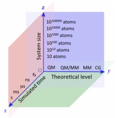
ADMET (absorption, distribution, metabolism, excretion and toxicity) properties; all along this timeline, target dynamics must be factored in. This rapid evolution has led to increasing the size of the systems under study, the level of theoretical detail in their representation (not only of the target but also of its environment, especially the solvent) and the simulated time per system. Computational modeling must move up and down these coordinates (Figure 2) depending on the problem to be solved and this choice has a direct impact on how target flexibility is modeled. Typically, the goal is to quickly and reliably predict: i) geometries of ligand-‐receptor interactions; ii) the energetics (thermodynamics) of complex formation; and only recently iii) the prediction of the kinetics of binding.16
At the lowest end of the scale, there are static methods that describe the system in a simplified form, neglecting target dynamics (conformational sampling) completely. This is the case for the rigid docking (rigid receptor and flexible ligand) approach. Although it is one of the most widely used techniques for structure-‐based pose prediction and for virtual screening (VS) of compound catalogues, it handles the receptor as a rigid object. This leads to the appearance of many false negatives, as a particular target conformation can only match certain ligands while others require rearrangements at the binding site. This can partly be alleviated by the so-‐ called “soft-‐docking” approach, where the van der Waals radii of target atoms, ligand atoms or both are reduced to diminish steric clashes. Scaling the van der Waals, however, comes with the drawback of generating more false positives, as the receptor-‐binding site then becomes artificially too slack. Rigid docking can be applied not to one, but to many structures such as an ensemble derived from modeling, NMR or several X-‐ray (hence its name “ensemble docking”).17 However, target flexibility is only modeled before the ligand is docked. Of note, rigid docking only makes crude estimates of the thermodynamics of binding, enabling, however, a high throughput performance.
At the middle of the complexity scale, there are methods that take into account the flexibility of targets and ligands simultaneously, but which typically represent the solvent only implicitly. One such method relies on the iterative combination of rigid docking with side chain and (slight) backbone sampling.18 Medusadock is an example of a docking program that samples the conformations of both interacting partners on the fly,19 which has given good results. RosettaLigand also samples ligand and target side chain and backbone flexibility simultaneously via a Monte Carlo (MC) algorithm.20 Another such method is CDOCKER,21 which has been shown to outperform rigid receptor docking tools. The MC program PELE (Protein Energy Landscape Exploration) also explores all degrees of freedom simultaneously while simulating a binding event.22 It has been benchmarked as a very efficient tool for binding mode prediction, both in the CSAR competition23 and in an industrial setting.24 These methods, although representing bulk solvent implicitly, can handle a few explicit water molecules in an active site, if they are bridging target and ligand via H-‐bonds. In general, this group of techniques is more reliable at pose prediction and ranking than rigid (receptor) docking approaches, as they account for target flexibility. However, they are computationally more demanding, so that their throughput is lower.
Arguably, the gold standard for computational exploration of target flexibility is molecular dynamics (MD) and all its varying forms and derivatives.25 Depending on the force field used, system description can be at the coarse-‐grained or atomic level. The system can even be partitioned such that a small portion is studied at the quantum mechanics (QM) level while the rest is modeled via molecular mechanics, opening a way to study covalent docking26 and ligand polarization.27 The solvent can be represented implicitly or explicitly, consisting of water or a combination of different solvents. Given enough computational resources, MD simulations can in principle be capable of fully describing target-‐ligand binding in explicit solvent. In fact, this has been the case for the most powerful MD machine described (Anton),28 which has been
used to simulate from scratch the binding of drugs to receptors.29 However, limitations inherent to the force fields as well as restrictions on the simulated times (as of today, usually from hundreds to just a few thousand nanoseconds) often preclude sampling transitions over longer timescales. Because of this, a series of MD-‐based enhanced sampling techniques have been developed, such as steered MD (sMD),30 replica-‐exchange MD (REMD)31 and metadynamics.32 These developments, along with high performance computing in the form of, for example, graphical processing units (GPUs) and cloud computing, open great perspectives to map target flexibility.
Computational chemists have found a way to exploit and combine MD-‐based techniques for studying conformational transitions of targets when engaged in drug recognition.33 The particular choice of system description, level of theory and simulation time dictates their eventual throughput, which is usually rather low for MD-‐based techniques. However, they are routinely applied for the prediction of ligand binding thermodynamics with greater success than low level flexibility descriptions. In fact, atomistic simulations can be used for estimating both absolute and relative binding free energies (DGs).34 Absolute DGs can be calculated by applying “end-‐point” methods, where receptor and ligand are only simulated in isolation or in closed complex, such as MMPB(SA) and MMGB(SA),35, 36 or they can be calculated based on reproducing the whole binding event, such as those that apply Markov State Models (MSM).37 In the former methods, flexibility is explored partially, as the intermediate states of recognition are neglected, while the latter exhaustively explore how the conformational landscape of target and ligand vary all along binding, opening up a way to study association mechanisms and kinetics. Relative DGs can be calculated thanks to the application of thermodynamic cycles by way of applying Free Energy Perturbation (FEP) or Thermodynamic integration (TI),38 which are considered some of the most reliable methods.39
The next sections will review recent studies involving receptor flexibility in 3D structural drug design. We divide them in i) high-‐medium throughput flexible techniques, ii) low throughput flexible techniques, and iii) the PELE technique. While we discuss success cases, it should be mentioned that introducing protein flexibility might sometimes degrade the modeling, as a result of incomplete sampling, poor energy functions, inaccuracy of solvent models, etc. Assessment of the modeling results with higher-‐level techniques and more sampling should be a common practice.
HIGH-‐MEDIUM THROUGHPUT FEXIBLE PROTEIN-‐LIGAND TECHNIQUES
In this section we will review methods and applications capable of screening thousands of compounds in a fast manner. As stated, the lack of flexibility in the receptor is one of the main sources of error. In parallel, however, there is a real interest in developing faster approaches capable of screening millions of compounds.40, 41 Thus, a compromise between speed (number of compounds) and accuracy is necessary, which is forcing, more and more, the use of hierarchical approaches: an overall technique combining methods that increase conformational sampling only for a selected subset of the screening space. 42-‐45
The first important addition of flexibility in 3D docking was introduced at the ligand level; today almost all docking techniques include ligand flexibility to some degree. Ligand flexibility is mainly introduced by an exhaustive search of possible conformations of the ligand, using stochastic or deterministic approaches, where possible conformations are pre-‐generated and selected before the docking step.46 Such deterministic generation has recently included mixed classic/quantum chemistry explorations, providing, not only an improved list of conformations for the ligand, but also a better estimation for the strain energy of the bound complex.47 While this review focuses on structural methods, we note also the importance in sampling ligand
conformations for quantitative structure–activity relationship (QSAR) descriptors, as shown recently with the xMaP (flexible MaP) descriptor.48
Sampling receptor flexibility is a significantly more complex process, which has centered a significant amount of methods development. The approaches for high-‐medium throughput VS can be divided in two main blocks: the ensemble docking technique and a plethora of protein rearrangement algorithms aiming at quickly sampling side chains and, less often, add into it the protein backbone as well. Here we will not consider soft-‐docking as a flexible method since it only allows for a deeper overlap of molecules, not describing conformation rearrangements. Nevertheless, its importance/use in the early steps of hierarchical VS should be seriously considered.
Ensemble methods
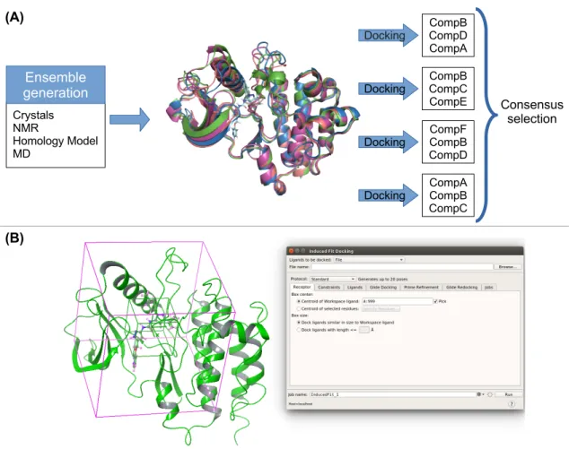
One straightforward approach to introduce flexibility into docking is by means of ensemble docking, that is, to use several (different) structures of the same target to dock ligands. These structures can be obtained from modeling and experimental sources: (i) receptors co-‐ crystallized with different ligands that induce distinctive conformational states; (ii) different models from an NMR ensemble; (iii) snapshots from an atomistic MD or MC simulation;49 (iv) snapshots from simpler modeling approaches such as using a normal mode technique (anisotropic network modeling, etc.) or homology modeling. Once the ensemble is generated, one can readily use rigid docking methods turning the overall VS simulation time directly proportional to the number of receptor conformations (Figure 3).
Several software packages provide means to perform ensemble docking. The OEDocking suite, for example, includes different options. Its HYBRID module is capable of using different receptors structures as input.50 A different module, POSIT, has an automatic procedure to select from a list of ligand-‐protein structures the best suited to guide ligand docking. They found its performance to be very close to that found by using ensemble docking, with the corresponding significant gain in performance (using one structure instead of many). We find multiple application studies of this suite of programs in the discovery of new anti-‐cancer molecules,51 in the development of new models to find new antiviral molecules against HIV52 and antibacterial activity.53
A 4D docking approach (the 4th dimension referring to the multiple receptor variable) is also implemented in ICM.54 In addition, different methods for the ensemble generation are provided, including a normal modes and a “fumigation” technique, which samples pocket torsion angles in the presence of a repulsive generic ligand. The 4D docking methodology developed by Bottegoni et al.55 optimized the efficiency of ensemble docking by integrating the procedure in one step. The user defines one structure from the ensemble as a template, to which all the other structures will be aligned, creating a grid with multiple conformations of the receptor that is sampled along rotations and translations of the ligand. This methodology has been used in the discovery of anticancer drugs, in particular cGMP efflux inhibitors.56 While some of the top commercial vendors, such as Schrodinger with the Glide rigid docking software, do not offer a ready to use ensemble docking tools, their graphical interfaces provide workflow solutions, like the Virtual Screening Workflow in Maestro, that largely facilitate this task. MOE, for example, also provides MD or normal modes tools to generate different receptor structures. Titan et al. used Schrodinger’s workflow in order to improve the docking results over one single structure-‐docking in kinase targets.57 Middendorp et al. used FlexX from MOE to dock several ligands into different homology models of GABA, finding new leads for this receptor.58 The ICM workflow was used to find novel inhibitors of the SERT protein, which could act as antidepressants or psychostimulants.59 The ensemble docking
methodology, however, is in high demand and many groups use their own approach to implement this technique manually, by combining different software for different steps.60 In some cases it is necessary to study the influence of large conformational changes such as the differences between active and inactive conformations. The presence of multiple protein-‐ ligand activation states opens new inhibition possibilities; this is the case, for example, of kinases and their activation loop, where computational modeling has difficulties to predict such large scale (and slow) conformational change. While below we review some attempts to such a modeling effort, the existence of crystallographic structures in different states introduces a ready to use ensemble. Meirson et al used 4 crystal structures of Pyk2 with different conformations induced by ligands with different selectivity profiles. To each of them they performed rigid docking, ADMET efficiency metrics filtering, and refined the best candidates using MD (a nice example of hierarchical VS).61 Another example of the potential of this technique (and an example of the heavy use of soft-‐docking techniques) is its use in the development of a new consensus scoring model for mTOR by Li et al., where they use six different crystal structures to perform soft docking with Glide in order to account for induced fit effects.62
Protein rearrangement algorithms
One of the most used approaches to provide induce fit conformational explorations is to perform binding pocket side chain sampling. Different variations of this approach are incorporated, for example, into Medusadock,19 GOLD,63 Molegro Virtual Docker (MVD),64 IFD,18 PLANTS,65 RosettaLigand,20 AutoDock4,66 CDOCKER,21 IMGdock67 and ICM68; these induced fit techniques have shown significant improvement in pose prediction when compared to rigid docking. In addition, some tools like IMGDock and PLANTS, also have algorithms that optimize crystal water molecules within the binding pocket along the flexible docking.
Matijsen et al. were able to define binding modes of benzimidazole to the CHK2 kinase thanks to the exploration of side chain flexibility. They combined the use of ensemble docking with a rigid body docking methodology and the flexible docking of GOLD.69 Gupta et al used a combination of rigid docking and flexible docking to study new putative drugs against malaria. They observed that flexible docking results differed from rigid docking ones in the same fashion for both MVD and Autodock, and that for both programs the flexible docking had a better performance than the rigid docking.70
Rosetta’s flexible docking was used by Brüser et al. to explain the differences in binding for UDP and PGE2-‐Gf to P2Y6 receptor.71 Luo et al. also used Rosetta’s flexible docking (along with MD simulations) to model the binding modes of allosteric inhibitors to an extracellular cap of the KP2 channel;72 the high flexibility of this region mandated the use of flexible docking.
Ding et al. stressed the importance of taking care of the receptor flexibility for the prediction of compounds during their participation on the DRD3 2 challenge.73 They found that the efficacy of rigid docking with CDOCKER was impaired by conformational changes of the protein upon binding of different ligands, and by steric clashes between the ligand and some residues with alternate positions. These problems could be solved by using a receptor with the right conformation or by using the flexible docking protocol.
Flexible docking with ICM was used to model the binding mode and induced-‐fit effect of a new drug against KEAP1, which has been patented in USA by the General Hospital Corp, University of California.74 Lane et al. also used ICM flexible docking to model dopamine binding to the apo dopamine D3 receptor, using the resulting model to perform ICM rigid docking of 4.1 million
compounds.75 Flexible docking with Autodock has been used to explain the inhibition mechanism of laccase by medicarpin.76 It also has been key in the study of hydrophobic surfaces on the cataract-‐related G18V variant of human γS-‐crystallint.77 IFD was used in the identification of active compounds to develop new kind of drugs against chronic infections.78 In the work of Chatzileontiadou et al. the IFD protocol was key for finding binding modes in human Angiogenin in agreement with the NMR structures.79
Adding protein backbone flexibility is a more complex and demanding (CPU time) task. Some of the programs providing side-‐chain rotamers treat backbone sampling by means of coupling the (side-‐chain) induced fit procedure with ensemble docking or with minimizations, as shown in Medusadock.80 The newest Schrodinger IFD protocol combines Glide rigid body docking with soft potentials and protein sampling using Prime. It is based on 4 steps: (i) a rigid docking of the ligand into the protein using soft-‐potentials; (ii) sampling protein conformations that may accommodate the ligand; (iii) rigid body docking of the ligand poses obtained in the first step with the protein conformations from the second step; (iv) scoring the receptor/ligand poses using a combination of Glide’s score and Prime’s energy. This protocol has been used to study new phitoconstituents derived from Silybum marianum, which can have an antiamnesic effect.81 Most of these methods, however, are not used as a stand-‐alone approach, the lack of a robust backbone sampling drives users to combine them with more computationally expensive techniques, such as metadynamics, as in the work of Clark et al..82
LOW THROUGHPUT FLEXIBLE PROTEIN-‐LIGAND TECHNIQUES
In this section we turn into techniques that are capable of introducing flexibility in a more accurate manner, but at the expense of screening only dozens (low hundreds in the best case scenario) compounds. We first center on MD techniques analyzing, afterwards, MC contributions.
Molecular Dynamics
As stated, MD simulations are currently the gold standard for exploring target flexibility in drug design.43, 83, 84 We explore their contribution by grouping them in three main areas: target treatment, mechanistic studies, and pose refinement (Figure 4); the separation between these groups, however, is often narrow and the same simulation might address multiple aspects.
Target treatment
Besides providing diversity for the ensemble docking procedure,85 adding flexibility to a target by means of MD simulations can reveal surface properties not observed in the available crystal structures, such as the presence of cryptic transient open binding pockets or subpockets,86-‐88 or the role of solvation effects through identifying/mapping key water molecules.89 For example, Wassman et al. described a transiently binding pocket between loop L1 and sheet S3 of the tumor suppress p53 core domain. Virtual screening against this revealed pocket identified a compound capable of reactivating mutated forms of p53 in human cell, demonstrating its potential as a pharmaceutical target.90 Related MD work also reported a narrow crevice on the surface of the p53 Y220C mutant, pinpointing key interactions for developing stabilizing small molecules.91, 92
Hagler et al. studied the effect of protein plasticity on the ability to identify active compounds through VS. They performed small-‐molecule docking on the ensemble structures generated by MD and REMD on the androgen receptor (AR), the HIV protease, and CDK2. Their results showed that flexibility increased significantly the enrichment and enhanced the diversity of hits.93, 94 Similarly, Hou et al. reported better predictive accuracies when using MD structures for three kinases: ALK, CDK2 and VEGFR2.57 Antolin et al. applied REMD simulations to sample the conformational space of the catalytic domain of PARP-‐1 in the ligand-‐bound and unbound
forms, assessing how enzyme flexibility affects the docking of a library of PARP-‐1 inhibitors. They pinpointed a key role of Leu324, Tyr325 and Lys242 in opening an additional binding site pocket with implications on ligand binding enrichment factors.95
McCammon and co-‐workers introduced the "relaxed complex scheme" (RCS) for receptor flexibility, which combines all-‐atom nanosecond MD simulations with small molecule docking to representative snapshots. RCS has shown to be successful to a variety of docking studies, demonstrating its potential for discovering new inhibitors and characterization of local-‐ induced and global effects on ligand binding.96-‐98 For instance, Schames et al. identified a novel binding cavity in HIV integrase using RCS with the 5CITEP inhibitor,99 which inspired the discovery of FDA-‐approved drug raltegravir.100 Cheng et al. have extended RCS by the efficient use of RMSD-‐based clustering on the study of avian influenza N1 neuraminidase in the apo form and in complex with oseltamivir. They showed a wide opening from the closed crystallographic structure (PDB:2HU4) and, by docking the National Cancer Institute Diversity Set 1 (NCIDS1), identified new hits.101
Recently, Valant et al. applied accelerated MD (aMD) to generate a receptor ensemble of the M2 muscarinic acetylcholine receptor (mAChR). Then, through iterative molecular docking and experimental testing, they successfully identified positive and negative allosteric modulators of M2 mAChR with remarkable chemical diversity, showing that aMD simulations combined with Glide IFD provided much-‐improved enrichment factors when compared with standard Glide.102 The mixed Essential Dynamics/Molecular dynamics technique has also recently been described for quickly generating alternative images of a receptor cavity.103 This technique uses a previously existing protein ensemble to describe its essential space of deformation. This space is perturbed by the presence of a small molecule, so that new, perturbed images of the receptor cavity can be used in the rigid body approximation. The technique was shown to outperform not only docking based on a single conformation, but also docking based on an unperturbed ensemble of the same protein.
Zacharias performed principal component analysis (PCA) on a MD trajectory and extracted its "soft" flexible modes that were used later in the docking process, implemented in the "PCRELAX" program.104 They showed that rigid docking of the FK506 ligand to an unbound FKBP conformation failed to identify a pose close to the experimental structure; inclusion of the flexible soft modes provided a native-‐like structure as the lowest energy conformation. Louet et al. applied Normal Mode Analysis (NMA) with conventional MD simulation to investigate the conformational dynamics of the hetero-‐trimeric G-‐ protein. Their results showed that G-‐proteins undergo large conformational changes without energy penalties. More interestingly, one of the lowest-‐frequency representative motions was able to open the GDP binding site and was consistent with experimental data. They proposed that GDP release and subsequent GDP/GTP exchange mainly involves an inter-‐domain motion between the ras-‐ like and the helical domains of Gα, together with an uprising of both αG and α4 helices.105, 106
Pande et al. introduced a computational paradigm coupling transition pathway with MSM techniques in the frame of massively distributed simulations, mapping the conformational landscape of a c-‐Src tyrosine kinase. This approach modeled the thermodynamics and kinetics of kinase activation for the first time, identifying key structural intermediates, and providing a database of c-‐Src conformations for future design of novel kinase inhibitors.107
Importantly, Barril et al. raised concern on the extent at which protein flexibility should be considered. Using MD simulations of hen egg-‐white lysozyme (HEWL) with explicit aqueous/organic solvent mixtures (MDmix method) and a range of restraint conditions, they observed how artificially restricted mobility affects binding hot spots. The authors suggested
that using carefully selected experimental structures may be more realistic and productive than a MD ensemble. Ultimately, choosing the right level of flexibility will depend on the goal of each individual investigation and the nature of each particular system.108
Finally, within the target treatment, we want to underline MD studies addressing the role of water molecules109 which, on top of mediating in the protein-‐ligand interactions, might affect the receptor flexibility. Different techniques, mostly based in inhomogeneous solvation theory, have been recently developed for this purpose, including WaterMap89 and STOW.110 A deeper analysis of these methods is out of the scope of this review; we refer the reader to recent studies benchmarking these techniques and comparing them with quicker grid based methods.111, 112
Mechanistic studies
Flexibility studies through MD have also emphasized on mapping protein-‐ligand association mechanisms, ligand entry and exit and kinetics; all these being key aspects for rational drug design. We refer here to the term “dynamic docking” introduced by De Vivo et al. in their recent review.113 Due to the (very) large timescale associated to some of these processes, the use of enhanced techniques has been significant. Parrinello and co-‐workers, for example, applied metadynamics,32, 114 a technique based on penalizing visited phase space points along a collective variable, to investigate the dissociation process of a nonsteroidal anti-‐flammatory ligand (SC-‐558) to COX-‐1 and COX-‐2 isoforms. In this way, they found a novel alternative binding mode to COX-‐2, identified key residues along the binding path and built a kinetic model for the binding mechanism.115 Incerti et al. also used metadynamics for building the unbinding free energy potential surface of the antagonist UniPR129 from its EphA2 receptor. They reported a binding mode that agreed with structure-‐activity relationship data and proposed, synthesized and tested new compounds for their ability to displace ephrin-‐A1 from the EphA2 receptor.116 Recently, Berne and co-‐workers have combined metadynamics with IFD aiming for accuracy and efficiency (reducing the overall computational cost). The method significantly increased the performance of the underlying IFD protocol across a large data set comprising 42 different ligand–receptor complexes.82
sMD, based on imposing an external potential to the ligand, have made important contributions to study binding/unbinding pathways, including its free energy estimation by means of the Jarzynski equality. Thus, one might use it for ranking of ligand affinities as well as elucidation of key residues along the exit/entrance pathway. For instance, we find research involving ligands dissociation from malaria parasite Plasmodium falciparum FabZ protein,117 CDK5 protein kinase,118 and GPCRs.119, 120 Moreover, sMD was used to study the conformational transition on GC-‐T4P using the 18 subunits long cryo-‐EM reconstruction to probe dynamics under tension, and to gain insights about the response of GC-‐T4P to external force at atomistic detail.121
REMD uses several copies of the system evolving in parallel under different simulating conditions, such as temperature or even the Hamiltonian, exchanging coordinates among the replicas at regular intervals with a MC criterion.122, 123 In several studies with disordered proteins, associated with diseases such as diabetes, Parkinson's and Alzheimer's, REMD was used to characterize the conformational states.124 In Alzheimer's disease, disordered β amyloid monomers are postulated to form toxic fibrils. While fibrils can be resolved in X-‐ray crystallography, the conformation of amyloid monomers and their oligomerization process remain elusive. REMD simulations studied this transition and addressed if the disordered monomer structure is pre-‐folded and contains information on the well defined oligomeric state.125 Kokubo et al. applied their replica-‐exchange based technique for exploring the ligand binding to two kinase systems (p38 and JNK3) with two different ligand molecules for each
kinase. They illustrated that protein flexibility was essential to predict the correct binding structure for one of the systems, where dihydroquinolinone was bound to p38 alpha kinase.126
In order to enhance the cavity opening and its exploration, some methods include a biasing repulsive potential (repulsive probes, etc.) in the binding site, in a similar fashion to the ICM fumigation technique for docking (Figure 5). This bias tends to perturb the active site, allowing it to readapt better to a given ligand. The Laughton group introduced in 2008 the Active Site Pressurization (ASP) technique,127 where uncharged Lennard-‐Jones particles are injected in the active site, and applied it later on to map kinases128 and GPCR.129 More recently, Zacharias et al. introduced the repulsive potential as a simulation condition in REMD.123
Finally, hardware and software development has allowed, in the past few years, to perform complex protein-‐ligand binding mechanism studies using standard MD approaches. Besides the seminal work by the Shaw lab, partly introduced above,29, 130 we find several studies using GPU computing power.131, 132 Buch et al. performed the full binding mechanism of the trypsin-‐ benzamidine complex, including a kinetic model by means of a MSM analysis. Decherchi et al. combined microsecond-‐long MD with machine learning algorithms to estimate the thermodynamics and kinetic values of a transition state analogue molecule to PNP.133 WExplore, a technique based on ensemble trajectories combined with residence time calculations, has also been recently introduced by Dickson et al. to describe ligand binding kinetics on the TPPU receptor.134 While still being slightly off from experimental values, these studies constitute impressive simulations aiming at modeling ligand dynamics and kinetics.135 Alternatively, computer power (including GPUs) can be provided by distributed computing, such as in the Folding@home project. Multiple studies from the Pande group, present clear examples of how by adding flexibility we have advanced in the understanding of biomedical research, including glycan binding in the NMDA receptors,136 kinases,107, 137 and GPCRs.138 In this regard, distribute and cloud computing have the potential to turn low into med/high throughput techniques.
Pose refinement
MD can be used as a post processing tool to validate and/or refine docking results, typically implemented in the drug design lead optimization phase.139 From MD refinement, the docking poses should display more stable and specific interactions, as the result of the induced-‐fit effect. Moreover, MD postprocessing can provide clues on the specific role played by solvent and allow for better estimates to the binding free energy, both in absolute and relative terms.
MM/GB(PB)SA is probably the most practical and widely used approach to estimate the binding energy of small molecules in low throughput virtual screening. It basically rescores multiple complex snapshots, typically derived from an MD simulation, by means of single point all-‐atom force field interaction energies with the addition of implicit solvent (GB or PB). Numerous, and somehow contradictory, benchmark studies have assessed its performance. Based on the PDBbind data set several authors have shown its ability to yield higher enrichment factors and to discriminate/sort by affinity a series of small molecules.140-‐142 However, the success of this approach seems to be system dependent and the entropic term to be crucial for an accurate prediction (these are normally estimated from normal modes). For further detail, a nice review from Ulf Ryde on the MMGB/SA method is recommended.143 The message from the author concludes that, while this approach is useful and easy to implement, one should be careful when applying it. Higher accuracy techniques, involving more expensive quantum mechanics are under investigation.144, 145 Nevertheless, these deal with rescoring matters rather than adding additional flexibility to the system.
The Linear Interaction Energy (LIE) method, proposed by Åqvist, has shown good compromise between speed and accuracy when estimating absolute ligand-‐binding affinities.146, 147 This method is based on force field estimations of the receptor-‐ligand interactions and thermal conformational sampling. A notable feature is that the binding energy can be predicted from intermolecular interactions (electrostatic and van del Waals interaction energies) ensemble differences.148 Vermeulen, et al. described the combination of rigid docking, MD simulations and LIE, to quantitatively evaluate the set of small molecules binding affinity to human Cytochrome P450 with estimated errors of less than 1 kcal/mol.149 LIE performance however, has been shown to be slightly worst than MM/BGSA in some benchmarks.150 Finally, we should state that the main limitations of the LIE and MMGBSA techniques, when considering the number of compounds that are capable of studying, resides in the length of the MD trajectory performed for each compound. While performing longer simulations might seem an advantage (in terms of adding conformational sampling), it could degrade numerical convergence, as discovering new conformations might change significantly the interaction energies.143, 151
FEP methods are the standard when aiming for accuracy in low-‐throughput VS refinement. While this group of studies could be introduced in the previous mechanistic block, we prefer to add them here since they are typically used as a post docking refinement phase. FEP convergence requires extensive sampling and, in contrast to the previous methods, it requires pair-‐ wise interactions to be calculated with a higher frequency. This fact together with the requirement that the transformations must be “smooth”, i.e., small perturbations, often make the method quite computationally demanding. In early 1990, Merz and Kollman employed FEP within an MD framework to correctly predict the binding free energy of a previously unreported inhibitor of the thermolysin endopeptide, and to predict the affinity of a novel HIV-‐ 1 peptide inhibitor.152, 153 Today, FEP calculations have demonstrated astonishing potential for driving lead optimization campaigns. Especially in the recent year, the implementation of FEP+ from Schrödinger has made the method accessible and attractive for the pharmaceutical industry.39, 154 When combined with an extensive ligand dihedral parameterization, Wang et al. demonstrated that FEP+ enables highly accurate affinity predictions across a broad range of target classes and ligands, many of which involve significant changes in ligand chemical structures.39 In a recent study, Lenselink et al. predicted relative binding free energies of congeneric ligands binding to GPCRs using the FEP+ package, with successful predictions of binding affinities for 39 of 45 compounds (investigated on four GPCRs).155 Moreover, FEP+ can also predict the affinity of novel and potent adenosine A2A receptor (A2AR) antagonists. Four novel compounds were synthesized and tested and the affinity of two out of the four was correctly predicted (within 1 kcal/mol), including one compound with approximately a tenfold increase in affinity compared to the starting compound.155 Extensive test by pharmaceutical companies, however, have raised concerns on the efficacy of the technique, which still seems to be case dependent.156
Monte Carlo
Stochastic MC techniques offer a valuable alternative to deterministic MD methods. Since the study of bovine pancreatic trypsin by Wako et al. in 1981,157 multiple methods aimed at mapping protein (and ligand) flexibility by MC techniques. The difficulties in combining meaningful perturbations with a large enough acceptance ratio, however, drove the development of the MC-‐minimization technique by Li and Scheraga.158 An important contribution was then introduced by Still and coworkers: the MacroModel program,159 combining random moves, minimizations and a GBSA implicit solvent model; this technique is still part of the Schrödinger suite, being one of the most used MC software for drug design. Recent implementations of a MC minimization scheme include our PELE software, which combines random movements with protein structure prediction algorithms to enhance the exploration.22
An important group of MC techniques centered on obtaining protein-‐ligand binding free energies,160, 161 mainly using alchemical methods such as FEP. An example is the pioneering BOSS (Biomolecular and Organic Simulation System) and MCPRO (MC for Proteins)162 software from the Jorgensen group. Other free energy methods, such as thermodynamic integration, have also been used with MC, as seen in different works of the Essex’ group163 with their software package, ProtoMS.164 Aplication studies of MCPRO include the discovery of anti-‐HIV agents165 and the inhibitor design on tyrosine kinase.166 In addition, MCPRO+, a version under Schrödinger commercial use, has been used in the drug design studies for the actylcholine receptor167 and the Bcr-‐Abl kinase.168 We also find several studies using ProtoMS, more focused in obtaining free energies163 and the role of water molecules in drug recognition and binding.169, 170
Efforts have also been made to combine MC with MD. Recently, Chodera and coworkers presented Nonequilibrium candidate Monte Carlo (NCMC),171 where proposal moves are constructed via nonequilibrium dynamics, in a series of perturbation mixed with propagation moves. This technique showed an enhancing of two orders of magnitude in finding binding modes of toluene in a mutated T4 lysozyme.172
As a rule of thumb, MC techniques have a harder time mapping backbone reorganization than MD, but can perform local side-‐chains adjustments more efficiently.
THE PELE TOOL FOR FLEXIBLE PROTEIN-‐LIGAND SAMPLING
In this section, first we give a brief introduction to the method and then we summarize representative applications in drug design. As stated, the method was highlighted as a “outstanding breakthrough” in the latest CSAR blind competition.23
The PELE Method
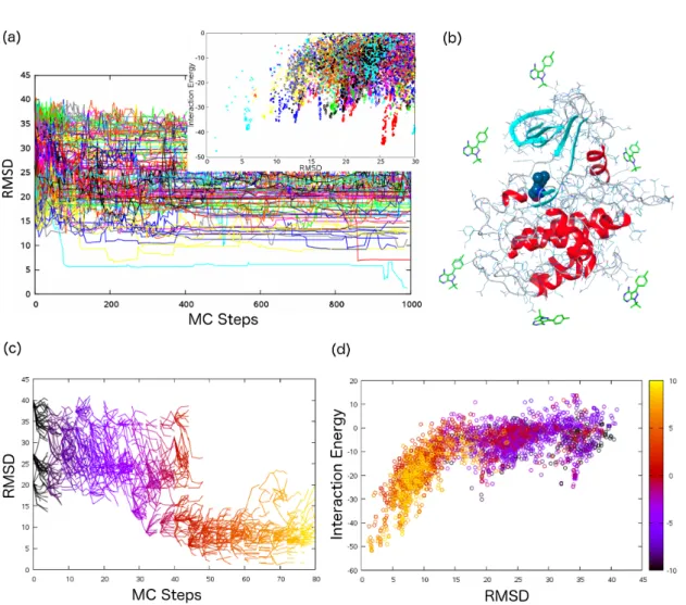
PELE follows a heuristic MC approach, generating conformational proposals by means of protein structure prediction techniques coupled to a system perturbation, so that the probability of acceptance in the Metropolis test remains high. The procedure is divided in two blocks: i) a ligand plus receptor perturbation, aimed at providing a conformational change in the overall system; ii) a relaxation step, comprising a side-‐chain sampling and a minimization step, aimed at driving the system to a local minima. A typical simulation involves tens to hundreds of processors (explorers) for hundreds of MC steps.
Ligand Perturbation. The ligand is perturbed by translating and rotating it within a user-‐ defined box (limiting the exploration space). After several perturbation trials (typically between 1 and 20), PELE chooses the one with the lowest system total energy. Each trial involves clash relieving using internal ligand degrees of freedom and rotamers from side chains around the ligand. When searching for the binding site, large translations are usually selected (up to 6 Å), while once in the binding site, small adjustments of ~1Å (or less) are used; a combination of translation ranges is often applied depending on the ligand solvent accessible surface area (SASA). In addition, to enhance the sampling of rare events, the (random) translational vector may be kept for a given number of MC simulation steps.
Receptor Perturbation. The receptor backbone is perturbed following normal modes calculated using the Anisotropic Network Model (ANM, see173) or PCA from different structures. It is possible to use a single mode, or to mix them randomly, with a preferred mode being given more weight, or weighting each mode according to its frequency (simulations typically use only the 6 lowest modes). As in the ligand translation, a perturbation direction can be kept during