0095-1137/10/$12.00 doi:10.1128/JCM.00917-10
Copyright © 2010, American Society for Microbiology. All Rights Reserved.
Agar Block Smear Preparation: a Novel Method of Slide Preparation
for Preservation of Native Fungal Structures for Microscopic
Examination and Long-Term Storage
䌤
Patrick C. Y. Woo,
1,2,3,4† Antonio H. Y. Ngan,
4† Hon-Kit Chui,
4Susanna K. P. Lau,
1,2,3,4and Kwok-Yung Yuen
1,2,3,4*
State Key Laboratory of Emerging Infectious Diseases,1Research Centre of Infection and Immunology,2Carol Yu Centre of
Infection,3and Department of Microbiology,4The University of Hong Kong, Hong Kong
Received 6 May 2010/Returned for modification 21 June 2010/Accepted 9 July 2010
We describe a novel method of fungal slide preparation named “agar block smear preparation.” A total of 510 agar block smears of 25 fungal strains obtained from culture collections, 90 QC fungal strains, and 82 clinical fungal strains from our clinical microbiology laboratory, which included a total of 137 species of yeasts, molds, and thermal dimorphic fungi, were prepared and examined. In contrast to adhesive tape preparation, agar block smears preserved the native fungal structures, such as intact conidiophores ofAspergillusspecies and arrangements of conidia inScopulariopsis brevicaulis. Furthermore, agar block smears allowed examination of fungal structures embedded in the agar, such as the ascomata with ascomal hairs inChaetomium funicola; pycnidium of Phoma glomerata; the intercalary ovoidal chlamydospores arranged in chains of Fusarium
dimerum; and the lateral, spherical chlamydospores arranged in pairs of Fusarium solani. After 1 year of
storage, morphological integrity was found to have been maintained in 459 (90%) of the 510 agar block smears. After 3 years of storage, morphological integrity was found to have been maintained in 72 (71%) of the 102 smears prepared in 2006. Agar block smear preparation preserves the native fungal structures and allows long-term storage and examination of fungal structures embedded in the agar, hence overcoming the major drawbacks of adhesive tape preparation. The major roles of agar block smear should be diagnosis for difficult cases, accurate identification of fungal species for clinical management of patients and epidemiological studies, and long-term storage for transportation of slides and education purposes.
Accurate identification of fungi is the cornerstone of select-ing and prescribselect-ing appropriate antifungal drugs to patients with fungal infections. In contrast to bacteria, for which anti-biotic susceptibility testing is routinely performed when a clin-ically significant bacterium is isolated, antifungal susceptibility testing is not commonly performed due to the relatively poor correlation betweenin vitro susceptibility results and clinical response. On the other hand, the choice of antifungal drugs for patients with fungal infections relies heavily on the identifica-tion of the corresponding isolates. Although molecular meth-ods, such as those using the internal transcribed spacer region and 18S rRNA sequencing, have been increasingly used for fungal identification, these technologies are still expensive and require corresponding expertise for laboratory technicians (5– 7). Therefore, most clinical microbiology laboratories still rely on phenotypic methods for identification of fungi.
Identification of molds in clinical microbiology laboratories is most commonly performed by the use of culture on agar plates followed by microscopic examination of lactophenol cot-ton blue-stained adhesive tape preparations of the fungal col-onies for direct visualization of characteristic microscopic
mor-phological features. However, adhesive tape preparations are associated with three major drawbacks. First, fungal structures may be crushed and damaged during the preparation proce-dures, affecting the accurate identification of the fungus. Sec-ond, as a result of drying of the smear, adhesive tape prepa-rations can be used for microscopic examination only within a few hours of the time of the initial preparation. Third, fungal structures embedded in the agar cannot be observed. To over-come these and other drawbacks, approaches such as the mi-croslide method are sometimes employed when greater mor-phological detail is necessary. However, microslide cultures are also not suitable for long-term storage. Throughout the years, modifications of microslide cultures have been suggested, but these methods have still been far from ideal (1, 2). Although an improvement to adhesive tape preparations, “double-layer tape prep,” which extends the useful life of the tape prepara-tion to several weeks, was suggested recently, this is still not suitable for long-term storage, for sending the slide out for consultation, and for examination of fungal structures embed-ded in agar (3). In this article, we describe a novel method of slide preparation, named “agar block smear preparation,” which preserves the native structures of molds and yeasts and allows long-term storage and examination of fungal structures embedded in the agar, hence overcoming all three drawbacks of adhesive tape preparation.
MATERIALS AND METHODS
Strains.Twenty-five fungal strains obtained from culture collections, 90 qual-ity control (QC) fungal strains, and 82 fungal strains isolated from patients in the * Corresponding author. Mailing address: State Key Laboratory of
Emerging Infectious Diseases, Department of Microbiology, The Uni-versity of Hong Kong, UniUni-versity Pathology Building, Queen Mary Hospital, Hong Kong. Phone: (852) 22554892. Fax: (852) 28551241. E-mail: [email protected].
† P. C. Y. Woo and A. H. Y. Ngan contributed equally to the manuscript.
䌤Published ahead of print on 21 July 2010.
3053
on May 16, 2020 by guest
http://jcm.asm.org/
TABLE 1. Fungal species used in the present study
Fungal species
Sourcea
NEQAS UK QC
CAP USA QC
Our clinical microbiology laboratory
Culture collection
Hyaline molds (nondermatophytes)
Aspergillus candidus 公 公
Aspergillus clavatus 公
Aspergillus flavipes 公
Aspergillus flavus 公 公 公(ATCC 204304)
Aspergillus fumigatus 公 公
Aspergillus glaucus 公
Aspergillus niger 公 公
Aspergillus restrictus 公
Aspergillus sydowii 公
Aspergillus terreus 公 公 公
Aspergillus ustus 公
Aspergillus versicolor 公 公 公
Emericella nidulans 公 公 公
Eurotium chevalieri 公(CBS 522.65)
Eurotium cristatum 公(CBS 123.53)
Eurotium rubrum 公
Neosartorya fischeri 公
Neosartorya pseudofischeri 公
Acremonium strictum 公 公
Arthrinium phaeospermum 公
Beauveria bassiana 公
Botrytis cinerea 公
Cephalotheca foveolata 公
Chrysosporium keratinophilum 公
Fusarium chlamydosporum 公
Fusarium dimerum 公 公
Fusarium nygamai 公(FRC-M7492)
Fusarium oxysporum 公 公(NRRL-28973)
Fusarium proliferatum 公 公(FRC-M6992)
Fusarium solani 公 公 公(CBS 109028)
Fusarium verticillioides 公 公(ATCC MYA 3629)
Gliocladiumspecies 公
Lecythophora hoffmannii 公
Malbrancheaspecies 公
Moniliaspecies 公
Nectria haematococca 公
Paecilomyces lilacinus 公 公
Paecilomyces variotii 公 公(ATCC MYA 3630)
Penicilliumspecies 公
Scopulariopsis brevicaulis 公 公
Thermomycesspecies 公
Trichodermaspecies 公
Trichothecium roseum 公
Hyaline molds (dermatophytes)
Epidermophyton floccosum 公 公
Microsporum canis 公 公(CBS 113480)
Microsporum ferrugineum 公
Microsporum gypseum 公
Microsporum nanum 公
Microsporum persicolor 公
Trichophyton erinacei 公
Trichophyton fischeri 公
Trichophyton mentagrophytes 公 公 公
Trichophyton rubrum 公 公 公 公(CBS 118892)
Trichophyton schoenleinii 公
Trichophyton soudanense 公
Trichophyton tonsurans 公 公 公
Trichophyton verrucosum 公
Trichophyton violaceum 公 公
Dematiaceous molds
Alternariaspecies 公
Continued on following page
on May 16, 2020 by guest
http://jcm.asm.org/
TABLE 1—Continued
Fungal species
Sourcea
NEQAS UK QC
CAP USA QC
Our clinical microbiology laboratory
Culture collection
Aureobasidium pullulans 公
Bipolaris hawaiiensis 公
Chaetomium funicola 公
Chaetomium globosum 公 公
Cladophialophora bantiana 公
Cladophialophora boppii 公
Cladosporium carrionii 公
Cladosporium cladosporioides 公
Cladosporium herbarum 公
Curvulariaspecies 公 公
Cyphellophora pluriseptata 公
Epicoccumspecies 公
Exophiala dermatitidis 公 公
Exophiala jeanselmei 公
Exophiala moniliae 公
Exserohilumspecies 公
Fonsecaea pedrosoi 公
Hormonema dematioides 公
Hortaea werneckii 公
Lasiodiplodia theobromae 公
Nigrosporaspecies 公
Ochroconis constricta 公
Ochroconis gallopavum 公
Phaeoacremonium parasiticum 公
Phialemonium curvatum 公
Phialophora fastigiata 公
Phialophora richardsiae 公 公
Phialophora verrucosa 公
Phoma glomerata 公
Pithomycesspecies 公
Pseudallescheria boydii 公 公 公(CBS 101.22)
Pyrenochaeta unguis-hominis 公
Rhinocladiella aquaspersa 公
Scedosporium prolificans 公 公
Scytalidium dimidiatum 公 公
Scytalidium hyalinum 公
Ulocladiumspecies 公
Mucoralesspecies
Absidia coerulea 公(MUCL10045)
Cunninghamella bertholletiae 公 公
Cunninghamellaspecies 公
Lichtheimia corymbifera 公 公 公(MUCL10046)
Lichtheimia blakesleeana 公(CBS 100.28)
Lichtheimia hyalospora 公(CBS 173.67)
Mucorspecies 公 公
Rhizomucor pusillus 公
Rhizopus azygosporus 公
Rhizopus microsporusvar.microsporus 公
Rhizopus microsporusvar.oligosporus 公(CBS 112586)
Rhizopus microsporusvar.chinensis 公 公(CBS 631.82)
Rhizopus microsporusvar.
rhizopodiformis
公 公(CBS 343.29)
Rhizopus oryzae 公 公 公(CBS 112.07)
Rhizopus stolonifer 公
Syncephalastrum racemosum 公
Yeasts
Blastoschizomyces capitatus 公
Candida albicans 公(ATCC 90028)
Candida dubliniensis 公 公
Candida glabrata 公 公
Candida guilliermondii 公
Continued on following page
on May 16, 2020 by guest
http://jcm.asm.org/
clinical microbiology laboratory of Queen Mary Hospital in Hong Kong were included in the present study (Table 1). A total of 137 species of yeasts, molds, and thermal dimorphic fungi were included.
Culture medium.A transparent, extremely low-nutrition culture medium for detection ofAcanthamoebaspecies in ophthalmologic specimens (Acanthamoeba agar) that contained 12% (wt/vol) NaCl, 4% (wt/vol) MgSO4䡠7H2O, 14.2% (wt/vol)
Na2HPO4, 13.6% (wt/vol) KH2PO4, and 4% (wt/vol) CaCl2䡠2H2O was used for
agar block smear preparation unless otherwise stated (4).
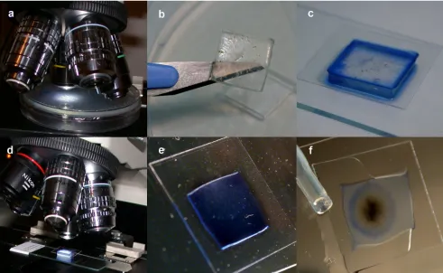
Agar block smear preparation method.Fungi were inoculated onto Acan-thamoeba agar in a class II biological safety cabinet and incubated under optimal conditions for the specific fungus species. Spore formations and other charac-teristic structures were checked every 2 to 7 days, depending on the growth rate of the fungus, by agar block examination using agar plates under a light micro-scope (⫻40 or ⫻100 magnification) (Fig. 1a). When the fungus colony was mature, photos were taken directly using the microscope camera and the plates were used for subsequent agar block cutting. For known or suspected biosafety level 3 fungi such asCoccidioides immitis, the agar plate was fumigated using 37% formaldehyde for 48 h before agar block cutting was performed.
In a class II biological safety cabinet, an agar block (15 by 15 mm) was cut using a sterile dissecting knife and placed on a glass slide (Fig. 1b). After a drop of lactophenol cotton blue stain or of another stain such as calcofluor white stain was added, a coverslip (18 by 18 mm) was put onto the agar block (Fig. 1c). The agar block was examined under a light microscope (⫻400 or⫻1,000 magnifica-tion) (Fig. 1d). For long-term storage, the block was dried in air at room temperature in a class II biological safety cabinet until the thickness of the agar block reached 0.5 mm, which usually took about 48 h (Fig. 1e). The four sides of the agar block under the coverslip were filled with Permount mounting medium (Fisher Scientific, NJ) (Fig. 1f). After the mounting medium was dried, the agar block smear was examined under a light microscope (⫻400 or⫻1,000 magnifi-cation).
RESULTS
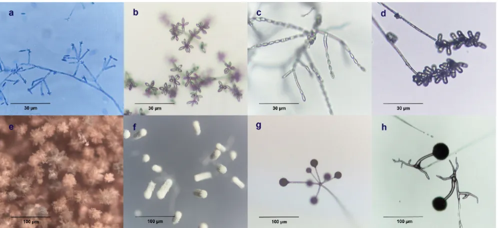
Direct examination of agar plates under a microscope be-fore cutting of the agar block.This allows observation of very delicate fungal structures of diagnostic significance. It should be noted that for very fragile and delicate conidial structures, their arrangements on conidiophores may still be destroyed
even when the agar block smear preparation method is used. For example, all the conidia ofFusarium chlamydosporumwere dislodged from the polyphialides during agar block smear preparation (Fig. 2a), whereas their flower-like arrangement on the polyphialides was well preserved when the whole agar plate was observed under a light microscope before cutting of the agar block was performed (Fig. 2b). Similarly, the conidial chains of Fusarium verticillioides that were attached to the phialides (Fig. 2c) and the unique conidial chains resulting from basipetal growth inTrichothecium roseum(Fig. 2d) were well preserved when the whole agar plate was observed under a light microscope, but they were destroyed by the agar block smear preparation. Additionally, direct examination of the agar plate under a microscope was particularly useful for ob-servation of the conidial head ofAspergillusspecies and of the arrangement of sporangiophores in members of theMucorales
order, both being key features for species identification. Observed with a lateral light source, the conidial heads of
Aspergillus nigerwere radiate (Fig. 2e) whereas those of Aspergil-lus fumigatus were columnar (Fig. 2f). As for theMucorales
species, the sporangiophores ofLichtheimia corymbiferawere branched in an umbel formation (Fig. 2g) whereas the un-branched sporangiophores of members of theRhizopus micro-sporusgroup were found directly above the rhizoids (Fig. 2 h).
[image:4.585.43.544.80.330.2]Microscopic examination of slides prepared by the agar block smear preparation method. A total of 510 agar block smears of 137 fungal species were prepared (Table 1), with 102 smears prepared in 2006, 153 in 2007, and 255 in 2008. Slides that were prepared using the agar block smear preparation method were compared to those prepared using the adhesive tape method.
TABLE 1—Continued
Fungal species Sourcea NEQAS UK QC CAP USA QC Our clinical microbiology laboratory Culture collection
Candida kefyr 公
Candida krusei 公 公(ATCC 6258)
Candida lusitaniae 公 公
Candida parapsilosis 公 公(ATCC 22019)
Candida tropicalis 公
Cryptococcus albidus 公
Cryptococcus neoformans 公 公 公 公(CBS 132)
Cryptococcus uniguttulatus 公
Geotrichum candidum 公 公
Malassezia pachydermatis 公
Malassezia furfur 公 公 公(CBS 1878)
Prototheca wickerhamii 公
Rhodotorula rubra 公
Saccharomyces cerevisiae 公
Trichosporonspecies 公 公 公
Ustilagospecies 公
Dimorphic fungi
Blastomyces dermatitidis 公
Coccidioides immitis 公
Penicillium marneffei 公
Sporothrix schenckii 公
a
NEQAS UK QC, National External Quality Assessment Service (United Kingdom) quality control; CAP USA QC, College of American Pathologists (United States) quality control; FRC, Fusarium Research Center; MUCL, Mycothe`que de l’Universite´ catholique de Louvain.
on May 16, 2020 by guest
http://jcm.asm.org/
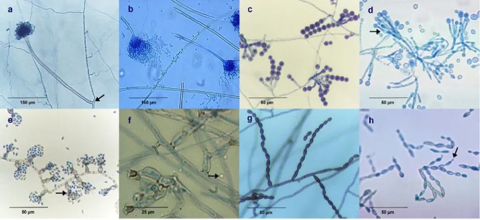
Preservation of native structural features. Intact conidio-phores with foot cells ofAspergillus versicolor were well pre-served in agar block smears. As a result, the length of conid-iophores, which is one of the key features for Aspergillus
species identification (Fig. 3a), could be measured easily. On the other hand, conidiophores, especially the longer ones, were usually broken due to the tearing force exerted during prepa-ration of adhesive tape smear (Fig. 3b).
For Scopulariopsis brevicaulis, the conidiogenous cells (an-nelids) were well preserved in agar block smears (Fig. 3c). They were found either singly arranged or in brush-like clus-ters with short conidiophores. From the annelids, truncated conidia in chains were basipetally produced. On the other hand, the annelids were overlapped with each other and the native state was destroyed as a result of the adhesive tape smear preparation method (Fig. 3d).
In the agar block smear of Phialophora verrucosa, its phi-alides were distributed alongside the vegetative hyphae like a dozen vases of flowers (Fig. 3e). The phialides are shaped like flasks with funnel-like collarettes. Round-to-oval conidia accu-mulated in clusters at their apices. However, in the adhesive tape smear ofP. verrucosa, artificial overlapping of phialides was observed (Fig. 3f).
For Cladophialophora bantiana, the elliptical conidia ar-ranged in chains and their origins were well preserved in agar block smears (Fig. 3g). On the other hand, many acropetal
conidial chains, with the youngest conidia at the tip, were detached, and conidiophores were not observed in the adhe-sive tape smears (Fig. 3h).
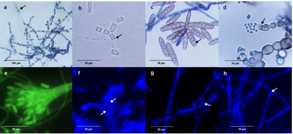
Observation of fungal structures embedded under the agar surface.ForChaetomium funicola, the ascomata with typical ascomal hairs that were partially embedded in agar were well shown by the agar block smear technique (Fig. 4a). The ar-rangements of the straight, stiff ascomal hairs that were repeat-edly dichotomously branched were better preserved in the agar block smears (Fig. 4a) than in tease mounts. They differed from the nonbranched, undulate ascomal hairs of a more com-monChaetomiumspecies,C. globosum.Under high-powered magnification, the limoniform ascospore and the distinctly ver-rucose ascomal hairs were visible (Fig. 4b).
[image:5.585.46.537.70.372.2]For Phoma glomerata, a coelomycete which produces conidia by the activity of conidiogenous cells lining the inner cavity of its asexual fruiting bodies (i.e., its pycnidia), a spher-ical pycnidium embedded partially under the agar surface with two ostioles in the agar block smear was observed (Fig. 4c). The pycnidium is darkly pigmented around the ostioles from which conidia were released. A brown chlamydospore with longitudinal and transverse septa (muriform), an additional key structure for identification ofP. glomerata, was also well demonstrated (Fig. 4d). Pyrenochaeta unguis-hominis, also a coelomycete but differing fromP. glomerataby its setose pyc-nidium, was also well shown in the agar block smear (Fig. 4e).
FIG. 1. Preparation of agar block smears. (a) Spore formations and other characteristic structures were checked by agar block examination of agar plates under a light microscope. (b) An agar block (15 by 15 mm) was cut using a sterile dissecting knife. (c) A coverslip (18 by 18 mm) was put onto the agar block after lactophenol cotton blue staining was performed. (d) The agar block with the coverslip was examined under a light microscope. (e) The block was dried in air until the thickness of the agar block reached 0.5 mm. (f) The agar block under the coverslip was filled with mounting medium.
on May 16, 2020 by guest
http://jcm.asm.org/
Unlike otherPyrenochaetaspecies with setae tapering toward the tip, the setae observed inP. unguis-hominiswere obtuse at their apices (Fig. 4f).
The arrangements and positioning of chlamydospores are among the critical characteristics forFusariumspecies identi-fication. ForF. dimerum, the intercalary chlamydospores ar-ranged in a chain were ovoidal in shape (Fig. 4g), but forF. solani, the lateral chlamydospores were usually arranged in pairs and spherical in shape (Fig. 4 h). These chlamydospores were often found on the submerged hyphae.
Microscopic examination of agar block smears after long-term storage.After 1 year of storage, 459 (90%) out of 510 agar block smears were in a good state. After 3 years of stor-age, 72 (71%) out of the 102 smears prepared in 2006 were still in a good state.
Figure 5a and b show an agar block smear ofC. immitisthat had been stored for more than 3 years. Under low-power magnification, chains of arthroconidia with alternate disjunctor cells were well displayed (Fig. 5a). Raquet hyphae were also visible. Under high-power magnification, conidiogenesis of al-ternate arthroconidial chains was clearly demonstrated (Fig. 5b). The chains were formed by the fragmentation of hyphae through the dissolution of disjunctor cells. Barrel-shaped arthroconidia were usually attached with an annular frill on each end (Fig. 5b).
Figure 5c shows an agar block smear ofBipolaris hawaiiensis
stored for more than 1 year. In contrast to those of other
Bipolaris species, its macroconidia showed more than three distosepta. They were pseudosepta and were differentiated from the true septa by the cytoplasm contraction of distoseptate conidia and by their having become angular, as demonstrated by
lactophenol cotton blue staining. Figure 5d shows an agar block smear ofExophiala dermatitidis that had been stored for more than 1 year. Torulose hyphae with rare spherical phialides were demonstrated. Since this species is a dematiaceous fungus, dark pigment of melanin was obvious on its cell walls.
Figure 5e, f, g, and h show agar block smears that had been stained with calcofluor white and stored for more than 6 months. The fluorescence did not fade; the different colors of fluorescence were due to the use of different barrier wave-lengths in the fluorescent microscope (430 nm for blue fluo-rescence and 520 nm for green fluofluo-rescence). Figure 5e shows an agar block smear of Graphium eumorphum, the asexual synanamorph of Pseudallescheria boydii.Conidiophores were aggregated into a compound stalk (i.e., a synnemata). A sterile basal part and a fertile head producing cylindrical terminal conidia were revealed using green fluorescence. Figure 5f shows an agar block smear ofExophiala moniliaein blue flu-orescence. Annellated taping, which protruded and became rather long with age, is clearly observable as a deeply pig-mented tip due to melanin deposits. Figure 5g shows an agar block smear ofPhialemonium curvatumin blue fluorescence. Short adelophialides without a basal septum, a characteristic of
P. curvatum, were clearly observed, unlike the results seen with
Acremonium strictum, which showed long phialides with a basal septum (Fig. 5h).
DISCUSSION
[image:6.585.44.542.68.296.2]The marked increase in the density of intact native struc-tures of fungi observed in agar block smears greatly improved the accuracy of laboratory identification of pathogenic fungi.
FIG. 2. Direct examination of an agar plate under a microscope before agar block cutting. (a and b)Fusarium chlamydosporum, showing the conidia dislodged from the polyphialides during agar block smear preparation (a) and the well-preserved flower-like arrangement of the polyphialides when the whole agar plate was observed under a light microscope before the agar block was cut (b). (c)Fusarium verticillioides, showing the conidial chains attaching to the phialides. (d)Trichothecium roseum, showing the unique conidial chains resulting from basipetal growth. (e)Aspergillus niger, showing the radiating conidial heads. (f)Aspergillus fumigatus, showing the columnar conidial heads. (g) Lichtheimia corymbifera, showing the sporangiophores that are branched in umbel. (h)Rhizopus microsporus, showing the unbranched sporangiophores directly above the rhizoids.
on May 16, 2020 by guest
http://jcm.asm.org/
The integrity and the native arrangements of conidiophores and sporulating structures are of paramount importance for microscopic species identification of pathogenic fungi. The length of conidiophores is important for species identification of some fungi, such as the Aspergillus species, whereas the arrangement of conidia is crucial to identification of others, such as S. brevicaulis. During the process of adhesive tape smear preparation, which primarily demonstrates fungal struc-tures on the surface of agar plates, many native strucstruc-tures of fungi were crushed and torn, leading to breaking of most co-nidiophores and loss of the native arrangement of the sporu-lating structures. As for fungal structures partially or com-pletely embedded inside the agar, such as the fruiting bodies of some ascomycetes and coelomycetes, although they could be dug out by the use of tease mounts, the native orientation of the fruiting structures was often destroyed. On the other hand, when agar block smears are prepared, the whole thickness of agar is gradually compressed to 0.5 mm. This allows the pres-ervation and examination of the native structures of most fungi, both those formed on the surface and those embedded in the agar. Furthermore, it also markedly increases the chance of recognizing rare structures, such as the spherical phialides ofExophiala dermatitidis. As for the medium, when the agar block smear technique was first conceived in 2006, a number of transparent culture media, including Sabouraud dextrose agar, carnation leaf medium, and Acanthamoeba medium, were em-ployed in efforts to grow the fungi for agar block smear prep-arations. Our preliminary study showed that Acanthamoeba medium was best for promoting conidiogenesis of most molds
because of its extremely low level of available nutrition. For slow-growing dermatophytes, a longer incubation time was re-quired for spore formation. For example, at least 3 weeks of incubation was necessary for the observation of the macro-conidia ofMicrosporum canisandMicrosporum gypseum.
Good preservation of the native fungal structures in agar block smears over long time periods allows slides to be sent out for consultation and education purposes. In July 2006, the concept of agar block smear was conceived and the first agar block smear was made from a strain ofFusarium solani.Up to the time of writing, more than 500 smears had been prepared from 137 fungal species of yeasts, hyaline molds with septate hyphae, dematiaceous molds with septate hyphae,Mucorales
species with rarely septate hyphae, and thermal dimorphic fungi. In our experience, more than 90% of the smears were still in good condition after the first year. After 1 year of storage, the most serious problem that could affect the quality of the smears was air leakage through the mounting medium, with the resultant air bubbles and drying effect jeopardizing the spore structures. Other minor problems include fading of stain coloring and degeneration of the mycelium.
[image:7.585.57.527.71.286.2]The major roles of the agar block smear technique should be diagnosis of difficult cases, accurate identification of fungal species for epidemiological and clinical studies, and long-term storage for transportation of slides and education purposes. Despite the advantages of the agar block smear technique, it cannot replace the adhesive tape smear technique. The adhe-sive tape smear technique is easy, quick to perform, and inex-pensive. With an agar plate culture, preliminary identification
FIG. 3. Preservation of native structural features of fungi by agar block smears. (a)Aspergillus versicolor, prepared by the agar block smear method, showing intact conidiophores with well-preserved foot cells (arrow). (b)A. versicolor, prepared by the adhesive tape method, showing broken conidiophores. (c)Scopulariopsis brevicaulis, prepared by the agar block smear method, showing its annelids arranged either singly or in brush-like clusters and truncated conidia basipetally produced in chains. (d)S. brevicaulis, prepared by the adhesive tape method, showing annelids artificially overlapping each other (arrow). (e)Phialophora verrucosa, prepared by the agar block smear method, showing its flask-shaped phialides well distributed along the vegetative hyphae with funnel-like collarettes (arrow). (f)P. verrucosa, prepared by the adhesive tape method, showing artificial overlapping of philalides and air bubbles (arrow). (g)Cladophialophora bantiana, prepared by the agar block smear method, showing elliptical conidia arranged in chains from indistinct conidiophores. (h)C. bantiana, prepared by the adhesive tape method, showing many detached acropetal conidial chains with the youngest conidia at the tip (arrow) and the presence of tape ripples, making it impossible to take photographs at the same focal plane.
on May 16, 2020 by guest
http://jcm.asm.org/
of molds to the genus level can often be achieved within 15 min using the adhesive tape smear technique. This preliminary identification of clinically significant molds is of paramount importance in choosing antifungal agents and has a great
im-pact on patient management. On the other hand, agar block smear preparation requires subculturing of the fungus and the time for preparation of the smear. Therefore, the agar block smear technique cannot replace the adhesive tape smear
[image:8.585.57.527.70.285.2]tech-FIG. 4. Observation of fungal structures embedded under the agar surface in agar block smears. (a)Chaetomium funicola, showing the ascomata with typical straight, stiff, dichotomously branched ascomal hairs partially embedded in agar. (b)Chaetomium globosum, showing the distinctly verrucose ascomal hairs (arrow) and its limoniform ascospore (arrow). (c and d)Phoma glomerata, showing a spherical pycnidium partially embedded under the agar surface with two ostioles (arrows) and a brown chlamydospore with longitudinal and transverse septa. (e and f)Pyrenochaeta unguis-hominis, showing the characteristic setose pycnidium and setae with obtuse apices (arrow). (g)Fusarium dimerum, showing the intercalary ovoidal chlamydospores arranged in chains. (h)Fusarium solani, showing the lateral spherical chlamydospores arranged in pairs.
FIG. 5. Microscopic examination of agar block smears after long-term storage. (a and b)Coccidioides immitis, showing chains of arthroconidia with alternate disjunctor cells, racquet hyphae (arrow), and alternate arthroconidial chains with barrel-shaped arthroconidia, usually with an annular frill (arrow) on each end. (c)Bipolaris hawaiiensis, showing macroconidia with more than three distosepta (arrow). (d) Exophiala dermatitidis, showing the torulose hyphae with rare spherical phialides (arrow) and the dark pigment of melanin on its cell walls. (e)Graphium eumorphumstained with calcofluor white, showing conidiophores aggregated into a compound stalk (synnemata) and cylindrical terminal conidia. (f)Exophiala moniliaestained with calcofluor white, showing annellated taping (arrow), which protruded and became rather lengthy with age. (g)
Phialemonium curvatumstained with calcofluor white, showing the short adelophialides without a basal septum (arrow). (h)Acremonium strictum
stained with calcofluor white, showing the long phialides with a basal septum (arrow).
on May 16, 2020 by guest
http://jcm.asm.org/
[image:8.585.45.543.422.651.2]nique in clinical microbiology laboratories for use on a day-to-day basis because of the turnaround time and economic con-siderations. On the other hand, for accurate identification at the species level to elucidate, for example, the differential susceptibilities of different species to antifungal agents, the agar block smear technique would be preferred to the adhesive tape smear technique.
ACKNOWLEDGMENTS
This work was partly supported by a Research Grants Council grant; the University Development Fund, The University of Hong Kong; and the Hong Kong Special Administration Region (HKSAR) Research Fund for the Control of Infectious Diseases of the Health, Welfare and Food Bureau.
REFERENCES
1.Connell, S. L., and D. E. Padgett.1988. An improved technique for making permanent slide cultures of fungi. Mycopathologia101:165–166.
2.Harris, J. L.1986. Modified method for fungal slide culture. J. Clin. Micro-biol.24:460–461.
3.Hughes, A. D., G. D. Lorusso, and D. L. Greer.2004. The ‘double-layer tape prep’: an improvement to a standard technique. J. Med. Microbiol.53:455. 4.Isenberg, H. D (ed.).2004. Clinical microbiology procedures handbook, 2nd
ed. ASM Press, Washington, DC.
5.Lau, S. K., P. C. Woo, S. K. Chiu, K. W. Leung, R. W. Yung, and K. Y. Yuen. 2003. Early diagnosis ofExophialaCAPD peritonitis by 18S ribosomal RNA gene sequencing and its clinical significance. Diagn. Microbiol. Infect. Dis. 46:95–102.
6.Woo, P. C., S. K. Lau, A. H. Ngan, H. Tse, E. T. Tung, and K. Y. Yuen.2008.
Lasiodiplodia theobromaepneumonia in a liver transplant recipient. J. Clin. Microbiol.46:380–384.
7.Woo, P. C., S.-Y. Leung, K. K. To, J. F. Chan, A. H. Ngan, V. C. Cheng, S. K. Lau, and K.-Y. Yuen.2010. Internal transcribed spacer region sequence het-erogeneity inRhizopus microsporus: implications for molecular diagnosis in clinical microbiology laboratories. J. Clin. Microbiol.48:208–214.