0095-1137/09/$08.00
⫹
0
doi:10.1128/JCM.00001-09
Copyright © 2009, American Society for Microbiology. All Rights Reserved.
Development and Evaluation of a Sensitive and Specific Assay for
Diagnosis of Human Toxocariasis by Use of Three Recombinant
Antigens (TES-26, TES-30USM, and TES-120)
䌤
Suharni Mohamad, Norhaida Che Azmi, and Rahmah Noordin*
Institute for Research in Molecular Medicine (INFORMM), Universiti Sains Malaysia, 11800 Penang, Malaysia
Received 1 January 2009/Returned for modification 16 March 2009/Accepted 7 April 2009
Diagnosis of human toxocariasis currently relies on serologic tests that use
Toxocara
excretory-secretory
(TES) antigen to detect immunoglobulin G (IgG) antibodies to the larvae. In general, however, these assays do
not have adequate specificity for use in countries in which other soil-transmitted helminths are endemic. The
use of recombinant antigens in these assays, however, is promising for improving the specificity of the diagnosis
of toxocariasis. Toward this goal, we developed an IgG4 enzyme-linked immunosorbent assay (ELISA)
involv-ing three recombinant antigens: rTES-30USM (previously produced), rTES-26, and rTES-120. The latter two
antigens were produced by reverse transcription-PCR cloning; subcloned into glutathione
S
-transferase
(GST)-tagged and His-(GST)-tagged prokaryotic expression vectors, respectively; and expressed in
Escherichia coli
. The
recombinant proteins were subsequently purified by affinity chromatography using GST and His-Trap resins.
The diagnostic potential of each purified recombinant antigen was tested with various immunoglobulin classes
(IgG, IgM, and IgE) and IgG subclasses. The IgG4 ELISA was determined to have the highest specificity and
was further evaluated using a panel of serum samples. The rTES-26 IgG4 ELISA showed 80.0% (24/30 samples
positive) sensitivity, and both the rTES-30USM IgG4 ELISA and rTES-120 IgG4 ELISA had 93.0% (28/30)
sensitivity. Combined use of rTES-120 and rTES-30 IgG4 ELISA for the diagnosis of toxocariasis provided
100% sensitivity. The specificities of rTES-26, rTES-30USM, and rTES-120 antigens were 96.2%, 93.9%, and
92.0%, respectively. These results indicate that the development of a diagnostic test using the three
recombi-nant antigens will allow for more-accurate detection of toxocariasis.
Human toxocariasis is a worldwide parasitic zoonosis, caused
most commonly by the intestinal parasites the dog roundworm
(
Toxocara canis
) and also the cat roundworm (
Toxocara cati
)
(2). It commonly manifests as visceral larva migrans, ocular
larva migrans, and covert toxocariasis. Toxocariasis is probably
one of the most common zoonotic helminthiases in temperate
climates and developed countries (20). In Malaysia, the
sero-prevalence rate is approximately 20% (6).
Definitive diagnosis of toxocariasis is based on the detection
of
Toxocara
larvae from biopsy tissues, but this test is
time-consuming and difficult to perform. Therefore, diagnosis is
commonly based on clinical and serologic diagnosis. Currently,
common routine serology tests are applied for detection, such
as commercial immunoglobulin G (IgG) enzyme-linked
immu-nosorbent assay (ELISA) kits in which
Toxocara
excretory-secretory (TES) antigens obtained from culture of
T. canis
second-stage (L2) larvae are used (22). The use of the native
TES antigen for serodiagnosis of human toxocariasis is a
labo-rious and time-consuming technique, and the production
ca-pacity is limited by the culture volume (22). Further, the
spec-ificity is often low, especially in developing countries, where
infections with helminths that cause cross-reactions,
particu-larly soil-transmitted helminths, are prevalent (10, 15).
The use of recombinant antigens offers significant benefits
for detection because their production is basically limitless,
and assays using recombinant antigens have increased
sensitiv-ity and specificsensitiv-ity compared to those of assays using native TES
antigens. Several investigators have reported recombinant
an-tigens that are potentially useful for the serodiagnosis of
toxo-cariasis, namely, TES-30 (16, 27, 28) and TES-120 (3, 4), but
more validation studies are needed to establish their specificity
and sensitivity for use as diagnostic reagents.
Accurate diagnosis is important for patient management,
understanding the epidemiology of toxocariasis, and
establish-ing preventive measures. Thus, the aim of this study was to
develop a robust diagnostic test for human toxocariasis based
on three recombinant antigens, rTES-30USM (previously
pro-duced), rTES-26, and rTES-120.
MATERIALS AND METHODS
Isolation and cultivation ofT. caniseggs.Live adult femaleT. canisworms were obtained postmortem from the small intestinal tracts of naturally in-fected puppies and dogs. The adult worms were washed in normal saline solution, the uteri of gravid female worms were dissected, and fertile eggs were collected.
The in vitro cultivation of the L2 larvae was performed according to the method described previously (1, 13) with some modifications. Briefly, eggs were digested in acid-pepsin solution for 30 min and then incubated in 2% neutral formalin in a sterile 250-ml flask for 14 days at 30°C with occasional inspection. To the 10% egg suspension, an equal volume of⬃4% sodium hypochlorite solution (Sigma-Aldrich; ready solution) was added for 30 min until the eggs lost their outer pitted shells. A hand tissue homogenizer (B. Braun) was used to disrupt the decoated eggs. The egg suspension was then placed in RPMI 1640 medium containing 100 IU/ml penicillin, 100g/ml streptomycin, and 2.5g/ml amphotericin B (Fungizone). The suspension was then incubated in a 37°C CO2
incubator with a mixture of 5% CO2and 95% N2bubbled through the
suspen-sion. After 1 h, the mixture was transferred onto two layers of gauze in a modified
* Corresponding author. Mailing address: Institute for Research in
Molecular Medicine (INFORMM), Universiti Sains Malaysia, 11800
Penang, Malaysia. Phone: 604-653 4802. Fax: 604-653 4803. E-mail:
rahmah8485@gmail.com.
䌤
Published ahead of print on 15 April 2009.
1712
on May 16, 2020 by guest
http://jcm.asm.org/
Baermann apparatus. The live L2 larvae were collected from the bottom of the apparatus after approximately 8 h. The larvae were then homogenized in lysis buffer. The mRNA was extracted fromT. canisL2 larvae using the Straight A’s mRNA isolation system in accordance with the manufacturer’s instructions (No-vagen, Germany). The yield/concentration and purity of the mRNA sample were then quantified by spectrophotometry (Eppendorf) at wavelengths of 260 and 280 nm.
Human serum samples.A total of 242 serum samples, collected after obtain-ing informed consent from the subjects and approval from the institutional ethics review board, were used in this study. Thirty serum samples were obtained from patients with clinical and hematologic evidence of toxocariasis and were sero-positive by a commercial IgG ELISA kit (CypressToxocaraIgG; Cypress, Bel-gium); 28 samples came from patients with soil-transmitted helminth infections, namely,Ascarisworms,Trichurisworms, and/or hookworm (STH); 30 samples came from patients with serologic diagnosis of extraintestinal amoebiasis; 20 samples came from patients with a serologic diagnosis of toxoplasmosis; 28 samples came from patients withBrugia malayimicrofilaremia; five samples came from patients withStrongyloides stercoralisin their stool; one sample came from a patient withGnathostoma spinigerumin the eye; and 100 serum samples came from healthy individuals.
Oligonucleotide design. The complete coding sequences (open reading frames) of the TES-26 and TES-120 genes were obtained from GenBank (ac-cession numbers U29761 and U39815, respectively). Primers were designed and analyzed for high-efficiency amplification with OligoPerfect Primer Designer and Vector NTi version 6.0 software (Informac Inc., Invitrogen).
RT-PCR.Reverse transcription-PCR (RT-PCR) was performed by using the commercial StrataScript One-Tube RT-PCR System with the Easy-A high-fidel-ity PCR cloning enzyme kit (Stratagene), following the manufacturer’s instruc-tions. The specific primers for each DNA sequence were as follows: TES26F, 5⬘-CACCATGTCAGTTGTACACAAAGCTTGC-3⬘; TES26R, 5⬘-TTAGGCC TGCGATCGATAGA-3⬘; TES120F, 5⬘-ATGCACGTCCTTACCGTCGCT-3⬘; TES120R, 5⬘-ACAGAAGCCGCACGTCAGTGG-3⬘. The PCR mixture com-prised 39.5l RNase-free water, 5l of 10⫻RT-PCR buffer, 1l of forward and reverse primers (20 pmol/l each), 1l of 40 mM deoxynucleoside triphosphate mix, 1l of mRNA sample, 1l of diluted StrataScript reverse transcriptase (2.5 U/l), and 0.5l of Easy-A HiFi PCR cloning enzyme. These were added sequentially to an 0.2-ml PCR tube in a total reaction volume of 50l. The amplification process was then performed as follows: first-strand synthesis at 42°C for 15 min; StrataScript reverse transcriptase inactivation at 95°C for 1 min; 40 cycles of denaturation at 95°C for 30 s, template-primer annealing at 60°C for 30 s, and extension at 68°C for 2 min; and final extension at 68°C for 5 min.
Cloning of genes encoding TES-26 and TES-120.The A-tailed freshly purified RT-PCR products were cloned into a TOPO TA cloning vector (PCR2.1 TOPO TA; Invitrogen), followed by transformation into the TOP10Escherichia colihost (Invitrogen). The orientation of the recombinant plasmids was confirmed by PCR screening using both gene-specific primers (TES26F and TES26R or TES120F and TES120R) in tandem with a vector-specific primer (M13R) and a gene-specific primer (TES26R or TES120R), followed by DNA sequencing. Sequences of the engineered genes were then compared with the appropriate published sequences using Vector NTi software version 6.0.
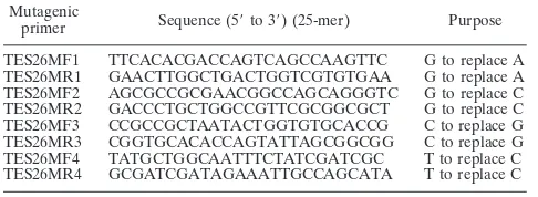
Repair of base mutations.Four DNA base errors were detected in TES-26/ TOPO, and no base errors were detected in TES-120/TOPO. Thus, in vitro PCR-based site-directed mutagenesis was performed to correct the four base errors (124, 502, 613, and 768 bp) in TES-26/TOPO by using a commercially available kit (QuikChange XL; Stratagene). Four specific forward and reverse primers (Table 1) at the particular regions were designed according to the kit manufacturer’s instructions. Plasmid DNA template (4l) was added to a PCR mixture consisting of 5l of 10⫻mutagenesis buffer, 2l each of 1M forward
and reverse mutagenic primers, 1l of 10 mM deoxynucleoside triphosphates, 1 l ofPfuDNA polymerase (2.5 U/l), and water (DNase and RNase free; Sigma) to a final volume of 50l. PCR was performed using the following parameters: one cycle at 95°C (5 min); 12 cycles at 95°C (30 s), 60°C (1 min), and 72°C (5 min); and finally one cycle at 4°C (5 min). The PCR product was then digested with 1l of 10 U/l DpnI enzyme (Fermentas) at 37°C for 1 h, followed by transformation into the TOP10E. colihost (Invitrogen). The corrected DNA sequences of the recombinant plasmids were then verified by sequencing.
Subcloning into bacterial expression vectors.All recombinant plasmids and expression vectors were digested with EcoRI enzyme (Fermentas). After diges-tion, TES-26 and TES-120 recombinant plasmids were subcloned into pET42 version b (Novagen, Germany) and pPROExHT version a (Life Technologies), respectively, using a T4 rapid DNA ligation kit (Roche Diagnostics). After the construct was verified by sequencing, the recombinant plasmids were trans-formed into an expression host, BL21(DE3) (Novagen, Germany).
Expression and purification of TES-26 and TES-120 recombinant proteins.
Each of the recombinant bacteria was cultured in Terrific broth containing 30 g/ml kanamycin (for rTES-26) or 50g/ml ampicillin (for rTES-120) and incubated at 37°C until the optical density (OD) at 600 nm reached mid-log phase (OD at 600 nm of 0.5). The expression was then induced with isopropyl--D-thiogalactopyranoside to a final concentration of 1 mM at 30°C in an
incu-bator shaker. The culture was harvested after 3 h (for rTES-26) or 5 h (for rTES-120). The affinity purification of TES-26 glutathioneS-transferase (GST)-tagged and TES-120 histidine-(GST)-tagged recombinant proteins was performed under native conditions, as described by the manufacturer, because both recombinant proteins were present in sufficient amounts in soluble form. The cells of rTES-26 were lysed in buffer (pH 7.3) containing 43 mM NaH2PO4, 14.7 mM KH2PO4,
1.37 M NaCl, and 27 mM KCl by using a French press (Thermo Spectronic) and purified using GST resin (Novagen, Germany). The cells of rTES-120 were lysed in buffer (pH 7.0) containing 50 mM NaH2PO4, 500 mM NaCl, and 10 mM
imidazole using the same method as that described above. rTES-120 was then purified with a His-Trap column (GE Healthcare) using an AKTAprimemachine (GE Healthcare). A restriction-grade site-specific protease, factor Xa enzyme, was used for specific cleavage/removal of the GST tag in TES-26 GST-tagged recombinant protein by using a commercial kit (factor Xa cleavage capture kit; Novagen, Germany). After cleavage of the target protein, factor Xa was removed by affinity chromatography using Xarrest agarose (Novagen, Germany). The sizes of the expressed target proteins were determined by sodium dodecyl sul-fate-polyacrylamide gel electrophoresis (SDS-PAGE) analysis, and the immuno-reactivities of these recombinant proteins were analyzed by Western blotting using serum samples and IgG4 antibody detection.
Western blotting.The rTES-26 and rTES-120 antigens (20g/ml) were sep-arated by 10% SDS-PAGE and transferred onto a nitrocellulose membrane (Osmonic) by using a semidry transblot apparatus (Bio-Rad). The membrane was cut into strips and blocked with 1% casein blocking solution (Roche Diag-nostics) for 1 h. The strips were then incubated with serum samples (diluted 1:100 in 0.5% blocking solution) at 4°C overnight, followed by monoclonal anti-human IgG4-horseradish peroxidase (Zymed) at 1:2,000 (in 0.5% blocking solution) for 30 min. BM chemiluminescence blotting reagent (Roche Diagnos-tics) and X-ray films (Kodak) were used to develop the blots.
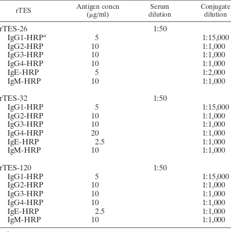
[image:2.585.41.284.90.179.2]ELISA.ELISAs based on rTES-26 and rTES-120 and the previously produced rTES-30USM recombinant antigens were developed, and the diagnostic value of the assays was evaluated using monoclonal anti-human IgG subclasses (IgG1, IgG2, IgG3, and IgG4), IgE, and IgM. The final assay was then evaluated using a panel of sera from patients with positiveToxocaraserology results or other helminth-related infections and from healthy individuals to determine its sensi-tivity and specificity. Each well of the 96-well flat-bottomed microtiter plate (Nunc Immuno Maxisorp) was coated with 100l of each recombinant antigen at the optimum concentration for each antigen in 0.02 M bicarbonate buffer, pH 9.6 (Table 2). The plate was then covered and incubated in a humid chamber at 4°C overnight followed by 2 h at 37°C. The plate was washed in phosphate-buffered saline (PBS), pH 7.2, containing 0.05% (vol/vol) Tween 20, pH 7.2, to remove unadsorbed antigen. After a washing step of five washes for 5 min each with PBS with Tween 20, each well was blocked with 1.0% blocking reagent (Roche Diagnostics) for 1 h at 37°C. The plate was again washed as previously described, followed by the addition of serum samples (100l, 1:50 in PBS, duplicate wells), and incubated at 37°C for 2 h. After the excess serum samples were washed off, mouse monoclonal anti-human IgG1- to IgG4-, IgE-, and IgM-horseradish peroxidase (Zymed) were added at an optimized dilution in PBS (Table 2) and incubated at 37°C for 30 min. Following a final washing step, 2,2⬘-azinobis(3-ethylbenzthiazoline-6-sulfonic acid-diammonium salt) substrate (Roche Diagnostics) was added and the ODs were measured after 30 min as absorbance at 405 nm (reference, 490 nm) using an ELISA spectrophotometer
TABLE 1. Mutagenic primers used to repair base errors in TES-26
recombinant plasmids
Mutagenicprimer Sequence (5⬘to 3⬘) (25-mer) Purpose
TES26MF1 TTCACACGACCAGTCAGCCAAGTTC G to replace A TES26MR1 GAACTTGGCTGACTGGTCGTGTGAA G to replace A TES26MF2 AGCGCCGCGAACGGCCAGCAGGGTC G to replace C TES26MR2 GACCCTGCTGGCCGTTCGCGGCGCT G to replace C TES26MF3 CCGCCGCTAATACTGGTGTGCACCG C to replace G TES26MR3 CGGTGCACACCAGTATTAGCGGCGG C to replace G TES26MF4 TATGCTGGCAATTTCTATCGATCGC T to replace C TES26MR4 GCGATCGATAGAAATTGCCAGCATA T to replace C
on May 16, 2020 by guest
http://jcm.asm.org/
(Tecan, Germany). The OD readings were blanked with the PBS, and an OD reading of 0.200 was used as the cutoff value to discriminate between the positive and negative results. This cutoff value was based on the mean OD reading plus three standard deviations of 30 serum samples from healthy individuals.
Statistical analysis.Analyses of the differences in sensitivity and specificity be-tween ELISAs with the different recombinant antigens were performed using Pear-son’s chi-square test. One-way analysis of variance was used to compare the ODs obtained among the IgG4 ELISAs using rTES-26, rTES-30USM, and rTES-120. Unless otherwise indicated, aPvalue of 0.05 or less was considered to be significant.
RESULTS
Gel electrophoresis of the amplified RT-PCR products of
the DNA sequence encoding TES-26 and TES-120 proteins
produced bands at the expected sizes of 793 and 528 bp,
re-spectively (Fig. 1). All four mutations that occurred at 124, 502,
613, and 789 bp in TES-26/TOPO were successfully corrected
(data not shown).
The SDS-PAGE profiles of the purified TES-26 GST fusion
and TES-120 His-tagged proteins showed distinctive and thick
bands at molecular masses of approximately 72 kDa and 26
kDa, respectively (Fig. 2). The histidine tag is a very small
peptide (approximately 0.8 kDa), is not immunogenic, and is
rarely disruptive to the properties of the proteins on which it is
attached. Therefore, the His tag was not removed. The
TES-120 His-tagged protein was thus used directly for developing
the immunoassay to detect
Toxocara
infection. The GST tag,
on the other hand, is a large molecule (approximately 26 kDa)
that affects the immunogenicity of the protein; thus, the GST
tag was removed prior to developing the immunoassay. After
removal of the GST tag, the molecular mass of the TES-26
recombinant protein was approximately 31 kDa.
The antigenicity study by Western blot analysis of the cleaved
TES-26 and TES-120 recombinant proteins showed reactivity
with serum samples from only toxocariasis patients, as indicated
by the presence of bands with molecular masses of approximately
31 and 26 kDa, respectively. Sera from healthy individuals and
patients with other helminthic infections showed no reactivity
(Fig. 3a and b).
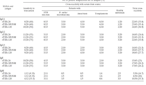
The sensitivity and cross-reactions of sera from patients with
other helminthic infections in rTES-ELISA with IgG
sub-classes (IgG1 to IgG4), IgE, and IgM are shown in Table 3.
The IgG4 ELISA demonstrated the least cross-reactivity, and
hence the highest specificity, whereas the IgG1 ELISA
dem-onstrated the lowest specificity. The decreasing order of the
assay specificities was as follows: IgG4
⬎
IgG3
⬎
IgG2
⬎
IgG1. The greatest assay sensitivity was also demonstrated by
IgG4, and the decreasing order of the assay sensitivities was as
follows: IgG4
⬎
IgG2
⬎
IgG1
ⱖ
IgG3.
The sensitivity evaluation results of the final developed IgG4
ELISA are shown in Table 4. Of 30 positive human serum
samples examined, 28 (93.3%) were reactive for anti-
Toxocara
IgG4 antibodies with rTES-30USM and rTES-120; the IgG4
ELISA using rTES-26 antigen gave 80.0% (24/30) sensitivity.
The increased level of sensitivity of 30USM and
rTES-120 IgG4 ELISA was significant in comparison with the
rTES-26 IgG4 ELISA (
P
⬍
0.001, as determined by Pearson
chi-square test). However, there was no significant difference
in mean ODs obtained with 30
Toxocara
-infected serum
sam-ples among the three tests (
P
⫽
0.76; determined by one-way
analysis of variance).
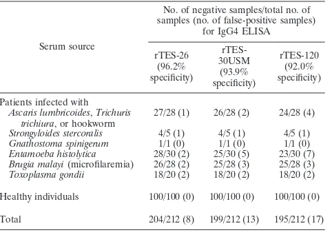
[image:3.585.371.471.69.185.2]The specificity evaluation of each of the assays is shown in
Table 5. The specificity values obtained were as follows: 96.2%
by rTES-26, 93.9% by rTES-30USM, and 92.0% by rTES-120.
At an alpha level of 0.05, there was no significant difference
TABLE 2. Optimum conditions for each rTES ELISA
rTES Antigen concn (g/ml)
Serum dilution
Conjugate dilution
rTES-26
1:50
IgG1-HRP
a5
1:15,000
IgG2-HRP
10
1:1,000
IgG3-HRP
10
1:1,000
IgG4-HRP
10
1:1,000
IgE-HRP
5
1:2,000
IgM-HRP
10
1:1,000
rTES-32
1:50
IgG1-HRP
5
1:15,000
IgG2-HRP
10
1:1,000
IgG3-HRP
10
1:1,000
IgG4-HRP
20
1:1,000
IgE-HRP
2.5
1:1,000
IgM-HRP
10
1:1,000
rTES-120
1:50
IgG1-HRP
5
1:15,000
IgG2-HRP
10
1:1,000
IgG3-HRP
10
1:1,000
IgG4-HRP
10
1:1,000
IgE-HRP
2.5
1:1,000
IgM-HRP
10
1:1,000
a
[image:3.585.43.283.80.317.2]HRP, horseradish peroxidase.
FIG. 1. Agarose gel electrophoresis of the amplified RT-PCR products
of genes encoding TES-26 and TES-120. Lane 1, 100-bp DNA ladder; lane 2,
amplified product of genes encoding TES-26; lane 3, amplified product of
genes encoding TES-120; lane 4, positive control; lane 5, negative control.
FIG. 2. SDS-PAGE of the purified 26 GST fusion and
TES-120 His-tagged proteins. Lane 1, protein marker; lane 2, rTES-TES-120
antigen (26 kDa); lane 3, rTES-26 antigen (72 kDa).
on May 16, 2020 by guest
http://jcm.asm.org/
[image:3.585.102.224.588.688.2]between the specificities of rTES-26 and rTES-120 (
P
⫽
0.059), rTES-26 and rTES-30USM, or rTES-30USM and
rTES-120. Minimal cross-reactivities were observed with serum
samples from patients with ascariasis, trichuriasis, amoebiasis,
filariasis, toxoplasmosis, and strongyloidiasis; none of the sera
from healthy individuals were reactive. Meanwhile, when 30
in-dividuals were used as healthy controls, the specificities (number
of positive samples/total number of samples) were as follows:
94.4% (134/142), 88.0% (125/142), and 90.8% (129/142) for
rTES-26, rTES-120 and rTES-30USM, respectively. These data
show high values for all parameters and show also that the
spec-ificity values of the tests were still high even when a much-reduced
number of healthy individuals was considered.
DISCUSSION
The development of a highly specific, sensitive, and reliable
[image:4.585.93.235.70.261.2]assay to detect the presence of anti-
Toxocara
antibodies is an
TABLE 4. Sensitivity evaluations of rTES-26, rTES-30USM, and
rTES-120 IgG4 ELISAs
IgG4 ELISA type
No. of samples (n⫽30)
Sensitivity (%) Positive Negative
rTES-26
24
6
80.0
rTES-30USM
28
2
a93.3
rTES-120
28
2
a93.3
a
Two serum samples that were negative by rTES-30USM were positive by rTES-120 and vice versa. Thus, the combination of rTES-30USM and rTES-120 provided 100% sensitivity.
FIG. 3. (a) Western blot analysis of rTES-26 antigen probed with
various categories of infected and noninfected serum. Lane 1, protein
marker; lanes 2, 3, and 4, sera from three different
Toxocara
-infected
patients; lane 5, serum from trichuriasis patient; lane 6, serum from
toxoplasmosis patient; lanes 7 and 8, sera from apparently healthy
people. (b) Western blot analysis of rTES-120 antigen probed with
sera from variously infected and healthy individuals. Lane 1, protein
marker; lanes 2, 3, and 4, sera from three different
Toxocara
-in-fected patients; lane 5, serum from trichuriasis patient; lane 6,
serum from toxoplasmosis patient; lanes 7 and 8, sera from
appar-ently healthy people.
TABLE 3. Sensitivities and cross-reactions of sera from patients with other helminthic infections in rTES ELISAs using
IgG subclasses, IgE, and IgM
ELISA and antigen
No. of positive samples/total no. of samples (%)
Sensitivity to toxocariasis
Cross-reactivity with serum from source
Total cross-reactivity Patients with:
Healthy individuals STH
infection
B. malayi
microfilaremia Amoebiasis Toxoplasmosis
IgG1
rTES-26
8/20 (40)
8/15
3/10
4/10
6/10
1/20
22/65 (33.8)
rTES-30USM
8/20 (40)
8/15
3/10
5/10
6/10
1/20
23/65 (35.4)
rTES-120
10/20 (50)
8/15
3/10
5/10
6/10
2/20
24/65 (36.9)
IgG2
rTES-26
11/20 (55)
5/15
2/10
3/10
3/10
3/20
16/65 (24.6)
rTES-30USM
11/20 (55)
8/15
2/10
5/10
3/10
3/20
21/65 (32.3)
rTES-120
15/20 (75)
8/15
2/10
5/10
4/10
3/20
22/65 (33.8)
IgG3
rTES-26
8/20 (40)
4/15
2/10
2/10
3/10
3/20
14/65 (21.5)
rTES-30USM
8/20 (40)
5/15
2/10
4/10
5/10
2/20
18/65 (27.7)
rTES-120
9/20 (45)
5/15
2/10
4/10
4/10
3/20
18/65 (27.7)
IgE
rTES-26
10/20 (50)
4/15
3/10
3/10
2/10
3/20
15/65 (23)
rTES-30USM
11/20 (55)
5/15
3/10
3/10
3/10
3/20
17/65 (26.2)
rTES-120
11/20 (55)
5/15
3/10
3/10
3/10
3/20
17/65 (26.2)
IgM
rTES-26
1/12 (8.33)
2/11
0/5
0/5
1/4
2/5
5/30 (16.7)
rTES-30USM
1/12 (8.33)
2/11
1/5
0/5
1/4
2/5
6/30 (20)
rTES-120
4/12 (33.3)
3/11
3/5
1/5
0/4
3/5
10/30 (33.3)
on May 16, 2020 by guest
http://jcm.asm.org/
[image:4.585.301.541.89.153.2] [image:4.585.43.541.436.727.2]important goal toward improving the diagnosis of human
toxo-cariasis. TES antigen derived from
T. canis
L2 maintained in
defined medium in vitro has been extensively used for the
immunodiagnosis of human toxocariasis. Serum samples from
patients with filariasis and with STH infections such as
ascar-iasis and strongyloidascar-iasis, however, cross-react with the native
TES antigen in immunoassays (10, 12, 15, 16, 27, 28). This may
not be a major problem in developed countries where STH
in-fections are not prevalent, but it is a significant problem in
trop-ical countries where STH infections are endemic (7, 10). Thus,
native TES antigen is useful only for differential diagnosis, and
test interpretation is problematic when the result is positive (15).
The diagnosis of human toxocariasis is currently performed
by detecting IgG in an ELISA format. The IgG assay, however,
provides false-positive reactions with other parasitic helminths,
whereas the IgG4 antibody greatly increased the specificity of
the assay for toxocariasis (15, 16, 26). Similarly, in the diagnosis
of lymphatic filariasis, antifilarial IgG4 is often used as a
marker of active infection (8, 9, 19, 25), but to date, there is no
commercially available IgG4 test for the detection of
Toxocara
infection.
Previously rTES-30USM has been successfully cloned by
assembly PCR and expressed in the prokaryotic expression
vector by our group (16). rTES-120 has been cloned and
ex-pressed as insoluble protein in the pTrcHis2 prokaryotic vector
and yeast expression vectors (3, 4). Both of these recombinant
antigens have potential in the diagnosis of toxocariasis in an
IgG ELISA (3, 4, 28). Evaluation of the diagnostic values of
both recombinant antigens, however, requires further validation.
In addition, previous studies indicate that TES-30 recombinant
antigens demonstrated high sensitivity and specificity for the
de-tection of anti-
Toxocara
IgG4 subclass antibodies (16).
A 26-kDa antigen is being used as one of six serodiagnostic
markers for confirmation of toxocariasis in a commercial
West-ern blot IgG kit that uses native TES antigen (Testline,
France). Furthermore previous studies have shown that
low-molecular-mass bands (24 to 35 kDa) of native TES antigen
were more specific for toxocariasis, while the
high-molecular-weight TES antigen bands showed reactivity with sera from
various helminth infections (11, 17). Therefore, we
hypothe-sized that rTES-26 might be useful as a serodiagnostic marker
for toxocariasis. However, it is interesting that previously
rTES-26 was reported to be of poor diagnostic value when
tested with serum from toxocariasis patients (18/118; 11.5%)
(5). The investigators themselves thought the low reactivity was
unexpected since the gene selection was based on reactivity to
antisera from TES antigen-immunized mice. Their results are
in apparent contradiction to the results of the present study;
the reasons could be due to differences in expression vectors,
purification methods, and the secondary antibodies employed.
In this study, the genes encoding TES-26 and TES-120 were
successfully cloned via RT-PCR and expressed in the
appro-priate prokaryotic expression vectors for the expression of the
recombinant proteins. The target proteins proved to be
immu-nologically reactive. Because both recombinant proteins were
well expressed in soluble form, the proteins were purified
un-der native conditions. The predicted molecular masses of
rTES-26 and rTES-120 recombinant proteins were 70 and 26
kDa, respectively. These values corresponded with the sizes of
the proteins observed using SDS-PAGE/Western blotting. The
purified recombinant proteins were then used to develop an
ELISA for the detection of specific antibody in sera from
patients infected with
T. canis
by using various antibody classes
and subclasses.
Increased levels of total IgE in many cases of toxocariasis
indicated that IgE specific for TES antigen was present. High
levels of IgE specific for TES antigen are also observed in the
serum of patients with clinical signs suggestive of
Toxocara
infection (22, 23). Not all patients with elevated total IgE
levels, however, demonstrated specific anti-
Toxocara
IgE.
Pawlowski (18) reported that
Toxocara
-specific IgE was
present in half of the patients in that study, a finding consistent
with the results in the present study, where only half of the
patients with toxocariasis had increased IgE levels.
The demonstration of increased
Toxocara
-specific IgM has
been considered indicative of acute infection (21). In our study,
however, IgM was not a good marker for
Toxocara
infection
because only 1 of 30 samples was positive by the assay. This result
is not surprising, because toxocariasis is often not an acute
dis-ease.
When the three recombinant antigens were tested in ELISAs
using all the IgG subclasses (IgG1 to IgG4), the results clearly
indicated that only the IgG4 assay displayed good specificity.
Thus, IgG4 ELISA was employed in the final development of
the diagnostic assay for toxocariasis. The use of rTES-26 IgG4
ELISA alone gave a sensitivity of 80.0%, and rTES-30USM
and rTES-120 IgG4 ELISA gave similar sensitivities of 93.3%.
When both rTES-30USM and rTES-120 were used in separate
wells, 100% sensitivity was obtained. The three recombinant
antigens were found not to be different in terms of the mean
ODs of 30 samples from patients with toxocariasis.
The results obtained differed from those obtained in a study
by Watthanakulpanich et al. (24). They reported that with
native TES antigen, anti-
Toxocara
IgG2 gave the greatest
sen-sitivity (98%) (IgG2
⬎
IgG3
⬎
IgG4
⬎
IgG1
⬎
IgG) whereas
anti-
Toxocara
IgG3 gave the greatest specificity (81%) (IgG3
⬎
IgG1
⬎
IgG
⬎
IgG2 and IgG4). These differences may be due
[image:5.585.43.284.88.260.2]to the fact that native antigen was employed in their study
whereas recombinant antigens were used in our study. The
result of the present study, however, is in agreement with other
TABLE 5. Specificity evaluations of rTES-26, rTES-30USM, and
rTES-120 IgG4 ELISAs
Serum source
No. of negative samples/total no. of samples (no. of false-positive samples)
for IgG4 ELISA
rTES-26 (96.2% specificity)
rTES-30USM (93.9% specificity)
rTES-120 (92.0% specificity)
Patients infected with
Ascaris lumbricoides,Trichuris trichiura, or hookworm
27/28 (1) 26/28 (2) 24/28 (4)
Strongyloides stercoralis 4/5 (1) 4/5 (1) 4/5 (1)
Gnathostoma spinigerum 1/1 (0) 1/1 (0) 1/1 (0)
Entamoeba histolytica 28/30 (2) 25/30 (5) 23/30 (7)
Brugia malayi(microfilaremia) 26/28 (2) 25/28 (3) 25/28 (3)
Toxoplasma gondii 18/20 (2) 18/20 (2) 18/20 (2)
Healthy individuals 100/100 (0) 100/100 (0) 100/100 (0)
Total 204/212 (8) 199/212 (13) 195/212 (17)
on May 16, 2020 by guest
http://jcm.asm.org/
previous studies that reported a significant increase in the
specificity for detection of toxocariasis when an IgG4 assay
(instead of an IgG assay) was used (15, 16, 26).
In terms of specificity, all three recombinant antigens
dis-played high specificities. Some of the false-positive cases
re-corded in the present study may have been due to coinfections
with
Toxocara
. The high specificities obtained can be explained
by the fact that the recombinant antigens (unlike native TES
antigen) are single or homogenous molecules. In addition, the
recombinant antigens are not glycosylated because they are
expressed by bacteria, which reduces the cross-reactivities with
antibodies that recognize sugar moieties (14).
Previous reports showed that the low-molecular-mass bands
(24 to 35 kDa) were more specific for toxocariasis, while
higher-molecular-mass bands showed reactivity with sera
from patients with various helminth infections (11, 17).
These findings are thus consistent with the pattern of
spec-ificities obtained in the present study, namely, rTES-26
⬎
rTES-30USM
⬎
rTES-120.
Although the sensitivity of rTES-26 was lower than that of
rTES-30USM/rTES-120, it is still important to include it in the
panel of recombinant antigens in the final assay because
TES-26 displayed a very high specificity of 96.2%.
Further-more, the additional infection marker will increase the
robust-ness of the assay, as the final assay remains to be tested with a
larger number of samples and in different settings where the
organism is endemic.
When a patient’s sample is tested using the final assay
de-veloped in this study, the results can be interpreted as follows.
When all three recombinant antigen IgG4 ELISAs are positive
and the clinical symptoms are consistent with
Toxocara
infec-tion, there is high likelihood that it is a true-positive case. The
same applies for when rTES-26 and rTES-120 or rTES-26 and
30USM ELISAs are positive. When only the
rTES-30USM or rTES-120 IgG4 ELISA is positive, there is good
evidence that the patient is infected.
Conclusion.
A sensitive and specific IgG4 ELISA for
detect-ing toxocariasis was successfully developed usdetect-ing three
recom-binant antigens: rTES-26, rTES-30USM, and rTES-120 IgG4
ELISA antigens. The assay enabled serodiagnosis of human
toxocariasis with high sensitivity and specificity. The use of
three recombinant antigens instead of a single recombinant
antigen will provide a more robust assay for use in different
parts of the world.
ACKNOWLEDGMENTS
This study was funded by a research grant from the Malaysian
Ministry of Science and Innovation (06-02-05-4261 EA019).
We thank the USM Health Center, Penang; Department of
Micro-biology and Parasitology, School of Medical Sciences, USM; and R. M.
Rohela from the Faculty of Medicine, University of Malaya, Kuala
Lumpur, for providing serum samples. We also thank Seberang Prai
City Council, Penang, and the Veterinary Laboratories at Bukit
Ten-gah, Penang; Kota Bharu, Kelantan; and Kangar, Perlis, for assistance
in the collection of adult
T. canis
worms.
REFERENCES
1.Chung, L. Y., B. H. Fang, J. H. Chang, S. M. Chye, and C. M. Yen.2004. The infectivity and antigenicity of Toxocara caniseggs can be retained after long-term preservation. Ann. Trop. Med. Parasitol.98:251–260.
2.Despommier, D.2003. Toxocariasis: clinical aspects, epidemiology, medical ecology, and molecular aspects. Clin. Microbiol. Rev.16:265–272. 3.Fong, M. Y., and Y. L. Lau.2004. Recombinant expression of the larval
excretory-secretory antigen TES-120 ofToxocara canisin the methylotropic yeastPichia pastoris. Parasitol. Res.92:173–176.
4.Fong, M. Y., Y. L. Lau, I. Init, I. Jamaiah, A. K. Anuar, and N. Rahmah.
2003. Recombinant expression ofToxocara canisexcretory-secretory anti-gens TES-120 inEscherichia coli. Southeast Asian J. Trop. Med. Public Health34:723–726.
5.Gems, D., C. J. Ferguson, B. D. Robertson, R. Nieves, A. P. Page, M. L. Blaxter, and R. M. Maizels.1995. An abundant, trans-spliced mRNA fromToxocara canisinfective larvae encodes a 26-kDa protein with ho-mology to phosphatidylethanolamine-binding proteins. J. Biol. Chem.
270:18517–18522.
6.Hakim, S. L., J. W. Mak, and P. L. W. Lam.1993. ELISA seropositivity for
Toxocara canisantibodies in Malaysia, 1989–1991. Med. J. Malaysia48:303–307. 7.Jacquier, P., B. Gottstein, Y. Sringelin, and J. Eckert.1991. Immunodiag-nosis of toxocariasis: evaluation of a new enzyme-linked immunosorbent assay. J. Clin. Microbiol.29:1831–1835.
8.Klion, A. D., A. Vijaykumar, T. Oei, B. Martin, and T. B. Nutman.2003. Serum immunoglobulin G4 antibodies to the recombinant antigen, LI-SXP-1, are highly specific forLoa loainfection. J. Infect. Dis.187:128–133. 9.Kwan-Lim, G. E., K. P. Forsyth, and R. M. Maizels.1990. Filarial-specific IgG4 response correlates with activeWuchereria bancroftiinfection. J. Im-munol.145:4298–4305.
10.Lynch, N. R., L. K. Wilkes, A. N. Hodgen, and K. J. Turner.1988. Specificity ofToxocaraELISA in tropical population. Parasite Immunol.10:323–337. 11.Magnaval, J. F., L. T. Glickman, P. Dorchies, and B. Morassin.2001.
Highlights of human toxocariasis. Korean J. Parasitol.39:1–11.
12.Magnaval, J. F., R. Fabre, P. Maurieres, J. P. Charlet, and B. De Larrard.
1991. Application of the western blotting procedure for immunodiagnosis of human toxocariasis. Parasitol. Res.77:697–702.
13.Maizels, R. M., D. H. de Savigny, and B. M. Oglivie.1984. Characterization of surface and excretory-secretory antigens ofToxocara canisinfective lar-vae. Parasite Immunol.6:23–27.
14.Maizels, R. M., M. W. Kennedy, M. Meghji, B. D. Robertson, and H. V. Smith.1987. Shared carbohydrate epitopes on the secreted and surface antigens ofToxocara canis. J. Immunol.139:207–214.
15.Noordin, R., H. V. Smith, S. Mohamad, R. M. Maizels, and M. Y. Fong.2005. Comparison of IgG-ELISA and IgG4-ELISA forToxocaraserodiagnosis. Acta Trop.93:57–62.
16.Norhaida, A., M. Suharni, A. T. Liza Sharmini, J. Tuda, and N. Rahmah.2008. rTES-30USM: cloningviaassembly PCR, expression and evaluation of useful-ness in the detection of toxocariasis. Ann. Trop. Med. Parasitol.102:151–160. 17.Park, S. P., I. Park, H. Y. Park, S. U. Lee, S. Huh, and J. F. Magnaval.2000.
Five cases of ocular toxocariasis confirmed by serology. Korean J. Parasitol.
38:267–273.
18.Pawlowski, Z.2001. Toxocariasis in humans: clinical experiment and treat-ment dilemma. J. Helminthol.75:299–305.
19.Rahmah, N., B. H. Lim, A. Khairul Anuar, R. K. Shenoy, V. Kumaraswami, S. Lokman Hakim, P. Chotechuang, K. Kanjanopas, and C. P. Ramachan-dran.2001. A recombinant antigen-based IgG4-ELISA for the specific and sensitive detection ofBrugia malayiinfection. Trans. R. Soc. Trop. Med. Hyg.95:280–284.
20.Schantz, P. M.1989.Toxocaralarva migrans now. Am. J. Trop. Med. Hyg.
41:21–34.
21.Shetty, A. K., and D. H. Aviles.1999. Nephrotic syndrome associated with
Toxocara canisinfection. Ann. Trop. Paediatr.19:297–300.
22.Smith, H., and N. Rahmah.2006. Diagnostic limitations and future trends in the serodiagnosis of human toxocariasis, p. 93–102.InC. V. Holland and H. V. Smith (ed.),Toxocara: the enigmatic parasite. CABI Publishing, Wall-ingford, United Kingdom.
23.Smith, H. V., and M. W. Kennedy.1993. Significance and quantification of antigen-specific IgE in helminthic infections of humans. J. Clin. Immunol.
16:131–143.
24.Watthanakulpanich, D., H. V. Smith, G. Hobbs, A. J. Whalley, and D. Billington.2008. Application ofToxocara canisexcretory-secretory antigens and IgG subclass antibodies (IgG1-4) in serodiagnostic assays of human toxocariasis. Acta Trop.106:90–95.
25.Weil, G. J., C. Stell, F. Liftis, B. W. Li, G. Mearns, E. Lobos, and T. B. Nutman.2000. A rapid-format antibody card test for diagnosis of onchocer-ciasis. J. Infect. Dis.182:1796–1799.
26.Wiechinger, W.1998. Diagnosticher Wert der Spezifischen IgG4antiko
¨rper-bestimmung bei der toxocariasis. Ph.D. thesis. Medizinischen Fakulta¨t der Ludwig-Maximilians-Universita¨t zu Mu¨nchen, Munich, Germany. http: //edoc.ub.unimuenchen.de/archive/00000178/.
27.Yamasaki, H., K. Araki, P. K. C. Lim, N. Zasmy, J. W. Mak, R. Taib, and T. Aoki.2000. Development of a highly specific recombinantToxocara canis
second-stage larva excretory-secretory antigen for immunodiagnosis of hu-man toxocariasis. J. Clin. Microbiol.38:1409–1413.
28.Yamasaki, H., R. Taib, Y. I. Watanabe, J. W. Mak, N. Zasmy, K. Araki, P. K. C. Lim, K. Kita, and T. Aoki.1998. Molecular characterization of a cDNA encoding an excretory-secretory antigen fromToxocara canissecond stage larvae and its application to the immunodiagnosis of human toxocari-asis. Parasitol. Int.47:171–181.