Infected Patients in Nigeria
Lana Dinic,aPatrick Akande,bEmmanuel Oni Idigbe,dAgatha Ani,cDan Onwujekwe,dOche Agbaji,cMaxwell Akanbi,cRita Nwosu,d Bukola Adeniyi,cMaureen Wahab,dChindak Lekuk,cChioma Kunle-Ope,dNkiru Nwokoye,dand Phyllis Kankia
Harvard School of Public Health, Immunology and Infectious Diseases, Boston, Massachusetts, USAa
; AIDS Prevention Initiative in Nigeria, Ltd., Abuja, Nigeriab ; Jos University Teaching Hospital, Jos, Nigeriac
; and Nigerian Institute of Medical Research, Lagos, Nigeriad
Tuberculosis (TB) is the most common opportunistic infection in human immunodeficiency virus (HIV)-infected patients and the emergence of drug-resistant tuberculosis (DR-TB) is a growing problem in resource-limited settings. Adequate infrastruc-ture for testing drug sensitivity and sufficient evidence of first-line resistance are currently unavailable in Nigeria. We collected sputum samples from HIV-infected patients enrolled in the Harvard PEPFAR/APIN Plus program over 12 months at two PEPFAR antiretroviral therapy (ART) clinics in the southwest and north central regions in Nigeria. Smear-positive sputum sam-ples were submitted for GenoType MTBDRplus testing (nⴝ415); mutations were confirmed through sequencing. Our results show high rates of DR-TB in Nigerian HIV-infected individuals (7.0% for rifampin [RIF] and 9.3% for RIF or isoniazid [INH]). Total RIF resistance indicative of MDR-TB in treatment-naive patients was 5.52%, far exceeding the World Health Organization predictions (0 to 4.3%). RIF resistance was found in 6/213 (2.8%) cases, INH resistance was found in 3/215 (1.4%) cases, and MDR-TB was found in 8/223 (3.6%) cases. We found significantly different amounts of DR-TB by location (18.18% in the south of the country versus 3.91% in the north central region [P<0.01]). Furthermore, RIF resistance was genetically distinct, sug-gesting possible location-specific strains are responsible for the transmission of drug resistance (P<0.04). Finally, GenoType MTBDRplus correctly identified the drug-resistant samples compared to sequencing in 96.8% of cases. We found that total DR-TB in HIV-infection is high and that transmission of drug-resistant TB in HIV-infected patients in Nigeria is higher than predicted.
H
uman immunodeficiency virus (HIV) greatly increases the risk for tuberculosis (TB), and the two epidemics continue to fuel one another (31). HIV-infected patients are significantly more likely to develop active TB diseases than non-HIV-infected people and are more likely to die from TB (13,27,28). In sub-Saharan Africa, 30% of HIV-infected patients who are diagnosed with TB die 12 months after the initiation of treatment (12,33). With an estimated national prevalence of HIV in Nigeria of 3.6% (7), the number of people living with HIV (3.3 million) represents the second largest burden of disease on the continent (32). Nigeria has the world’s third largest TB burden, with the prevalence of 830,000 cases. The World Health Organization (WHO) estimates that 26% of patients with TB infection in Nigeria are HIV infected (37).Multidrug-resistant TB (MDR-TB), defined by resistance to isoniazid (INH) and rifampin (RIF), is a growing global health problem (5,19,22). While MDR-TB emerges as a consequence of poor adherence to anti-TB treatment (34,35), it can also be trans-mitted. MDR-TB results in significantly higher mortality rates in HIV-infected patients than drug-susceptible TB (18). The esti-mates based on modeling predict MDR-TB prevalence in Nigeria to range from 1.8% (0.0 to 4.3%) for new cases up to 7.7% (0.0 to 18.0%) for previously treated patients (36). Currently in Nigeria, streptomycin is the only treatment available for patients previ-ously treated for TB or suspected of infection with MDR-TB. Fur-thermore, MDR-TB in HIV-infected individuals leads to higher mortality compared to mortality in non-HIV-infected patients or HIV-infected individuals with susceptible TB (18,24). These find-ings, combined with alarming evidence that MDR-TB can be transmitted, calls for close monitoring of the incidence of drug resistance, especially in HIV-infected populations (6).
The conventional methods of drug resistance testing involve growth ofMycobacterium tuberculosison liquid or solid culture medium (35). Culture methods are costly and time-consuming, thus limiting both utility for patient care and likelihood of timely treatment. Recently, several new commercial tests have been de-scribed that identify MDR-TB based on the genetic sequence; one is a line probe assay named GenoType MTBDRplus, which diag-noses TB and identifies drug resistance. It specifically examines samples for classic genetic mutations that confer resistance to both INH and RIF. Due to the reported efficiency and low cost of this test, it represents an alternative to conventional drug sensitivity testing through culture. The test has been successfully used in several locations worldwide with high sensitivity rates for RIF (⬎95.5%) and INH (⬎81.8%) resistance (2,4,15,17,21,23) and 100% specificity (2,4,15,17,21,23).
The GenoType MTBDRplus test is also reliable as a method for surveillance of drug resistance (26). The surveillance and moni-toring of both INH- and RIF-resistantM. tuberculosisis not only beneficial for an individual patient but also for the HIV-infected population as a whole, since they are more susceptible to M. tuberculosis infection (10). Implementation of the GenoType
Received23 April 2012Returned for modification28 May 2012
Accepted14 June 2012
Published ahead of print27 June 2012
Address correspondence to Phyllis Kanki, pkanki@hsph.harvard.edu.
Supplemental material for this article may be found athttp://jcm.asm.org/. Copyright © 2012, American Society for Microbiology. All Rights Reserved.
doi:10.1128/JCM.00982-12
on May 16, 2020 by guest
http://jcm.asm.org/
MTBDRplus test as a routine test can have a significant impact by improving the lives of HIV-infected patients with TB. It is there-fore imperative to identify the individuals at highest risk of acquir-ing the drug-resistantM. tuberculosisstrains in Nigeria in order to develop a programmatic policy to prevent further transmission.
MATERIALS AND METHODS
Two geographically distinct locations in Nigeria were chosen, the Nige-rian Institute for Medical Research (NIMR) in Lagos and the Jos Univer-sity Teaching Hospital (JUTH), located in the southwest and north central zones of the country, respectively. NIMR data were collected from June 2009 to June 2010, and JUTH data were collected between August 2009 and November 2010. The ethical approval was obtained from the institu-tional review boards at NIMR, JUTH, and Harvard School of Public Health (approval 16430-103).
At regular clinic visits, HIV-infected patients were screened for symp-toms of pulmonary TB, including chest pain, cough lasting more than 2 weeks, fever, night sweats, and weight loss. Upon identification, patients were asked to participate in the study and provided written informed consent. Consented patients were queried about their TB history to assess their treatment exposure prior to the study and asked to provide three sputum samples. All samples were decontaminated using the modified Petroff method and stained directly for acid-fast bacilli (AFB) using the Ziehl-Neelsen method (20). Patients with AFB-positive samples were en-rolled in the study for a 12-month period.
Samples identified as sputum smear positive (SS⫹) for AFB were in-cluded in the study. Crude DNA extraction was performed on site, fol-lowed by PCR and hybridization on test strips, according to the GenoType MTBDRplus (Hain Lifesciences, Nehren, Germany) protocol (11). Iso-lated DNA was stored at⫺20°C until the genotypic resistance testing was performed. GenoType MTBDRplus test instructions were followed forM. tuberculosisDNA amplification and hybridization (11). The tests strips were scored for resistance based on the presence of a mutant strain or the absence of wild-type DNA.
Multiplex-nested PCR and DNA sequencing of resistance genes.
Samples diagnosed as resistant with the GenoType MTBDRplus test and 16 susceptible samples from the same cohort were sequenced. Portions (5 to 10l) of crude lysate were used to amplify the four resistance-confer-ring genes (rpoB,katG, theinhApromoter, and theoxyR-ahpCintergenic regulatory region) using a multiplex PCR. The PCR was established using 1⫻ KOD polymerase buffer, 0.2 mM deoxynucleoside triphosphate (each), 1.5 mM MgSO4, 5% dimethyl sulfoxide, 100 nM concentrations of
each primer, and 1% KOD Hot Start polymerase (Toyobo, Osaka, Japan). Cycling consisted of 1 cycle of 3 min at 95°C, followed by 25 cycles of 20 s at 95°C, 10 s at 63°C, and 15 s at 72°C, and then 1 cycle of 2 min at 72°C. A nested PCR was performed individually on each gene using 5 to 10l of the multiplex sample. The reagent concentrations were identical to the multiplex PCR, except that 200 nM inner primer was used. The nested
PCR protocol consisted of 1 cycle of 3 min at 95°C, followed by 30 cycles of 20 s at 95°C and 10 to 15 s of annealing/extension at various tempera-tures (rpoBandinhA,10 s for 60°C and 5 s for 72°C;katG, 10 s for 65°C;
oxyR-ahpC, 10 s for 62°C and 5 s for 72°C).
The nested PCR products were then separated through electrophore-sis on a 2% NuSieve (Lonza, Rockland, ME) agarose gel. The bands of the appropriate sizes were excised and purified from the gel using a MinElute kit (Qiagen, Hilden, Germany) according to the manufacturer’s protocol. After the PCR fragment concentrations were evaluated using a Quant-iT PicoGreen (Invitrogen, Carlsbad, CA), 10 ng of the DNA was used for sequencing with a BigDye Terminator (v1.1; Applied Biosystems, Carls-bad, CA) cycle sequencing kit on an Applied Biosystems 3100 sequencing instrument. Primers used for sequencing were the inner primers of the multiplex-nested PCR, except for theinhApromoter region that used separate primers (see Table S1 in the supplemental material).
Data analysis.Experimentally obtained sequences were aligned with the known genetic sequence of a susceptibleM. tuberculosisstrain H37Rv using Lasergene (DNAStar, Madison, WI) and examined for previously reported resistance associated mutations. The sequence consensus of each sample was compared to the GenoType MTBDRplus results to assess the two genotypic evaluations of resistance. Groups of resistant samples were compared using the Fisher exact test, while patient characteristics at study entry were compared using the chi-square or Wilcoxon rank-sum test as appropriate. Significance threshold was set toP⬍0.05.
RESULTS
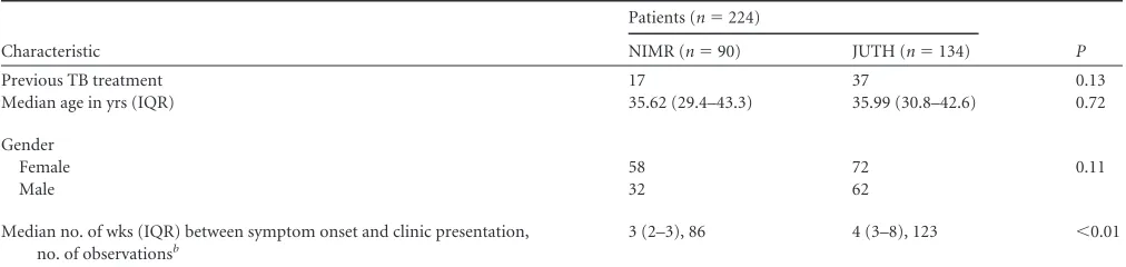
A total of 940 patients presented with signs and symptoms of TB, while 535 had at least one AFB⫹sputum. Of 415 available patient samples that were tested with GenoType MTBDRplus, genes rep-resentative of RIF susceptibility could be evaluated for 213 cases, INH susceptibility for 215 cases, and MDR-TB for 223 cases. RIF resistance with the GenoType MTBDRplus test was evaluated with the hybridized band profile of therpoBgene, while INH was eval-uated by hybridization to thekatGopen reading frame and the inhApromoter sequence. When we compared patients by site, there were no statistically significant differences in age (median, 36 years;P⫽0.72), evidence of previous TB treatment (JUTH, n⫽37; NIMR,n⫽17 [P⫽0.17]), or the percentages of female patients with 53.7% at JUTH and 64.4% at NIMR (P⫽0.11). There were differences in time from symptom onset to clinic pre-sentation (4 weeks at JUTH versus 3 weeks at NIMR;P⬍0.01)
(Table 1).
A greater number of resistant samples were observed at NIMR (n⫽14) than at JUTH (n⫽5) (P⫽0.001). The percentage of resistant samples for each drug, but not for both (MDR-TB), dem-onstrated significantly higher rates of resistance at NIMR
com-TABLE 1Patient characteristics at study entry by site locationa
Characteristic
Patients (n⫽224)
P
NIMR (n⫽90) JUTH (n⫽134)
Previous TB treatment 17 37 0.13
Median age in yrs (IQR) 35.62 (29.4–43.3) 35.99 (30.8–42.6) 0.72
Gender
Female 58 72 0.11
Male 32 62
Median no. of wks (IQR) between symptom onset and clinic presentation, no. of observationsb
3 (2–3), 86 4 (3–8), 123 ⬍0.01
aNIMR, Lagos state, South-west region; JUTH, Plateau state, North-central region. Data refer to numbers of patients except as noted in column 1. IQR, interquartile range. b
Data available for a subset of patients.
on May 16, 2020 by guest
http://jcm.asm.org/
[image:2.585.40.546.77.197.2]pared to JUTH (Table 2). Fifty-four patients had previous expo-sure to TB treatment, while 170 were treatment naive. The stratification based on patient’s prior treatment status showed no significant differences between the prevalence of drug resistance in naive versus pretreated patients (Table 3).
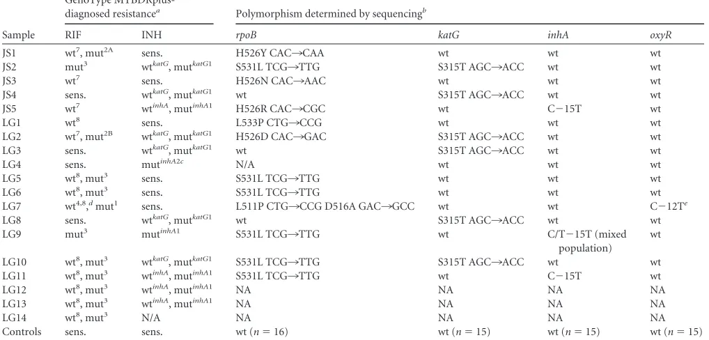
All of the polymorphisms found with the GenoType MTBDR-plus test, the codons they identify, and the numbers of samples with each profile by location are listed inTable 4. The GenoType MTBDRplus resistance profile for RIF differed by study site, whererpoBmutations from JUTH samples were predominantly found in codons 526 to 529 (wt7), while the NIMR samples showed mutations in codons 530 to 533 (wt8) (LG7 excluded,P⫽ 0.04). Genotype resistance profiles also indicate that mutations conferring INH resistance in Nigeria occur at a similar rate in the inhApromoter and thekatGS315T region, in contrast to previous reports (3,14, 16). Furthermore, all of the mutations found in katGwere the same point mutation, S315T1, and theinhA pro-moter was mutated at the C⫺15T position.
Using the multiplex/nested PCR technique, we amplified and sequenced most of the resistant samples (exceptions included LG12, LG13, and LG14), as well as 16 susceptible samples directly from sputum, to examine exact mutations found in resistance conferring genes. The GenoType MTBDRplus susceptible sam-ples showed no polymorphisms previously reported to be associ-ated with resistance (Table 4). Interestingly, each of the three sam-ples from JUTH with a mutation in codon 526 of therpoBgene had a different point substitution responsible for resistance. The most common mutation spanning therpoBwt8codon in NIMR samples was S531L.
Only 2 of the 16 resistant samples (LG7 and LG4) and none of 16 susceptible samples had discordant results obtained from the GenoType MTBDRplus test and sequencing. Sample LG7 was still resistant to RIF, and one of the three mutations indicated by GenoType MTBDRplus was concordant (rpoBwt4 D516A). In-stead of the mutations in therpoBmut1orrpoBwt8region, a muta-tion was observed in therpoBwt2region L511P.
In addition, although sample LG7 exhibited no mutations con-ferring INH resistance in thekatGorinhApromoter regions, it was the only sample that had a C¡T substitution at location⫺25 (in relation to theahpCtranscription start) of theoxyR-ahpC reg-ulatory region, previously described only ininhAresistant strains (14,16,25). Therefore, LG7 was not misdiagnosed for RIF resis-tance, but potentialinhAresistance could have been missed by not including theoxyR-ahpCregulatory region in the assay.
Sample LG4 was designated as INH resistant by the GenoType MTBDRplus test but was not confirmed by sequencing; instead, the sample appeared to be susceptible according to its sequence
analysis. With LG4 identified as susceptible, INH resistance still differed significantly by site (NIMR INH resistance⫽9.4% [P⬍ 0.03] versus JUTH; NIMR any resistance⫽16.9% [P⬍0.01] versus JUTH). There was not a significant difference in any resis-tance, including INH between treatment-naive and experienced individuals.
In total, 1 of 31 sequenced samples (3.2%) did not confirm the resistance diagnosis obtained with the GenoType MTBDRplus; therefore, the concordance rate for DR-TB diagnosis was 96.8% (oxyR-ahpC promoter region excluded). We sequenced a total of 93 regions (rpoB, katG, and inhA) that are analyzed by the GenoType MTBDRplus test. GenoType MTBDRplus misidenti-fied only four mutations and was therefore 95.7% specific.
DISCUSSION
In Nigeria, HIV/TB coinfection rates are as high as 30% in anti-retroviral therapy (ART) clinic settings, and the national preva-lence of MDR-TB is unknown. Our results show high rates of transmitted drug-resistant TB (5.5%), inferred by rates of rifam-pin resistance in treatment-naive patients. This rate exceeds the upper limit of the WHO MDR-TB models (4.3%). Furthermore, AFB sputum smears, using the Ziehl-Neelsen stain, lack sensitivity in identifying TB cases, and some cases ofM. tuberculosisinfection could have been missed. Since resistant bacteria are more likely to be less fit than sensitive bacteria (1,8,9,30) and therefore cause paucibacillary disease, our results may represent an underestimate of drug resistance. This indicates that transmission of drug-resis-tant TB is a more serious problem than previously anticipated.
[image:3.585.41.284.88.170.2]The GenoType MTBDRplus test correctly identified mutations with a high concordance rate. In recent literature, the gene-based identification of MDR-TB has gained prominence. The Gene-Xpert MTB/RIF is considered an appropriate new technology for diagnosing both TB and rifampin drug resistance. Although both GeneXpert and GenoType MTBDRplus work on a similar princi-ple— gene amplification and subsequent hybridization—Gene-Xpert MTB/RIF does not examine INH resistance. Mutational analysis of INH resistance is more complex than RIF because it requires evaluating more genes. Furthermore, the genotypic anal-ysis ofrpoBfor RIF resistance is thought to be sufficient for eval-uating the public health threat of drug-resistant TB. However, recent reports indicate that this remains controversial (29). In our study, we observed 1.4% INH monoresistance and 2.8% RIF monoresistance, highlighting the importance of evaluating both drug susceptibilities. Although one case of INH resistance was incorrectly identified with GenoType MTBDRplus, the inclusion ofinhAandkatGmutation analysis in this test correctly identified three INH-resistant, RIF-susceptible strains. This is particularly important in HIV-prevalent settings where INH prophylaxis is
TABLE 2M. tuberculosisdrug resistance in Nigerian HIV-infected patients by location as determined by GenoType MTBDRplusa
Resistance type
No. of patients/total no. of patients (%)
P
NIMR JUTH
[image:3.585.298.544.97.170.2]INH resistant 9/85 (10.59) 3/130 (2.31) ⬍0.02 RIF resistant 11/81 (13.58) 4/132 (3.03) ⬍0.01 MDR-TB 6/89 (6.74) 2/134 (1.49) ⬍0.07 Any resistance 14/77 (18.18) 5/128 (3.91) ⬍0.01 aNIMR, Lagos state southwest region; JUTH, Plateau state north central region.
TABLE 3M. tuberculosisdrug resistance in Nigerian HIV-infected patients by TB treatment history as determined by GenoType MTBDRplus
Resistance type
No. of patients/total no. of patients (%)
P
Previously treated Treatment naive
INH resistant 4/50 (8.00) 8/165 (4.85) 0.48 RIF resistant 6/50 (12.00) 9/163 (5.52) 0.12 MDR-TB 3/54 (5.56) 5/169 (2.96) 0.40 Any resistance 7/46 (15.22) 12/159 (7.55) 0.15
on May 16, 2020 by guest
http://jcm.asm.org/
being considered. Such preventative measures might not be effec-tive and could increase the rates of INH resistance, exacerbating the diagnostic challenges for MDR-TB. Furthermore, misdiag-nosing patients as MDR-TB when they are only RIF monoresis-tant would lead to inappropriate second-line treatment, when such treatment in resource-limited settings is already limited.
The baseline characteristics of age, gender, and prior treatment status of patients did not show major site differences, indicating that the sites were comparable. Although the duration of symp-toms prior to clinic visit differed, this difference might be due to the fact that patients visiting the clinic in Jos traveled longer dis-tances and therefore were at a disadvantage to be adherent to clinic visits. In these two geographically distinct study populations, a significantly higher number of drug-resistant TB was found in Lagos compared to Jos. This phenomenon could be explained by societal differences in the two cities, which might facilitate trans-mission, Lagos being more densely populated with crowded living arrangements and a congested public transportation system com-pared to Jos. Transmission of drug-resistant TB in Lagos is further suggested by the fact that 9 of 13 patients with drug-resistant TB were treatment naive (versus 2/5 in Jos). An alternative explana-tion is that theM. tuberculosisstrains differed by location and therefore so did their transmission and/or mutation rates. Al-though we were not able to evaluate theM. tuberculosisstrains or their characteristics in the present study, the mutations in therpoB gene are suggestive of such differences. All of the Jos mutations in therpoB gene were distinct, implying that they were acquired through separate mutagenesis events, whereas most of the Lagos
strains had the same mutation (S531L) belonging to a potentially more transmissible strain.
In summary, our study demonstrated high rates of drug-resis-tant TB in HIV-infected patients. We also showed that the cost-effective GenoType MTBDRplus test correctly identified resis-tance-conferring mutations in the majority of samples examined (95.7%). Although our sample size was small, this is the first mul-tisite study of MDR-TB in HIV-infected population in Nigeria. Consistent with WHO recommendations, our results support the urgent need for systematic drug resistance testing in all HIV-in-fected patients with symptoms suggestive of TB.
ACKNOWLEDGMENTS
We are deeply grateful to the patients for their willingness to participate in this study. Furthermore, we thank the clinical and laboratory staff of the NIMR and JUTH APIN Plus clinics. We especially thank Rosemary Adu, Favor Olatunbasun, Godwin Imade, Moses Adie, Nwanneka Tochukwu, Lauretta Efere, Peter Nwadike, Tope Abiodu, and Eucharia Obidike.
This study was funded in part by the U.S. Department of Health and Human Services, Health Resources, and Services Administration (U51HA02522-01-01). Research support to L.D. was provided by the Zlatko and Balokovic Scholarship, the Uwe Brinkmann Traveling Grant, and a Harvard University Science and Engineering Committee Scholar-ship.
The contents are solely the responsibility of the authors and do not represent the official views of the funding institutions.
REFERENCES
[image:4.585.39.548.85.329.2]1.Andersson DI.2006. The biological cost of mutational antibiotic resis-tance: any practical conclusions? Curr. Opin. Microbiol.9:461– 465.
TABLE 4Resistance profiles of samples from NIMR (LG) and JUTH (JS) in Nigeria
Sample
GenoType
MTBDRplus-diagnosed resistancea Polymorphism determined by sequencingb
RIF INH rpoB katG inhA oxyR
JS1 wt7, mut2A sens. H526Y CAC
¡CAA wt wt wt
JS2 mut3 wtkatG, mutkatG1 S531L TCG
¡TTG S315T AGC¡ACC wt wt JS3 wt7 sens. H526N CAC
¡AAC wt wt wt
JS4 sens. wtkatG, mutkatG1 wt S315T AGC
¡ACC wt wt
JS5 wt7 wtinhA, mutinhA1 H526R CAC
¡CGC wt C⫺15T wt
LG1 wt8 sens. L533P CTG
¡CCG wt wt wt
LG2 wt7, mut2B wtkatG, mutkatG1 H526D CAC
¡GAC S315T AGC¡ACC wt wt LG3 sens. wtkatG, mutkatG1 wt S315T AGC
¡ACC wt wt
LG4 sens. mutinhA2c N/A wt wt wt
LG5 wt8, mut3 sens. S531L TCG
¡TTG wt wt wt
LG6 wt8, mut3 sens. S531L TCG
¡TTG wt wt wt
LG7 wt4,8,dmut1 sens. L511P CTG
¡CCG D516A GAC¡GCC wt wt C⫺12Te
LG8 sens. wtkatG, mutkatG1 wt S315T AGC
¡ACC wt wt
LG9 mut3 mutinhA1 S531L TCG
¡TTG wt C/T⫺15T (mixed population)
wt
LG10 wt8, mut3 wtkatG, mutkatG1 S531L TCG
¡TTG S315T AGC¡ACC wt wt LG11 wt8, mut3 wtinhA, mutinhA1 S531L TCG
¡TTG wt C⫺15T wt
LG12 wt8, mut3 wtinhA, mutinhA1 NA NA NA NA LG13 wt8, mut3 wtinhA, mutinhA1 NA NA NA NA
LG14 wt8, mut3 N/A NA NA NA NA
Controls sens. sens. wt (n⫽16) wt (n⫽15) wt (n⫽15) wt (n⫽15) aGenoType MTBDRplus-diagnosed resistance (wt, hybridization to wild-type band missing; mut, hybridization to mutant band present); N/A, not available. sens., sensitive. b
“wt” refers to the H37Rv sequence. NA, not amplified in multiplex/nested PCR.
cMutation not observed by sequencing. d
Mutations on codon 530 –533(wt8
) and mut1
D516V were not confirmed by sequencing; however, the lack of a wt4
band was confirmed (D516A), and an additional mutation L511P (wt2) was found.
e
Mutation previously reported in resistant sample.
on May 16, 2020 by guest
http://jcm.asm.org/
2.Anek-Vorapong R, et al.2010. Validation of the GenoType MTBDRplus assay for detection of MDR-TB in a public health laboratory in Thailand. BMC Infect. Dis.10:123. doi:10.1186/1471-2334-10-123.
3.Baker LV, et al.2005. Molecular analysis of isoniazid-resistant Mycobac-terium tuberculosisisolates from England and Wales reveals the phyloge-netic significance of theahpC-46A polymorphism. Antimicrob. Agents Chemother.49:1455–1464.
4.Causse M, Ruiz P, Gutierrez JB, Zerolo J, Casal M.2008. Evaluation of new GenoType MTBDRplus for detection of resistance in cultures and direct specimens ofMycobacterium tuberculosis. Int. J. Tuberc. Lung Dis.
12:1456 –1460.
5.Chan ED, Iseman MD.2008. Multiresistant and extensively drug-resistant tuberculosis: a review. Curr. Opin. Infect. Dis.21:587–595. 6.Dorman SE, Chaisson RE.2007. From magic bullets back to the magic
mountain: the rise of extensively drug-resistant tuberculosis. Nat. Med.
13:295–298.
7.Federal Ministry of Health Nigeria.2011. Technical report: 2010 Na-tional HIV Sero-prevalance Sentinel Survey, p 1–110. Federal Ministry of Health, Abuja, Nigeria.
8.Gagneux S.2009. Fitness cost of drug resistance inMycobacterium tuber-culosis. Clin. Microbiol. Infect.15(Suppl 1):66 – 68.
9.Gagneux S, et al.2006. The competitive cost of antibiotic resistance in
Mycobacterium tuberculosis. Science312:1944 –1946.
10. Godfrey-Faussett P, et al. 2000. Tuberculosis control and molecular epidemiology in a South African gold-mining community. Lancet356: 1066 –1071.
11. Hain Lifescience.2009. GenoType MTBDRplus manual, p 23– 42. In
Molecular genetic assay for identification of resistance to rifampicin and/or isoniazid of the Mycobacterium tuberculosis complex. Hain Life-science GmbH, Nehren, Germany.
12. Harries AD, et al.2001. Deaths from tuberculosis in sub-Saharan African countries with a high prevalence of HIV-1. Lancet357:1519 –1523. 13. Harries AD, et al.1998. Treatment outcome of an unselected cohort of
tuberculosis patients in relation to human immunodeficiency virus se-rostatus in Zomba Hospital, Malawi. Trans. R. Soc. Trop. Med. Hyg.92: 343–347.
14. Hazbon MH, et al.2006. Population genetics study of isoniazid resistance mutations and evolution of multidrug-resistantMycobacterium tubercu-losis. Antimicrob. Agents Chemother.50:2640 –2649.
15. Huang WL, Chen HY, Kuo YM, Jou R.2009. Performance assessment of the GenoType MTBDRplus test and DNA sequencing in detection of mul-tidrug-resistantMycobacterium tuberculosis. J. Clin. Microbiol.47:2520 – 2524.
16. Kim SY, et al.2003. Molecular analysis of isoniazid resistance in Myco-bacterium tuberculosisisolates recovered from South Korea. Diagn. Micro-biol. Infect. Dis.47:497–502.
17. Lacoma A, et al.2008. GenoType MTBDRplus assay for molecular de-tection of rifampin and isoniazid resistance inMycobacterium tuberculosis
strains and clinical samples. J. Clin. Microbiol.46:3660 –3667.
18. Mannheimer SB, et al.1997. Risk factors and outcome of human immu-nodeficiency virus-infected patients with sporadic multidrug-resistant tu-berculosis in New York City. Int. J. Tuberc. Lung Dis.1:319 –325. 19. Maxmen A.2010. Fighting the monster. Nature466:S18 –S19.
20. Narvaiz de Kantor I, et al. 1998. Laboratory services in tuberculosis control. World Health Organization, Geneva, Switzerland.
21. Neonakis IK, et al.2009. Evaluation of GenoType mycobacteria direct assay in comparison with Gen-ProbeMycobacterium tuberculosis ampli-fied direct test and GenoType MTBDRplus for direct detection of Myco-bacterium tuberculosiscomplex in clinical samples. J. Clin. Microbiol.47: 2601–2603.
22. NIAID.2007. Scientific illustrations of drug-resistant TB. National Institute of Allergy and Infectious Diseases, Bethesda, MD.http://www.niaid.nih.gov /topics/tuberculosis/understanding/whatistb/scientificillustrations/pages /multidrugresistantillustration.aspx.
23. Nikolayevskyy V, et al.2009. Performance of the Genotype MTBDRPlus assay in the diagnosis of tuberculosis and drug resistance in Samara, Rus-sian Federation. BMC Clin. Pathol.9:2. doi:10.1186/1472-6890-9-2. 24. Park MM, Davis AL, Schluger NW, Cohen H, Rom WN.1996. Outcome
of MDR-TB patients, 1983–1993. Prolonged survival with appropriate therapy. Am. J. Respir. Crit. Care Med.153:317–324.
25. Ramaswamy S, Musser JM.1998. Molecular genetic basis of antimicro-bial agent resistance inMycobacterium tuberculosis: 1998 update. Tuberc. Lung Dis.79:3–29.
26. Rigouts L, et al.2011. Evaluation of the GenoType® MTBDRplus assay as a tool for drug resistance surveys. Int. J. Tuberc. Lung Dis.15:959 –965. 27. Selwyn PA, et al.1989. A prospective study of the risk of tuberculosis
among intravenous drug users with human immunodeficiency virus in-fection. N. Engl. J. Med.320:545–550.
28. Selwyn PA, et al.1992. High risk of active tuberculosis in HIV-infected drug users with cutaneous anergy. JAMA268:504 –509.
29. Smith SE, Kurbatova EV, Cavanaugh JS, Cegielski JP. 2012. Global isoniazid resistance patterns in resistant and rifampin-susceptible tuberculosis. Int. J. Tuberc. Lung Dis.16:203–205.
30. Strauss OJ, et al.2008. Spread of a low-fitness drug-resistant Mycobacte-rium tuberculosisstrain in a setting of high human immunodeficiency virus prevalence. J. Clin. Microbiol.46:1514 –1516.
31. Suchindran S, Brouwer ES, Van Rie A.2009. Is HIV infection a risk factor for multidrug resistant tuberculosis? A systematic review. PLoS One
4:e5561. doi:10.1371/journal.pone.0005561.
32. UNAIDS/World Health Organization.2009. AIDS epidemic update De-cember 2009. UNAIDS, Geneva, Switzerland.
33. Whalen CC, et al.2000. Impact of pulmonary tuberculosis on survival of HIV-infected adults: a prospective epidemiologic study in Uganda. AIDS
14:1219 –1228.
34. Global Tuberculosis Programme/WHO.2003. Treatment of tuberculo-sis: guidelines for national programs, 3rd ed. World Health Organization, Geneva, Switzerland.
35. World Health Organization.2007. The global MDR-TB and XDR-TB response plan. World Health Organization, Geneva, Switzerland. 36. World Health Organization.2011. Nigeria: tuberculosis profile 2010.
World Health Organization, Geneva, Switzerland.https://extranet.who .int/sree/Reports?op⫽Replet&name⫽%2FWHO_HQ_Reports%2FG2 %2FPROD%2FEXT%2FTBCountryProfile&ISO2⫽NG&outtype⫽html. 37. World Health Organization.2010. Global tuberculosis control: WHO
report 2010. World Health Organization, Geneva, Switzerland.