Obstructive Pulmonary Disease
Yvonne J. Huang,aSanjay Sethi,b,cTimothy Murphy,cSnehal Nariya,aHomer A. Boushey,aSusan V. Lynchd
Division of Pulmonary/Critical Care/Allergy/Sleep Medicine, Department of Medicine, University of California, San Francisco, San Francisco, California, USAa; Division of Pulmonary/Critical Care/Sleep Medicine, Veterans Western New York Health System, Buffalo, New York, USAb; Department of Medicine, University at Buffalo, State University of New York, Buffalo, New York, USAc; Division of Gastroenterology, Department of Medicine, University of California, San Francisco, San Francisco, California, USAd
Specific bacterial species are implicated in the pathogenesis of exacerbations of chronic obstructive pulmonary disease (COPD). However, recent studies of clinically stable COPD patients have demonstrated a greater diversity of airway microbiota, whose role in acute exacerbations is unclear. In this study, temporal changes in the airway microbiome before, at the onset of, and after an acute exacerbation were examined in 60 sputum samples collected from subjects enrolled in a longitudinal study of bacterial infection in COPD. Microbiome composition and predicted functions were examined using 16S rRNA-based culture-indepen-dent profiling methods. Shifts in the abundance (>2-fold,P<0.05) of many taxa at exacerbation and after treatment were ob-served. Microbiota members that were increased at exacerbation were primarily of theProteobacteriaphylum, including non-typical COPD pathogens. Changes in the bacterial composition after treatment for an exacerbation differed significantly among the therapy regimens clinically prescribed (antibiotics only, oral corticosteroids only, or both). Treatment with antibiotics alone primarily decreased the abundance ofProteobacteria, with the prolonged suppression of some microbiota members being ob-served. In contrast, treatment with corticosteroids alone led to enrichment forProteobacteriaand members of other phyla. Pre-dicted metagenomes of particular microbiota members involved in these compositional shifts indicated exacerbation-associated loss of functions involved in the synthesis of antimicrobial and anti-inflammatory products, alongside enrichment in functions related to pathogen-elicited inflammation. These trends reversed upon clinical recovery. Further larger studies will be necessary to determine whether specific compositional or functional changes detected in the airway microbiome could be useful indicators of exacerbation development or outcome.
A
cute exacerbations of chronic obstructive pulmonary disease(AECOPD) are a major source of morbidity, characterized by worsening of respiratory symptoms from the baseline normal
variation that require new treatment (http://www.goldcopd.org/).
Culture-based evaluations have implicated bacteria in⬃50%
of exacerbations (1). While acquisition of new strains of
Hae-mophilus influenzae, Streptococcus pneumoniae, Moraxella ca-tarrhalis, andPseudomonas aeruginosais associated with
exacer-bations (2,3), these represent relatively few species compared to
the overall diversity of airway microbiota recently described in
COPD (4–7).
Detection of bacteria in most prior studies was limited by the
use of culture-based methods (8,9). Application of more sensitive
molecular tools has consistently shown a greater diversity of air-way microbiota in COPD, despite differences in specimens and
platforms across studies (4–7,10). With increasing appreciation
of the fact that the microbial microenvironment can modify the
behavior of primary pathogens (11,12), this invites speculation
that other microbiota members may contribute to the pathogen-esis of AECOPD.
In this pilot study, we investigated the dynamics of the airway bacterial microbiome in the setting of AECOPD by examining a total of 60 sputum samples collected before, at the onset of, and after an exacerbation. Compositional and functional analyses were performed using 16S rRNA-based culture-independent and
in silicopredictive metagenomic approaches. The relationships between microbiome composition and clinical factors were exam-ined, including the type of treatment prescribed for exacerbation (antibiotics alone, systemic corticosteroids alone, or both).
MATERIALS AND METHODS
Subjects and sample collection.The sputum samples analyzed were col-lected from 12 subjects enrolled in a longitudinal study of bacterial infec-tion in COPD described extensively in previous publicainfec-tions (2,3,13). To briefly summarize, the parent study has been conducted at the Buffalo Veterans Affairs Hospital and approved by the VA Western New York Institutional Review Board since 1994. All participants gave written in-formed consent. Members of a cohort of approximately 50 participants with COPD (and without other lung disease) were followed monthly and whenever they experienced worsening symptoms suggestive of an exacer-bation. Expectorated sputum was collected at each visit and processed as previously described (2). This included homogenization with dithiothre-itol and plating of aliquots of serial dilutions on blood, chocolate, and MacConkey culture agar. Sputum cell pellets obtained after centrifuga-tion were stored long term at⫺80°C in a glycerol solution and used for microbiome analyses in this study. Molecular typing of isolates ofH. in-fluenzae,M. catarrhalis, andP. aeruginosawas performed as previously described (2) to determine whether a new strain, defined as one that had
Received7 January 2014 Returned for modification8 February 2014 Accepted17 May 2014
Published ahead of print21 May 2014
Editor:P. H. Gilligan
Address correspondence to Yvonne J. Huang, yvonne.huang@ucsf.edu.
Supplemental material for this article may be found athttp://dx.doi.org/10.1128 /JCM.00035-14.
Copyright © 2014, American Society for Microbiology. All Rights Reserved.
doi:10.1128/JCM.00035-14
on May 16, 2020 by guest
http://jcm.asm.org/
not been isolated from sputum samples obtained previously (⬎4 weeks) from an individual patient, was present at exacerbation (2).
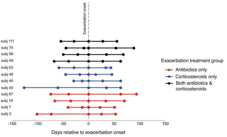
To investigate the exacerbation-related microbiome, subjects and samples were carefully selected for this study to include a diversity of exacerbations, those that were and were not associated with a new strain, and also treatment approaches commonly prescribed in clinical practice (antibiotics only, systemic corticosteroids only, or both). Specimens from five time points per subject were examined (Fig. 1). The specimens spanned an exacerbation-related time period occurring in a given year between 1999 and 2005 (with the specific year depending on the subject). Pre- and postexacerbation samples were those available closest to the ex-acerbation when the patients were at their baseline respiratory state and on their usual COPD treatment. All subjects were encouraged to seek treatment early for a suspected exacerbation; however, a fixed duration of symptoms was not proscribed. However, all exacerbation samples in-cluded in this study were obtained prior to initiation of any specific treat-ment for the exacerbation. Exacerbation events were determined using predefined criteria described previously (2) and involved evaluation of respiratory symptoms (dyspnea, cough, sputum production, viscosity, and purulence) and their change from the usual state. Pneumonia, if sus-pected, was ruled out by chest X ray. Decisions about treatments were made by study physicians (S. Sethi and T. Murphy) on the basis of symp-toms (especially sputum purulence) and prior to the availability of spu-tum culture data from the concurrent visit. Antibiotics were prescribed if bacterial infection was thought to be a cause of the exacerbation, whereas steroids only were prescribed if a nonbacterial etiology was suspected.
Sample preparations.DNA was extracted utilizing methods previ-ously described (14) and detailed further in the supplemental material. Briefly, universal 16S rRNA bacterial primers (Bact-27F, Bact-1492R) were used to generate amplicons, which were processed for hybridization to G2 PhyloChip microarrays (Second Genome, South San Francisco, CA) by methods previously described (14,15). Each sample was hybrid-ized to an individual array, resulting in 60 microbiome profiles. Bacterial taxa were defined as organisms sharingⱖ97% 16S rRNA gene sequence homology and classified according to the Hugenholtz taxonomy in the 2011 iteration of the Greengenes database (16).
Data analyses.Raw array data were processed as previously described (17–19), including correction for background and noise and scaling of fluorescence intensities to those for spiked-in quantitative standards,
fol-lowed by further data normalization on the basis of the mean intensity of all profiles. The presence of taxa was determined using published methods (19,20), with slightly modified scoring thresholds based on the scores for spiked-in quantitative standards being applied. Further information is detailed in the supplemental material. The initial normalized data set for all 60 samples combined included⬃4,800 taxa, and this data set was then filtered to include taxa present in at least 10% of samples, yielding for analysis a final data set of⬃3,300 taxa (available in Table S5 in the sup-plemental material). Log2-transformed intensities, which correlate with the relative abundance of taxa (21), were used in all primary analyses, which were performed using R (version 2.14.1).
Community richness (number of taxa detected), evenness (relative distribution of taxa in a community), and diversity (Shannon and inverse Simpson indices, which consider both the richness and the evenness in the calculation, as well as Faith’s phylogenetic diversity, which additionally incorporates phylogenetic information in this calculation [22]), were cal-culated using the R packages vegan and picante (23,24). Changes in these metrics were tested using repeated-measures analysis of variance (RM-ANOVA) and pairedttests. Bray-Curtis and Unifrac distance measures (25,26) were calculated and used in ordination analyses (e.g., nonmetric multidimensional scaling [NMDS]) to assess compositional differences and relationships to clinical factors, including the timing of pre- and postexacerbation sample collection relative to the exacerbation onset date, by distance matrix-based permutational multivariate analysis of variance (PERMANOVA) (27). Taxon-level analyses to identify specific compositional changes between time points were performed using linear models based on moderatedtstatistics and a paired test design in the R package limma (28). Adjustment for multiple comparisons was applied using the Benjamini-Hochberg method orqvalues (29). Changes of at least 2-fold (log2differenceⱖ1.0; unadjustedP⬍0.05) were considered notable and significant if the Benjamini-Hochberg-adjustedPvalue orq value was⬍0.05. These criteria were extrapolated from thresholds com-monly applied to screen for differentially expressed genes in microarray-based studies and that have also been used in prior PhyloChip-microarray-based studies (30,31). For the predictive functional analyses, the PICRUSt soft-ware package (32) was used to identify predicted gene families and asso-ciated pathways from inferred metagenomes of taxa of interest identified from the compositional analyses.
FIG 1Time points of sample collection before, at the onset of, and after an acute COPD exacerbation. The line coloring indicates the type of treatment for the
exacerbation that the subject received. Huang et al.
on May 16, 2020 by guest
http://jcm.asm.org/
[image:2.585.109.471.63.281.2]qPCR and sequencing.Total 16S rRNA gene copies in samples were determined using methods previously described (14). Species-specific quantitative PCR (qPCR) and Sanger sequencing were also performed to confirm, respectively, array-reported relative abundances and the identi-ties of representative organisms of interest. To further compare and vali-date the microarray data, 11 samples with sufficient remaining DNA that represented different sampling time points underwent 16S rRNA se-quencing using the Illumina MiSeq platform. Details of these experiments are described extensively in the supplemental material.
RESULTS
Relationships between bacterial community composition and clinical associations.For each subject, two samples from a clini-cally stable period prior to exacerbation (Pre1 and Pre2; range, 12 to 126 days before exacerbation), a single sample obtained at ex-acerbation onset before the start of new treatments (Exac), and two samples from after exacerbation when treatments were com-pleted (Post1 and Post2; range, 14 to 94 days after the date of
exacerbation onset) were collected (Fig. 1). The characteristics of
the subjects, most of whom had moderately severe airflow ob-struction (Global Initiative for Chronic Obstructive Lung Disease stage II by criteria published at the time that samples were
col-lected [33]), and the exacerbations are shown inTable 1.
Across all samples examined, bacterial community richness, evenness, and diversity did not differ significantly over time (RM-ANOVA) or when they were considered across pairwise compar-isons of sampling time points, e.g., Post1 versus Exac (paired
Stu-dent’s t test). However, the data indicated a trend toward a
decrease in diversity following treatment with antibiotics. In con-trast, diversity increased in the steroid only-treated group. Similar trends were observed for richness.
To further evaluate relationships between community compo-sition and clinical characteristics, multivariate analyses using PERMANOVA based on a constructed Bray-Curtis distance
ma-trix (27) were performed. No significant relationships were found
between community composition and parameters of lung func-tion, symptom severity score at exacerbafunc-tion, or the time points at which samples were collected (number of days relative to the ex-acerbation onset date). The latter results indicated that variation among subjects in the timing of sample collection before and after an exacerbation was not a significant factor in the observed com-positional differences.
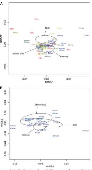
In clinical practice as well as in the parent study, clinical im-pression regarding the etiology of a COPD exacerbation deter-mines treatment choices. We therefore examined first, using all samples, whether an association existed between the type of exac-erbation treatment (antibiotics only, oral steroids only, or both;
n⫽4 subjects per treatment group) and differences in community
composition among samples. A statistically significant relation-ship between community composition and the treatment
admin-istered for exacerbation was observed (P⬍0.05;Fig. 2A). When
we limited this analysis to only postexacerbation samples, the
as-sociation remained close to statistically significant (P⫽0.08;Fig.
2B), indicating that the preexacerbation and exacerbation samples
contribute to the strength of this relationship. These results also suggest that the specific treatment administered for exacerbation has the potential to have a prolonged impact on community com-position.
Chronic use of inhaled corticosteroids (ICSs) also demon-strated a nearly statistically significant relationship with
differ-ences in community composition (P⫽0.06, based on analysis of
all samples;P⫽0.08 for analysis limited to only preexacerbation
samples;Fig. 2C), indicating that ICSs may impact the community
composition across the time points sampled in this study. We also observed a trend toward greater richness and diversity in samples from ICS-exposed subjects than those from non-ICS-exposed subjects, indicating that ICSs, perhaps through their
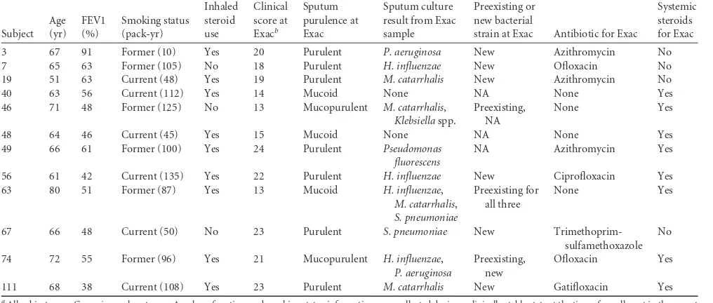
immunosup-TABLE 1Clinical characteristics of subjects and exacerbationsa
Subject Age (yr) FEV1 (%) Smoking status (pack-yr) Inhaled steroid use Clinical score at Exacb Sputum purulence at Exac Sputum culture result from Exac sample
Preexisting or new bacterial
strain at Exac Antibiotic for Exac
Systemic steroids for Exac
3 67 91 Former (10) Yes 20 Purulent P. aeruginosa New Azithromycin No
7 65 63 Former (105) No 18 Purulent H. influenzae New Ofloxacin No
19 51 63 Current (48) Yes 19 Purulent M. catarrhalis New Azithromycin No
40 63 56 Current (112) Yes 14 Mucoid None NA None Yes
46 71 48 Former (125) No 13 Mucopurulent M. catarrhalis, Klebsiellaspp.
Preexisting, NA
None Yes
48 64 46 Current (45) Yes 15 Mucoid None NA None Yes
49 66 61 Former (100) Yes 24 Purulent Pseudomonas
fluorescens
NA Azithromycin Yes
56 61 42 Current (135) Yes 22 Purulent H. influenzae New Ciprofloxacin Yes
63 80 51 Former (87) Yes 13 Mucoid H. influenzae,
M. catarrhalis, S. pneumoniae
Preexisting for all three
None Yes
67 66 48 Current (50) No 23 Purulent S. pneumoniae New
Trimethoprim-sulfamethoxazole No
74 72 55 Former (96) Yes 21 Mucopurulent H. influenzae, P. aeruginosa
Preexisting, new
Ofloxacin Yes
111 68 38 Current (108) Yes 23 Purulent M. catarrhalis New Gatifloxacin Yes
aAll subjects were Caucasian male veterans. Age, lung function, and smoking status information were collected during a clinically stable state at the time of enrollment in the parent
study. Inhaled corticosteroid use status was current for the analyzed time points. FEV1, forced expiratory volume in 1 s; pack-yr, number of packs of cigarettes smoked per day multiplied by the number of years that the person has smoked; NA, not applicable.
b
See reference13.
on May 16, 2020 by guest
http://jcm.asm.org/
[image:3.585.39.547.79.295.2]FIG 2(A) Nonmetric multidimensional scaling (NMDS) ordination demonstrates that samples from all subjects in the study segregate into distinct clusters (indicated by ellipses) on the basis of the type of treatment prescribed for exacerbation (antibiotics [Abx] only, oral steroids only, or both;n⫽4 subjects per treatment group; Bray-Curtis distance-based PERMANOVA,P⬍0.05). Ellipses represent the 95% confidence interval for the standard error of the distances of samples in each treatment group. (B) An NMDS plot of only postexacerbation treatment samples from all subjects demonstrates that the relationship between sample community composition and treatment type remains strong, though not statistically significant (P⫽0.08), when the analysis is limited to only these samples. (C) An NMDS plot of only preexacerbation samples from all subjects shows that chronic use of inhaled corticosteroids may be associated with differences in bacterial community composition (Bray-Curtis distance-based PERMANOVA,P⫽0.08).
Huang et al.
on May 16, 2020 by guest
http://jcm.asm.org/
[image:4.585.133.447.60.641.2]pressive activity, increase the airway microbiota diversity in COPD patients.
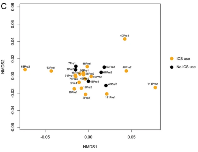
Taxon-level analyses of temporal changes in COPD airway microbiota composition.To determine the specific taxa that ex-hibited significant changes in relative abundance during the course of this study, we compared array-reported abundances for each taxon between sequential time points (R package limma). For this set of analyses, we first performed paired time point com-parisons using data from all subjects, followed by separate analyses for each exacerbation treatment group (antibiotics only, systemic steroids only, or both) limited to only the postexacerbation time points. Collectively, these results revealed several notable findings. During the clinically stable period before exacerbation
(Pre1-to-Pre2 comparison;Fig. 3A), relatively few taxa exhibited
differ-ential relative abundance. Only three taxa showed an increase of
ⱖ2-fold (the largest was aMoraxellaspecies, which showed a
2.6-fold increase;P⫽0.001), whereasNeisseriaceaemembers made up
many of the taxa that decreased byⱖ2-fold. Overall, these
obser-vations suggest relative stability in the airway microbiota compo-sition during a clinically stable period prior to AECOPD in this cohort.
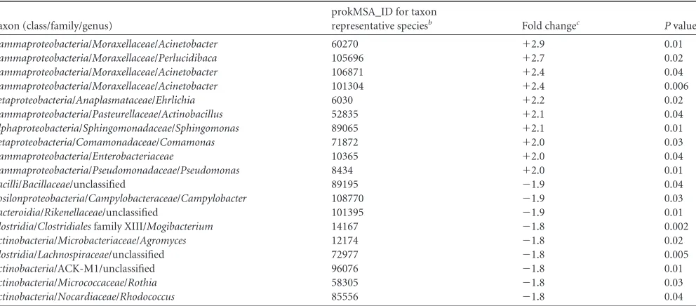
At exacerbation, it was predominantly members of the
Proteo-bacteria, such as theMoraxellaceae,Pasteurellaceae, Pseudomon-adaceae, andEnterobacteriaceae, that were enriched compared to
their levels in Pre2 (Table 2). These enriched taxa included other
potential respiratory pathogens, in addition to those typically as-sociated with AECOPD. In contrast, taxa that were decreased in abundance at exacerbation relative to their abundance in Pre2
were predominantly Actinobacteria, Clostridia, and Bacteroidia
(Table 2).Actinobacteriacomprise a large group of metabolically diverse organisms that are prolific producers of secondary
metab-olites, including those with antimicrobial activity (34), while clade
IV and XIVAClostridiaare known inducers of anti-inflammatory
T-regulatory cells (35). To speculate, these observations suggest
the possibility that members of the microbiome that are depleted
at the time of exacerbation may otherwise help maintain commu-nity and host immune homeostasis and potentially counteract the detrimental effects of respiratory pathogenic species.
The most dramatic changes in community composition were observed after treatment for exacerbation (Exac to Post1). This was supported by a significant difference in the mean Unifrac distance of communities between Exac and Post1 compared to that for communities between Pre2 and Exac, reflecting a
signifi-cant change in the phylogenetic makeup of the community (P⬍
0.01; see Fig. S1 in the supplemental material). Among those taxa exhibiting significant alterations in relative abundance, the vast majority decreased in abundance, were primarily members of the
Proteobacteria, and included organisms both associated with and
not traditionally associated with COPD (P⬍0.05,qⱕ0.12; see
Table S1 in the supplemental material).
Striking differences, however, were seen in the effects of
differ-ent treatmdiffer-ents on community composition (Fig. 3B). Treatment
with antibiotics alone reduced the abundance of many taxa
(mostlyProteobacteria), a number of which exhibited further
re-ductions in relative abundance at the second posttreatment time point (Post1 to Post2). The latter finding supports our data indi-cating a relationship between treatment type and community composition in posttreatment samples and provides evidence for the prolonged suppressive effects of antibiotics on some microbi-ota members. For subjects who received any antibiotics (either alone or with steroids), a similar pattern of reduction in microbi-ota members at Post1 was observed. However, in contrast to those treated with antibiotics only, those treated with both antibiotics and steroids showed a significant increase in abundance of
pri-marilyProteobacteriafrom Post1 to Post2. This was also reflected
in an increase in phylogenetic diversity at Post2 compared to that
at Post1 for this group (P⬍0.01) and suggests that the recovery of
airway community diversity following antibiotic treatment could be influenced by concomitant corticosteroid treatment.
In contrast to antibiotics, treatment with corticosteroids alone
FIG 2continued
on May 16, 2020 by guest
http://jcm.asm.org/
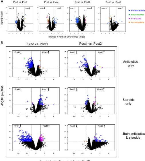
[image:5.585.134.452.63.307.2]FIG 3(A) Volcano plots indicating taxa that are significantly increased (upper right quadrant) or decreased (upper left quadrant) in the pairwise comparisons indicated, using moderatedt-test models (R package limma). Results shown are from analyses of all subjects. Dashed lines, significant false discovery rate-adjustedPvalues and changes in relative abundance of at least 2-fold, or log2equal to 1. Taxa exhibiting significant changes are colored by phylum-level
classification, as shown. Note that in addition to the highlighted taxa, many other microbiota members exhibited smaller-scale changes in abundance, which cumulatively may contribute importantly to microbiome community function, (B) Changes in the relative abundance of taxa from Exac to Post1 and Post1 to Post2, segregated by type of exacerbation treatment (antibiotics only, systemic corticosteroids only, or both;n⫽4 subjects in each treatment group). Huang et al.
on May 16, 2020 by guest
http://jcm.asm.org/
[image:6.585.38.544.69.631.2]led predominantly to enrichment for many taxa, mainly Proteo-bacteria, but alsoBacteroidetesandFirmicutesmembers (Fig. 3B).
Large increases were observed especially for theEnterobacteriaceae
(up to 16-fold),Lachnospiraceae,Burkholderiaceae, and
Neisseri-aceae(see Table S2 in the supplemental material). This was mir-rored by a significantly higher mean 16S rRNA copy number, a proxy for bacterial burden, in this group compared to the other
treatment groups (RM-ANOVA,Pⱕ0.02; for the steroids-only
group versus the antibiotics-only group,P⬍0.01; see Fig. S2 in
the supplemental material). This indicates that treatment with systemic corticosteroids alone can lead to an increased bacterial burden and an increased abundance of specific airway microbiota.
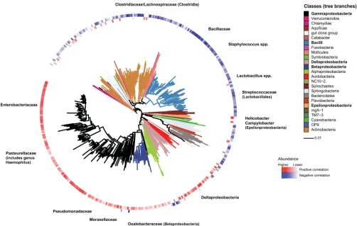
Relationships between COPD-associated pathogens and other microbiota members.Previous studies of the gut microbi-ota have shown that specific organisms in a niche can promote community enrichment for related species and influence
coloni-zation susceptibilities (36). To explore this in the airway
micro-biome context, we evaluated relationships between known COPD-related pathogens and other microbiota members by per-forming correlation analyses between the detected abundance of
taxa representingH. influenzae,P. aeruginosa, orM. catarrhalis
and that of all other taxa. Strong positive correlations were
ob-served betweenH. influenzaeand taxa belonging to the same or
related bacterial families for this species (Rⱖ 0.5,
Benjamini-Hochberg-adjustedP⬍0.05), e.g.,Pasteurellaceae,
Enterobacteri-aceae, andPseudomonadaceae(Fig. 4). Most belong to the larger
class ofGammaproteobacteria, as well as other classes of
Proteobac-teria. In contrast, taxa negatively correlated withH. influenzae
were primarily members of more phylogenetically distant groups.
Similar correlation patterns were observed forP. aeruginosaand
M. catarrhalis(data not shown). These observations suggest that enrichment of phylogenetically related organisms in a pathogen-rich airway milieu may represent an important factor in
micro-biome dynamics associated with the pathogenesis of exacerba-tions.
Quantitative PCR and sequencing validation studies.Total
16S rRNA gene copy numbers ranged from 1.9⫻105to 1.8⫻108
(see Fig. S2 in the supplemental material). While these did not differ significantly over time, the type of treatment for exacerba-tion was a significant factor in the observed differences
(RM-ANOVA,P⫽0.02), with the group treated with corticosteroids
alone demonstrating higher mean total 16S rRNA copy numbers
(for steroids only versus antibiotics only,P⬍0.01; for steroids
only versus both antibiotics and steroids,P⫽0.08).
Sanger sequencing using taxon-specific primers confirmed the
identity ofM. catarrhalisand organisms representative of other
taxons, including species of Corynebacteriaceae,
Porphyromon-adaceae, Enterobacteriaceae, and Bifidobacteria(see Table S3 in the supplemental material). Species-specific qPCR studies also showed excellent correlations between copy numbers and the
ar-ray-reported abundances ofH. influenzae(R⫽0.92,P⬍0.03)
andP. aeruginosa(R⫽0.95,P⬍0.01). To further validate array-reported taxon abundances, 16S rRNA sequencing using the Illumina MiSeq platform was performed on 11 samples. Strong correlations between the relative abundance of specific phyla and families detected by both sequencing and array analysis were
observed (i.e., Proteobacteria, Actinobacteria, Pasteurellaceae,
Moraxellaceae,Enterobacteriaceae; Spearman ⫽0.7 to 0.9,P⬍
0.001 to 0.01; see Table S4 in the supplemental material).
Functional analysis of COPD airway microbiome. Meta-genomic profiling of the microbiome affords insights into the functional capacity of the community. However, to do so at high resolution is both expensive and computationally intensive. To explore specifically the predicted functional capacity of the micro-biota involved in the changes observed at exacerbation and in response to treatment, we applied a recently developed
bioinfor-TABLE 2Bacterial taxa that demonstrated the largest changes in relative abundance at the onset of exacerbation compared to that at the most recent
preexacerbation time point (Pre2)a
Taxon (class/family/genus)
prokMSA_ID for taxon
representative speciesb Fold changec Pvalue
Gammaproteobacteria/Moraxellaceae/Acinetobacter 60270 ⫹2.9 0.01 Gammaproteobacteria/Moraxellaceae/Perlucidibaca 105696 ⫹2.7 0.02 Gammaproteobacteria/Moraxellaceae/Acinetobacter 106871 ⫹2.4 0.04 Gammaproteobacteria/Moraxellaceae/Acinetobacter 101304 ⫹2.4 0.006 Betaproteobacteria/Anaplasmataceae/Ehrlichia 6030 ⫹2.2 0.02 Gammaproteobacteria/Pasteurellaceae/Actinobacillus 52835 ⫹2.1 0.04 Alphaproteobacteria/Sphingomonadaceae/Sphingomonas 89065 ⫹2.1 0.01 Betaproteobacteria/Comamonadaceae/Comamonas 71872 ⫹2.0 0.03
Gammaproteobacteria/Enterobacteriaceae 10365 ⫹2.0 0.04
Gammaproteobacteria/Pseudomonadaceae/Pseudomonas 8434 ⫹2.0 0.01
Bacilli/Bacillaceae/unclassified 89195 ⫺1.9 0.04
Epsilonproteobacteria/Campylobacteraceae/Campylobacter 108770 ⫺1.9 0.03
Bacteroidia/Rikenellaceae/unclassified 101395 ⫺1.9 0.01
Clostridia/Clostridialesfamily XIII/Mogibacterium 14167 ⫺1.8 0.002 Actinobacteria/Microbacteriaceae/Agromyces 12174 ⫺1.8 0.02
Clostridia/Lachnospiraceae/unclassified 72977 ⫺1.8 0.005
Actinobacteria/ACK-M1/unclassified 96076 ⫺1.8 0.01
Actinobacteria/Micrococcaceae/Rothia 58305 ⫺1.8 0.03
Actinobacteria/Nocardiaceae/Rhodococcus 85556 ⫺1.8 0.04 a
Changes in relative abundance wereⱖ2-fold (P⬍0.05,qⱕ0.30) at the onset of exacerbation compared to that at the most recent preexacerbation time point (Pre2). The results are based on analysis of all subjects in the study (n⫽12 subjects).
b
prokMSA_ID is the identifier used by the Greengenes database. cFold change shows the increase or decrease at exacerbation.
on May 16, 2020 by guest
http://jcm.asm.org/
[image:7.585.47.543.86.304.2]matics tool, PICRUSt (32), which predicts the functional capacity of a community based on 16S rRNA data. PICRUSt has been used to predict the presence and relative abundance of gene families in different microbiomes and shown to recapitulate the results of actual metagenomic sequencing efforts from the Human
Micro-biome Project (32).
Representative 16S rRNA sequences for taxa that changed in
abundance byⱖ2-fold were used as PICRUSt inputs. The results
predicted a number of KEGG gene orthologs (KOs; Kyoto Ency-clopedia of Genes and Genomes, release 67.1) associated with known KEGG pathways to be enriched or depleted within the
bacterial community at these time points (Fig. 5). Predicted
func-tions enriched at exacerbation included pathways involved in viral
and bacterial infection (e.g., influenza A virus andStaphylococcus
aureusinfection), as well as those involved in apoptosis and p53 signaling. Conversely, functions predicted to be depleted involved pathways associated with the response to viral infection (e.g., RIG-I-like receptor signaling) and pathways involved in flavonoid and
steroid biosynthesis (known anti-inflammatory mediators [37]),
as well as betalain and indole alkaloid production (known
antimi-crobial properties [38,39]). These observations suggest that a loss
of community functions associated with maintenance of micro-bial homeostasis and regulation of the host immune response is associated with AECOPD. This is further supported by the
obser-vation that after treatment, reverse trends were observed. For ex-ample, predicted KOs that were enriched for after exacerbation included those involved in the betalain, indole alkaloid, and fla-vonol biosynthesis pathways. In addition, KOs involved in mac-rolide biosynthesis were also predicted to be enriched posttreat-ment. Thus, microbiome enrichment for taxa that encode these anti-inflammatory functional capacities may contribute to recov-ery from exacerbation.
DISCUSSION
In this study, we extensively examined the dynamics of the bacte-rial airway microbiome in the setting of AECOPD. In addition to assessment of compositional changes by multiple methods, we further explored the predicted functional capacities encoded by microbiota identified to be potentially important on the basis of observed compositional shifts. Thus, this study represents the most comprehensive effort to date to evaluate the exacerbation-associated microbiome in COPD and serves as a foundation for approaches that could be applied to study larger numbers of pa-tients. Collectively, our findings demonstrate that the COPD air-way microbiome is rich in both bacterial species and functional capacity and that identifiable changes in this microbiome are as-sociated with the development, treatment, and resolution of AECOPD.
FIG 4Correlations between the relative abundance ofH. influenzae(a member of thePasteurellaceae/Gammaproteobacteria) and that of all other identified taxa.
The analysis was performed using data from all 60 samples in this study. Significant positive and negative correlations are shown (PearsonRⱖ0.5, Benjamini-Hochberg-adjustedP⬍0.05). Positive correlations (red) occur predominantly with members ofPasteurellaceaeor closely related bacterial families and classes ofProteobacteria. Negative correlations (blue) occur with bacterial families and classes that phylogenetically are more distant toH. influenzae/Pasteurellaceae. Tree branches are color coded by bacterial class in the key on the right.
Huang et al.
on May 16, 2020 by guest
http://jcm.asm.org/
[image:8.585.40.542.64.383.2]An important limitation of our study is that the intensive mi-crobiome analyses were performed on 60 samples derived from only a small number of COPD patients. Offsetting this limitation is the longitudinal nature of the study, providing us with valid preexacerbation samples, as well as exacerbation samples prior to any specific treatment for exacerbation. Future studies involving larger patient numbers will be necessary, especially given the het-erogeneity of COPD and interest in identifying the best treatment
approaches for different phenotypes (40). Despite the cohort size,
we were able to capture some of the heterogeneity associated with exacerbations by intentionally including a proportionally even number of subjects treated for exacerbation using regimens pre-scribed in clinical practice. In this context, we were able to identify microbiome characteristics differentially associated with features of exacerbations in these subjects. Follow-up studies involving larger numbers of carefully phenotyped patients would be of in-terest.
Several specific findings from our study are important to em-phasize and have potential clinical implications to be further in-vestigated. An important overall observation from a microbial community perspective is that many bacterial groups not limited to typical COPD pathogens were among those contributing to the most salient compositional changes observed. These dynamics,
which involvedProteobacteriataxa, in addition to those
represen-tative ofH. influenzae,M. catarrhalis, andP. aeruginosa, suggest
potentially important contributions of related microbiota to the etiopathogenesis of AECOPD. Independent analyses confirmed that the abundance of these typical COPD pathogens was highly correlated with the presence of related bacterial phylotypes. While array cross-hybridization at the taxon level is a possible factor in these analyses, it unlikely accounts for the number of distinct groups, characterized at the family level and higher, found to be
correlated with these species. Our observations are consistent with previous demonstrations that organisms in an ecosystem can pro-mote enrichment for related species in a like will to like phenom-enon, as observed for gut microbiota and susceptibility to
patho-gen colonization (36). Moreover, quorum-sensing mechanisms
between organisms can influence bacterial pathogenicity or
resis-tance to antimicrobials (41,42). Overall, findings from this and
other studies (43) indicate that the context of the microbial
com-munity is important to consider in efforts to better understand the pathogenesis of AECOPD.
Another important observation is that intersubject variability in microbial community changes at exacerbation were seen. Sim-ilar observations were made in a study using a human model of
virus-induced COPD exacerbation (43). In our study,
composi-tional changes at exacerbation in general did not involve very large shifts, although this was not true of all subjects. This is similar to what has been described in exacerbations of cystic fibrosis and
non-cystic fibrosis bronchiectasis (44, 45). This suggests that
AECOPD could also result from the cumulative effects of smaller-scale changes in community composition that, on the basis of PICRUSt functional predictions, lead not only to an increase in pathogen-elicited inflammation but also the loss of potentially key protective functions in the microbiome. Moreover, organisms do not need to be highly prevalent in a community to have important effects on disease pathogenesis, as has been shown for
periodon-titis (46). However, two of our subjects (subjects 56 and 111) did
exhibit a greater compositional shift at exacerbation, determined on the basis of NMDS analysis (data not shown). Although new bacterial strains were identified in these patients, others in which new strains were also detected did not demonstrate large compo-sitional shifts at exacerbation. Collectively, these observations
FIG 5KEGG pathways associated with predicted gene functions (KOs) encoded by metagenomes of bacterial taxa that either were increased (red) or decreased
(blue) in relative abundance byⱖ2-fold at the time of and after exacerbations. Among the microbiome-related functions identified were those involved in the promotion of inflammation by pathogens (*) and the production of antimicrobial and anti-inflammatory compounds (**), functions that were differentially represented at exacerbation compared to postexacerbation.
on May 16, 2020 by guest
http://jcm.asm.org/
[image:9.585.139.450.68.303.2]suggest that there is likely interindividual heterogeneity in the microbiome associated with AECOPD.
Finally, our observations regarding the effects of COPD treat-ments on microbiota composition have potentially important clinical implications. Inhaled corticosteroids are commonly used in the management of COPD and other airway diseases, and our results suggest that the use of ICSs may alter the microbiome. Whether this has implications for susceptibility to exacerbations or their outcome will require additional study. Also, treatments for exacerbation led to significant changes in community compo-sition, some of which may last beyond the usual time frame of clinical recovery from exacerbations. In particular, oral steroid therapy alone led to community enrichment for many members of the microbiota. This suggests that steroids, even when they are administered systemically, may promote microbial colonization in the airways. Overall, the long-term effects of such treatments on the microbiome in COPD, particularly with recurrent administra-tions, are unknown.
Strengths of this study include the systematic collection of samples at times encompassing key periods before, at, and after AECOPD. Furthermore, we extended upon the compositional analyses to explore the functional capacity encoded by key micro-biome members. Weaknesses include the small number of sub-jects, the focus on only bacteria in examining the microbiome, and also the use of sputum as a representative specimen. Despite the potential for saliva admixture, collection of sputum represents the only practical approach for repeated airway sampling of COPD patients, particularly during exacerbations, when bronchoscopy may be unsafe. Important microbial insights, including several from this cohort, have been gleaned from analysis of sputum in
many studies of COPD (2,3,8,43).
In summary, the results of this study indicate that the context of the airway microbiome needs to be considered in efforts to improve the understanding of the pathogenesis of AECOPD. Given the heterogeneity of COPD, it will be important to study larger numbers of subjects to identify microbiome determinants associated with specific COPD phenotypes and associated exacer-bations.
ACKNOWLEDGMENTS
This work was supported by a University of California Tobacco-Related Disease Research Program grant (17FT-0040) and National Institutes of Health grant HL105572 to Y.J.H. and an American Lung Association award to S.V.L.
We gratefully acknowledge Ali Faruqi for guidance in performing PICRUSt analysis of microarray-identified taxa and Emily Cope for tech-nical assistance with Illumina MiSeq sequencing and guidance in the anal-ysis of the sequencing data.
REFERENCES
1.Sethi S, Murphy TF.2008. Infection in the pathogenesis and course of
chronic obstructive pulmonary disease. N. Engl. J. Med.359:2355–2365.
http://dx.doi.org/10.1056/NEJMra0800353.
2.Sethi S, Evans N, Grant BJ, Murphy TF.2002. New strains of bacteria
and exacerbations of chronic obstructive pulmonary disease. N. Engl. J. Med.347:465– 471.http://dx.doi.org/10.1056/NEJMoa012561.
3.Murphy TF, Brauer AL, Eschberger K, Lobbins P, Grove L, Cai X, Sethi
S.2008. Pseudomonas aeruginosa in chronic obstructive pulmonary dis-ease. Am. J. Respir. Crit. Care Med.177:853– 860.http://dx.doi.org/10 .1164/rccm.200709-1413OC.
4.Huang YJ, Kim E, Cox MJ, Brodie EL, Brown R, Wiener-Kronish JP,
Lynch SV.2010. A persistent and diverse airway microbiota present
dur-ing chronic obstructive pulmonary disease exacerbations. OMICS14:9 – 59.http://dx.doi.org/10.1089/omi.2009.0100.
5.Erb-Downward JR, Thompson DL, Han MK, Freeman CM, McCloskey
L, Schmidt LA, Young VB, Toews GB, Curtis JL, Sundaram B, Martinez
FJ, Huffnagle GB.2011. Analysis of the lung microbiome in the “healthy”
smoker and in COPD. PLoS One 6:e16384.http://dx.doi.org/10.1371 /journal.pone.0016384.
6.Pragman AA, Kim HB, Reilly CS, Wendt C, Isaacson RE.2012. The
lung microbiome in moderate and severe chronic obstructive pulmo-nary disease. PLoS One7:e47305.http://dx.doi.org/10.1371/journal .pone.0047305.
7.Cabrera-Rubio R, Garcia-Núñez M, Setó L, Antó JM, Moya A, Monsó
E, Mira A.2012. Microbiome diversity in the bronchial tracts of patients
with chronic obstructive pulmonary disease. J. Clin. Microbiol.50:3562– 3568.http://dx.doi.org/10.1128/JCM.00767-12.
8.Papi A, Bellettato CM, Braccioni F, Romagnoli M, Casolari P, Caramori
G, Fabbri LM, Johnston SL.2006. Infections and airway inflammation in
chronic obstructive pulmonary disease severe exacerbations. Am. J. Respir. Crit. Care Med.173:1114 –1121.http://dx.doi.org/10.1164/rccm .200506-859OC.
9.Rosell A, Monso E, Soler N, Torres F, Angrill J, Riise G, Zalacain R,
Morera J, Torres A.2005. Microbiologic determinants of exacerbation in
chronic obstructive pulmonary disease. Arch. Intern. Med.165:891– 897.
http://dx.doi.org/10.1001/archinte.165.8.891.
10. Sze MA, Dimitriu PA, Hayashi S, Elliott WM, McDonough JE,
Gosselink JV, Cooper J, Sin DD, Mohn WW, Hogg JC.2012. The
lung tissue microbiome in chronic obstructive pulmonary disease. Am. J. Respir. Crit. Care Med.185:1073–1080.http://dx.doi.org/10.1164 /rccm.201111-2075OC.
11. Duan K, Dammel C, Stein J, Rabin H, Surette MG.2003. Modulation of
Pseudomonas aeruginosa gene expression by host microflora through in-terspecies communication. Mol. Microbiol.50:1477–1491.http://dx.doi .org/10.1046/j.1365-2958.2003.03803.x.
12. Endt K, Stecher B, Chaffron S, Slack E, Tchitchek N, Benecke A, Van
Maele L, Sirard JC, Mueller AJ, Heikenwalder M, Macpherson AJ,
Strugnell R, von Mering C, Hardt WD.2010. The microbiota mediates
pathogen clearance from the gut lumen after non-typhoidal Salmonella diarrhea. PLoS Pathog. 6:e1001097. http://dx.doi.org/10.1371/journal .ppat.1001097.
13. Sethi S, Wrona C, Eschberger K, Lobbins P, Cai X, Murphy TF.2008.
Inflammatory profile of new bacterial strain exacerbations of chronic ob-structive pulmonary disease. Am. J. Respir. Crit. Care Med.177:491– 497.
http://dx.doi.org/10.1164/rccm.200708-1234OC.
14. Huang YJ, Nelson CE, Brodie EL, Desantis TZ, Baek MS, Liu J, Woyke
T, Allgaier M, Bristow J, Wiener-Kronish JP, Sutherland ER, King TS, Icitovic N, Martin RJ, Calhoun WJ, Castro M, Denlinger LC, Dimango E, Kraft M, Peters SP, Wasserman SI, Wechsler ME, Boushey HA,
Lynch SV.2011. Airway microbiota and bronchial hyperresponsiveness
in patients with suboptimally controlled asthma. J. Allergy Clin. Immu-nol.127:372–381.http://dx.doi.org/10.1016/j.jaci.2010.10.048.
15. Flanagan JL, Brodie EL, Weng L, Lynch SV, Garcia O, Brown R,
Hugenholtz P, DeSantis TZ, Andersen GL, Wiener-Kronish JP, Bristow J.2007. Loss of bacterial diversity during antibiotic treatment of intubated patients colonized with Pseudomonas aeruginosa. J. Clin. Microbiol.45: 1954 –1962.http://dx.doi.org/10.1128/JCM.02187-06.
16. McDonald D, Price MN, Goodrich J, Nawrocki EP, DeSantis TZ, Probst
A, Andersen GL, Knight R, Hugenholtz P.2012. An improved
Green-genes taxonomy with explicit ranks for ecological and evolutionary anal-yses of bacteria and archaea. ISME J.6:610 – 618.http://dx.doi.org/10 .1038/ismej.2011.139.
17. Brodie EL, Desantis TZ, Joyner DC, Baek SM, Larsen JT, Andersen GL,
Hazen TC, Richardson PM, Herman DJ, Tokunaga TK, Wan JM,
Firestone MK.2006. Application of a high-density oligonucleotide
mi-croarray approach to study bacterial population dynamics during ura-nium reduction and reoxidation. Appl. Environ. Microbiol.72:6288 – 6298.http://dx.doi.org/10.1128/AEM.00246-06.
18. DeSantis TZ, Brodie EL, Moberg JP, Zubieta IX, Piceno YM, Andersen
GL.2007. High-density universal 16S rRNA microarray analysis reveals broader diversity than typical clone library when sampling the environ-ment. Microb. Ecol.53:371–383. http://dx.doi.org/10.1007/s00248-006 -9134-9.
19. Hazen TC, Dubinsky EA, DeSantis TZ, Andersen GL, Piceno YM, Singh
N, Jansson JK, Probst A, Borglin SE, Fortney JL, Stringfellow WT, Bill Huang et al.
on May 16, 2020 by guest
http://jcm.asm.org/
M, Conrad ME, Tom LM, Chavarria KL, Alusi TR, Lamendella R, Joyner DC, Spier C, Baelum J, Auer M, Zemla ML, Chakraborty R, Sonnenthal EL, D’haeseleer P, Holman HY, Osman S, Lu Z, Van
Nostrand JD, Deng Y, Zhou J, Mason OU.2010. Deep-sea oil plume
enriches indigenous oil-degrading bacteria. Science330:204 –208.http: //dx.doi.org/10.1126/science.1195979.
20. Handley KM, Wrighton KC, Piceno YM, Andersen GL, DeSantis TZ,
Williams KH, Wilkins MJ, N=Guessan AL, Peacock A, Bargar J, Long
PE, Banfield JF.2012. High-density PhyloChip profiling of stimulated
aquifer microbial communities reveals a complex response to acetate amendment. FEMS Microbiol. Ecol. 81:188 –204.http://dx.doi.org/10 .1111/j.1574-6941.2012.01363.x.
21. DeSantis TZ, Stone CE, Murray SR, Moberg JP, Andersen GL.2005.
Rapid quantification and taxonomic classification of environmental DNA from both prokaryotic and eukaryotic origins using a microarray. FEMS Microbiol. Lett.245:271–278.http://dx.doi.org/10.1016/j.femsle.2005.03 .016.
22. Faith DP.1992. Conservation evaluation and phylogenetic diversity. Biol.
Conserv.61:1–10.http://dx.doi.org/10.1016/0006-3207(92)91201-3.
23. Oksanen J, Kindt R, Legendre P, O’Hara B, Simpson GL, Solymos P,
Henry M, Stevens H, Wagner H.2008.Vegan: community ecology
pack-age. R package, version 1.16-1. R Project for Statistical Computing, Vi-enna, Austria.
24. Kembel SW, Cowan PD, Helmus MR, Cornwell WK, Morlon H,
Ack-erly DD, Blomberg SP, Webb CO.2010. Picante: R tools for integrating
phylogenies and ecology. Bioinformatics26:1463–1464.http://dx.doi.org /10.1093/bioinformatics/btq166.
25. Gotelli N, Ellison A.2004. A primer of ecological statistics. Sinauer
As-sociates, Inc, Sunderland, MA.
26. Lozupone C, Knight R.2005. UniFrac: a new phylogenetic method for
comparing microbial communities. Appl. Environ. Microbiol.71:8228 – 8235.http://dx.doi.org/10.1128/AEM.71.12.8228-8235.2005.
27. Anderson MJ.2001. A new method for non-parametric multivariate
anal-ysis of variance. Austral Ecol.26:32– 46.http://dx.doi.org/10.1111/j.1442 -9993.2001.01070.pp.x.
28. Smyth GK.2004. Linear models and empirical Bayes methods for
assess-ing differential expression in microarray experiments. Stat. Appl. Genet. Mol. Biol.3:Article3.http://dx.doi.org/10.2202/1544-6115.1027.
29. Storey JD, Tibshirani R.2003. Statistical significance for genomewide
studies. Proc. Natl. Acad. Sci. U. S. A.100:9440 –9445.http://dx.doi.org /10.1073/pnas.1530509100.
30. Sunagawa S, DeSantis TZ, Piceno YM, Brodie EL, DeSalvo MK,
Vool-stra CR, Weil E, Andersen GL, Medina M.2009. Bacterial diversity and
white plague disease-associated community changes in the Caribbean coral Montastraea faveolata. ISME J.3:512–521.http://dx.doi.org/10.1038 /ismej.2008.131.
31. Ivanov II, Atarashi K, Manel N, Brodie EL, Shima T, Karaoz U, Wei D,
Goldfarb KC, Santee CA, Lynch SV, Tanoue T, Imaoka A, Itoh K,
Takeda K, Umesaki Y, Honda K, Littman DR. 2009. Induction of
intestinal Th17 cells by segmented filamentous bacteria. Cell139:485– 498.http://dx.doi.org/10.1016/j.cell.2009.09.033.
32. Langille MG, Zaneveld J, Caporaso JG, McDonald D, Knights D, Reyes
JA, Clemente JC, Burkepile DE, Vega Thurber RL, Knight R, Beiko RG,
Huttenhower C.2013. Predictive functional profiling of microbial
com-munities using 16S rRNA marker gene sequences. Nat. Biotechnol.31: 814 – 821.http://dx.doi.org/10.1038/nbt.2676.
33. Pauwels RA, Buist SA, Calverley PM, Jenkins CR, Hurd SS.2001. Global
strategy for the diagnosis, management, and prevention of chronic ob-structive pulmonary disease. Am. J. Respir. Crit. Care Med.163:1256 – 1276.http://dx.doi.org/10.1164/ajrccm.163.5.2101039.
34. Ventura M, Canchaya C, Tauch A, Chandra G, Fitzgerald GF, Chater
KF, van Sinderen D.2007. Genomics of Actinobacteria: tracing the
evo-lutionary history of an ancient phylum. Microbiol. Mol. Biol. Rev.71:495– 548.http://dx.doi.org/10.1128/MMBR.00005-07.
35. Atarashi K, Tanoue T, Shima T, Imaoka A, Kuwahara T, Momose Y,
Cheng G, Yamasaki S, Saito T, Ohba Y, Taniguchi T, Takeda K, Hori S,
Ivanov II, Umesaki Y, Itoh K, Honda K.2011. Induction of colonic
regulatory T cells by indigenous Clostridium species. Science331:337– 341.http://dx.doi.org/10.1126/science.1198469.
36. Stecher B, Chaffron S, Kappeli R, Hapfelmeier S, Freedrich S, Weber
TC, Kirundi J, Suar M, McCoy KD, von Mering C, Macpherson AJ,
Hardt WD.2010. Like will to like: abundances of closely related species
can predict susceptibility to intestinal colonization by pathogenic and commensal bacteria. PLoS Pathog.6:e1000711.http://dx.doi.org/10.1371 /journal.ppat.1000711.
37. Cushnie TP, Lamb AJ.2011. Recent advances in understanding the
an-tibacterial properties of flavonoids. Int. J. Antimicrob. Agents38:99 –107.
http://dx.doi.org/10.1016/j.ijantimicag.2011.02.014.
38. Vulic´ JJ, Cebovic´ TN, Canadanovic´ VM, Cetkovic´ GS, Djilas SM,
Canadanovic´-Brunet JM, Velic´anski AS, Cvetkovic´ DD, Tumbas VT. 2013. Antiradical, antimicrobial and cytotoxic activities of commercial beetroot pomace. Food Funct. 4:713–721. http://dx.doi.org/10.1039 /c3fo30315b.
39. Zoraghi R, Worrall L, See RH, Strangman W, Popplewell WL, Gong H,
Samaai T, Swayze RD, Kaur S, Vuckovic M, Finlay BB, Brunham RC, McMaster WR, Davies-Coleman MT, Strynadka NC, Andersen RJ,
Reiner NE.2011. Methicillin-resistant Staphylococcus aureus (MRSA)
pyruvate kinase as a target for bis-indole alkaloids with antibacterial ac-tivities. J. Biol. Chem.286:44716 – 44725.http://dx.doi.org/10.1074/jbc .M111.289033.
40. Miravitlles M, Soler-Cataluña JJ, Calle M, Soriano JB.2013. Treatment
of COPD by clinical phenotypes: putting old evidence into clinical prac-tice. Eur. Respir. J.41:1252–1256.http://dx.doi.org/10.1183/09031936 .00118912.
41. Armbruster CE, Hong W, Pang B, Weimer KE, Juneau RA, Turner J,
Swords WE.2010. Indirect pathogenicity of Haemophilus influenzae and
Moraxella catarrhalis in polymicrobial otitis media occurs via interspecies quorum signaling. mBio1(3):e00102–10.http://dx.doi.org/10.1128/mBio .00102-10.
42. Sibley CD, Duan K, Fischer C, Parkins MD, Storey DG, Rabin HR,
Surette MG.2008. Discerning the complexity of community interactions
using a Drosophila model of polymicrobial infections. PLoS Pathog. 4:e1000184.http://dx.doi.org/10.1371/journal.ppat.1000184.
43. Molyneaux PL, Mallia P, Cox MJ, Footitt J, Willis-Owen SA, Homola
D, Trujillo-Torralbo MB, Elkin S, Kon OM, Cookson WO, Moffatt MF,
Johnston SL.2013. Outgrowth of the bacterial airway microbiome after
rhinovirus exacerbation of chronic obstructive pulmonary disease. Am. J. Respir. Crit. Care Med.188:1224 –1231.http://dx.doi.org/10.1164/rccm .201302-0341OC.
44. Zhao J, Schloss PD, Kalikin LM, Carmody LA, Foster BK, Petrosino JF,
Cavalcoli JD, Vandevanter DR, Murray S, Li JZ, Young VB, Lipuma JJ. 2012. Decade-long bacterial community dynamics in cystic fibrosis air-ways. Proc. Natl. Acad. Sci. U. S. A.109:5809 –5814.http://dx.doi.org/10 .1073/pnas.1120577109.
45. Tunney MM, Einarsson GG, Wei L, Drain M, Klem ER, Cardwell C,
Ennis M, Boucher RC, Wolfgang MC, Elborn JS.2013. Lung microbiota
and bacterial abundance in patients with bronchiectasis when clinically stable and during exacerbation. Am. J. Respir. Crit. Care Med.187:1118 – 1126.http://dx.doi.org/10.1164/rccm.201210-1937OC.
46. Hajishengallis G, Liang S, Payne MA, Hashim A, Jotwani R, Eskan MA,
McIntosh ML, Alsam A, Kirkwood KL, Lambris JD, Darveau RP, Curtis MA. 2011. Low-abundance biofilm species orchestrates inflammatory periodontal disease through the commensal microbiota and complement. Cell Host Microbe10:497–506.http://dx.doi.org/10.1016/j.chom.2011.10 .006.