Submitted28 February 2018 Accepted 3 July 2018 Published6 August 2018
Corresponding author
Kwai Lin Thong, [email protected]
Academic editor Carlos Conte-Junior
Additional Information and Declarations can be found on page 15
DOI10.7717/peerj.5353
Copyright 2018 Tan et al.
Distributed under
Creative Commons CC-BY 4.0
OPEN ACCESS
Occurrence of virulent
multidrug-resistant
Enterococcus faecalis
and
Enterococcus faecium
in the pigs, farmers
and farm environments in Malaysia
Shiang Chiet Tan1, Chun Wie Chong2,3, Cindy Shuan Ju Teh4, Peck Toung Ooi5
and Kwai Lin Thong1
1Institute of Biological Science, Faculty of Science, University of Malaya, Kuala Lumpur, Malaysia
2Department of Life Sciences, School of Pharmacy, International Medical University, Kuala Lumpur, Malaysia 3Centre for Translational Research, Institute for Research, Development and Innovation, International
Medical University, Kuala Lumpur, Malaysia
4Department of Medical Microbiology, Faculty of Medicine, University of Malaya, Kuala Lumpur, Malaysia 5Department of Veterinary Clinical Studies, Faculty of Veterinary Medicine, Universiti Putra Malaysia,
Selangor Darul Ehsan, Malaysia
ABSTRACT
Background. Enterococcus faecalis andEnterococcus faecium are ubiquitous oppor-tunistic pathogens found in the guts of humans and farmed animals. This study aimed to determine the occurrence, antimicrobial resistance, virulence, biofilm-forming ability and genotypes of E. faecalisand E. faeciumfrom swine farms. Correlations between the genotypes, virulotypes, antibiotic resistance, and the environmental factors such as locality of farms and farm hygiene practice were explored.
Methods. E. faecalis and E. faecium strains were isolated from the oral, rectal and fecal samples of 140 pigs; nasal, urine and fecal samples of 34 farmers working in the farms and 42 environmental samples collected from seven swine farms located in Peninsular Malaysia. Antibiotic susceptibility test was performed using the disk diffusion method, and the antibiotic resistance and virulence genes were detected by Polymerase Chain Reaction. Repetitive Extragenic Palindromic-Polymerase Chain Reaction and Pulsed-Field Gel Electrophoresis were performed to determine the clonality of the strains. Crosstab/Chi-square test and DistLM statistical analyses methods were used to determine the correlations between the genotypes, virulence factors, antibiotic resistance, and the environmental factors.
Results. A total of 211E. faecalisand 42E. faeciumwere recovered from 140 pigs, 34 farmers and 42 environmental samples collected from seven swine farms in Peninsular Malaysia. Ninety-eight percent of the strains were multidrug-resistant (resistant to chloramphenicol, tetracycline, ciprofloxacin and erythromycin). Fifty-two percent of the strains formed biofilms. Virulence genesefa, asaI, gelE,esp,cylandacegenes were detected. Virulence genes efaandasaI were most prevalent inE. faecalis (90%) and
E. faecium(43%), respectively. Cluster analyses based on REP-PCR and PFGE showed the strains were genetically diverse. Overall, the strains isolated from pigs and farmers were distinct, except for three highly similar strains found in pigs and farmers. The strains were regional- and host-specific.
and the ability to form biofilm further enhance the persistence and pathogenicity of the strains. Although the overall clonality of the strains were regionals and host-specific, strains with high similarity were found in different hosts. This study reiterates a need of a more stringent regulation to ensure the proper use of antibiotics in swine husbandry to reduce the wide spread of multidrug-resistant strains.
SubjectsMicrobiology, Molecular Biology, Public Health
Keywords Enterococcus faecium,Enterococcus faecalis, MDR, Farmers, Virulence genes, Pigs
INTRODUCTION
Enterococci are Gram-positive facultative anaerobes and are part of microbiota of humans and animals (Lebreton, Willems & Gilmore, 2014;Kristich, Rice & Arias, 2014). Antibiotics such as macrolides, trimethroprim, fluoroquinolones and tetracyclines are commonly used for animal husbandry and human medicine in Southeast Asia, and resistances towards these antibiotics have been reported forEnterococcusspp. (Daniel et al., 2015;Health Action International Asia Pacific, 2013).
Livestock industry plays an important role in the transmission of multidrug-resistant (MDR)Enterococci strains due to the close interaction between farmers, livestock and the farm environment (Novais et al., 2013;Woolhouse et al., 2015). Antimicrobial agents are used as prophylactics in feeds and the pervasive selection of resistant bacteria in the swine gut facilitates the persistence and dissemination of MDR strains to other animals and humans (Woolhouse et al., 2015;Hwang et al., 2009). The emergence of MDR bacteria in the livestock industry is of public health concern as transmission of these bacteria to humans has been reported (Compassion in World Farming, 2011). The spread of such MDR strains can occur through direct (consumption of contaminated food, direct contact of farmers and veterinarians) or indirect (animal waste handling, contaminated ground water or surfaces) routes (Daniel et al., 2015). The resistance determinants carried by the MDR strains could also be transmitted to the other commensal strains in the host and cause further complications (Price et al., 2018). In addition, infections caused by MDR strains have been associated with long hospital stays, high morbidity and mortality rate (Beganovic et al., 2018).
The two most common enterococcal species, Enterococcus faecalisandEnterococcus faecium, present diverse virulence factors such as aggregation substance, gelatinase, cytolysin, enterococcal surface protein and collagen binding cell wall protein which are encoded by their respective genesasa1, gelE, cylA, esp andace (in plasmids) orhyl
(in chromosome) (Vidana et al., 2015). Enterococcal surface protein, encoded byesp
gene, is one of the factors associated with biofilm formation (Toledo-arana et al., 2001). The biofilm formation ability can enhance the persistence and endurance of the strains by providing extra protection to the bacteria cells and confers higher tolerance to the antibiotics (Holmberg & Rasmussen, 2016).
The objectives of the study were to determine the occurrence of MDR E. faecalisand
PF1
PF2
PF3
PF5 PF4
SF1 SF2
Terengganu Kelantan
Pahang Kedah
Perak
Selangor Kuala Lumpur
Johor Melacca
Negeri Sembilan Perlis
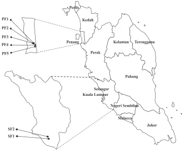
[image:3.612.184.548.87.391.2]Penang
Figure 1 The map of Peninsular Malaysia showing the locations of the seven swine farms.Number of strains in each farm: PF1 (nE.faecalis=40;nE.faecium=3); PF2 (nE.faecalis=31;nE.faecium=7); PF3 (nE.faecalis=
13;nE.faecium=0); PF4 (nE.faecalis=24;nE.faecium=4); PF5 (nE.faecalis=12;nE.faecium=1); SF1 (nE.faecalis=
43;nE.faecium=23); SF2 (nE.faecalis=48;nE.faecium=4).
Full-size DOI: 10.7717/peerj.5353/fig-1
Malaysia. The virulotypes, biofilm-forming ability, and the genotypes (based on PFGE and REP-PCR) were determined.
MATERIALS AND METHODS
Sample collection
farm location, hygiene practice, gender of the pigs, body temperature and health condition of the pigs (healthy/unhealthy) were collected and used for statistical analysis. The farm hygiene practices were arbitrarily divided into three categories: HP1 refers to application of fundamental hygiene practice (clean the pens with detergents only). HP2 refers to a better level of hygiene practice, includes cleaning the pens and disinfecting equipment. HP3 refers to the best hygiene practice, includes personal hygiene practice of farmers, foot-dip practices and all above mentioned hygiene practices. Physical examination was performed by the field veterinarian to determine the health status of pigs. Pigs that presented with abnormal clinical signs, behavior and elevated body temperature were categorized as unhealthy. This study was approved by the Animal Care and Use Committee (ACUC), Faculty of Veterinary Medicine, UPM (UPM/IACUC/FYP-AUP-T006/). The human samples collection was approved by Medical Ethics Committee, University Malaya Medical Centre (Ethic committee/IRB reference number: 1010.41), with written informed consent of the human subjects. All swabs were collected using sterile TranswabR
Amies Charcoal (Medical Wire & Equipment Co, Corsham, UK). All the samples were transported on ice and processed within 24 h upon collection.
Bacterial isolation and identification
All samples except fecal samples were enriched in Brain Heart Infusion (BHI) medium containing 6.5% NaCl followed by plating on the CHROMagar Orientation (CHROMagar Company, Paris, France). For fecal samples, one gram of feces was suspended in two ml of 0.85% saline before being streaked onto CHROMagar Orientation.
Approximately 3,450 presumptive colonies were obtained in the initial screening based on the manufacturer’s guidelines. Due to the large numbers of isolates, 10% of turquoise blue colored colonies (approximately 10–15 colonies per plate) were selected for further identification using Vitek-MS (Biomérieux, Marcy I’Etoile, France) according to the manufacturer’s protocol. TheEscherichia coli(ATCC 8739) strain was used as the control. PCR targetingsodAgenes was used to confirm the identity of the strains (Jackson, Fedorka-Cray & Barrett, 2004).
Antibiotic susceptibility test
antibiotics which are not intrinsically resistant byE. faecalis, according to the Clinical and Laboratory Standards Institute (CLSI) guidelines.
PCR detection of antibiotic resistance genes and virulence genes
Detection of antibiotic resistance genes and virulence genes were used to determine the resistance and virulence potential of the strains. Boiled cell lysates were used as DNA templates. Antibiotic resistance and virulence genes were identified by Polymerase Chain Reaction (PCR) using primers and condition as previously described (File S1).
Biofilm assay
To study the biofilm forming ability, crystal violet assay was performed using the protocol described by Baldassarri et al. (2001)with slight modifications. Biofilm was allowed to grow at 37 ◦C for 48 h and the optical density (OD) of eluted crystal violet stain was measured at OD570 nm. The true OD readings of each strain were acquired after deducting
the negative control, which contained only the growth medium. The biofilm forming ability of the studied strains was scored as previously described (Stepanovic et al., 2000). A strong biofilm forming strain VREr5 fromLim, Teh & Thong (2017)was used as a positive control.
Repetitive extragenic palindromic-PCR (REP-PCR)
REP-PCR was performed using primer (GTG)5as described byVersalovic et al. (1994).
DNA extraction was performed using Genomic DNA Mini kit (Yeastern Biotech Co., Taiwan) according to the manufacturer’s guidelines. PCR was carried out in a total volume of 25µl master mix containing 1X buffer, 2.5 mM of MgCl2, 0.8 mg/ml of bovine serum
albumin (BSA), 0.1 mM of dNTPs, 1.6 µM of primers and 1.0 U ofTaq polymerase (Promega, Madison, USA). The PCR protocol involved an initial denaturation of 7 min at 95◦C, followed by 30 cycles of 90◦C for 30 s, 40◦C for 1 min, 65◦C for 8 min and a final elongation of 16 min at 65◦C. The amplicons generated were electrophoresed on a 1.5% agarose gel at 100 V for 6 h. A 1 kb DNA marker (Promega, Madison, WI, USA) was used as the molecular size standard. The gels were stained with Gel-Red and visualized with Gel Doc XR imaging system (Bio-Rad, Hercules, CA, USA). The banding patterns generated were analyzed with BioNumerics 6.0 (Applied Maths, Kortrijk, Belgium). All the PCR fingerprints profiles were assigned an arbitrary designation. The quantitative difference among the profiles was defined by the Dice coefficient, Hierarchical clustering analysis was carried out based on unweighted pair group with arithmetric averages (UPGMA) using 1.5% tolerance. REP-PCR was repeated twice to ensure the reproducibility.
Pulsed-Field Gel Electrophoresis (PFGE)
were digested overnight withSmaI at 25◦C andXbaI at 37◦C. PFGE was run using pulse times 3.5 s to 25 s for 12 h (block 1, 6V, 120◦) and 1 s to 5 s for 10 h (block 2, 6V, 120◦).
XbaI-digestedSalmonella entericaserovar Braenderup H9812 was used as the size marker. PFGE was repeated once to ensure reproducibility. The PFGE profiles were analyzed by BioNumerics version 6.0 software (Applied Maths, Kortrijk, Belgium). A dendrogram was constructed based on Dice coefficient of similarity (F) and unweighted pair group method with arithmetic mean (UPGMA) algorithm with 1.5% position tolerance.
Statistical analysis
The environmental factors used for correlation study included sample subjects, sample matrix, sampling region and farm hygiene practice. The association of genotypes with resistotypes, virulotypes, biofilm-forming ability and the environmental factors were determined using distance-based linear model (DistLM) by PRIMER 6 with PERMANOVA add on package (PRIMER-E, UK). The correlation between the resistotypes and virulotypes with other factors were tested with crosstabs/chi-square analysis using SPSS version 21 (IBM SPSS Statistics; IBM corporation, Armonk, NY, USA).
RESULTS
Occurrence ofE. faecalisandE. faecium
A total of 289Enterococcusstrains (232 from pigs, 54 from farmers and three from farm environments) were isolated. These strains were affiliated with five species, which included
E. faecalis (n=211), E. faecium (n=42), E. hirae (n=15), E. gallinarium(n=11) and
E. casseliflavus (n=10). AllE. faecalisandE. faeciumstrains were confirmed with PCR targeting thesodAgenes (Jackson, Fedorka-Cray & Barrett, 2004).
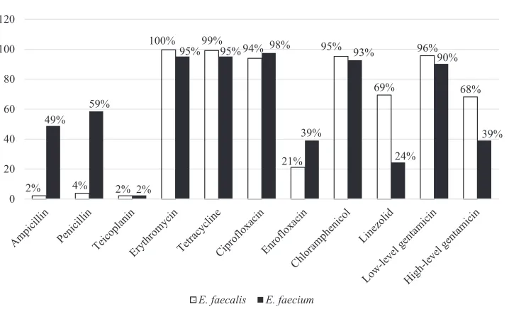
Eighty-seven pigs and 21 farmers harbored eitherE. faecalis,E. faeciumor both species ofEnterococcus.Figure 2shows the distribution ofE. faecalisandE. faeciumin each type of sample matrix. Higher occurrence of enterococci was observed in humans compared to pigs and only threeE. faecalisstrains were isolated from the environmental samples (Fig. 2).E. faecaliswas isolated from all participating farms butE. faeciumwas restricted to the swine samples collected from farms PF2, PF4, PF5, SF1 and SF2 as well as the human samples from PF1, PF4 and SF1.
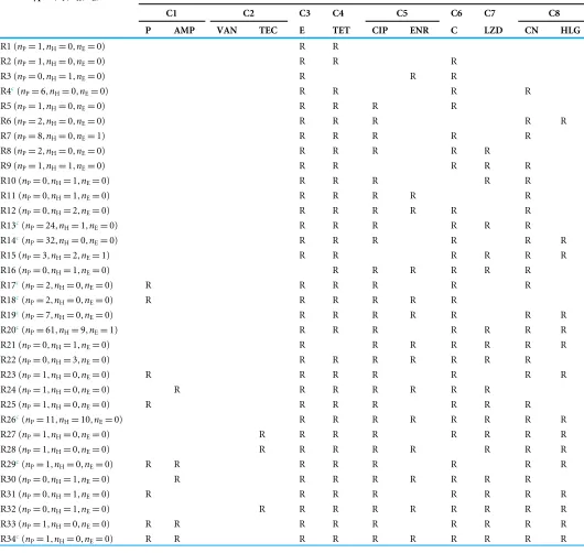
Antimicrobial susceptibility ofE. faecalisandE. faecium
Among the 211E. faecalisstrains, 98% and 96% of them were susceptible to ampicillin and penicillin, respectively. However,E. faeciumwere more resistant to ampicillin (49%) and penicillin (59%) compared toE. faecalis. All the strains were susceptible to vancomycin and only two percent of strains from each species were resistant to teicoplanin.E. faecalis
(68%) strains were relatively more resistant to high-level gentamicin (120µg) compared to
E. faecium(39%). Overall, more than 90% of both species were resistant to erythromycin, tetracycline, ciprofloxacin, chloramphenicol and low-level gentamicin (Fig. 3).
35% 47% 62% 32% 59% 49% 0% 7% 14% 6% 12% 54% 6% 8% 11%
0% 0% 0%
0 10 20 30 40 50 60 70 Nasal
(farmer) (farmer)Urine (farmer)Faecal (Pig)Oral Rectal(Pig) Faecal(Pig) Feed Water Fence
Pe rc en ta ge (%)
[image:7.612.189.545.88.292.2]E. faecalis E. faecium
Figure 2 Percentage of distribution ofE. faecalisandE. faeciumin each sample matrix.
Full-size DOI: 10.7717/peerj.5353/fig-2
2% 4% 2%
100% 99% 94%
21% 95% 69% 96% 68% 49% 59% 2%
95% 95% 98%
39% 93% 24% 90% 39% 0 20 40 60 80 100 120
E. faecalis E. faecium
Figure 3 Percentage of antibiotic resistance ofE. faecalisandE. faeciumin this study.
Full-size DOI: 10.7717/peerj.5353/fig-3
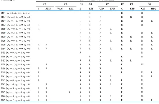
[image:7.612.188.547.340.565.2]Table 1 Resistotypes ofEnterococcus faeciumstrains based on the classes of antibiotics tested.
Resistotypes (nP,nH,nE)a Classes of antibiotic resistancesb
C1 C2 C3 C4 C5 C6 C7 C8
P AMP VAN TEC E TET CIP ENR C LZD CN HLG
R1 (nP=1,nH=0,nE=0) R R
R2 (nP=1,nH=0,nE=0) R R R
R3 (nP=0,nH=1,nE=0) R R R
R4c(n
P=6,nH=0,nE=0) R R R R
R5 (nP=1,nH=0,nE=0) R R R R
R6 (nP=2,nH=0,nE=0) R R R R R
R7 (nP=8,nH=0,nE=1) R R R R R
R8 (nP=2,nH=0,nE=0) R R R R R
R9 (nP=1,nH=1,nE=0) R R R R R
R10 (nP=0,nH=1,nE=0) R R R R R
R11 (nP=0,nH=1,nE=0) R R R R R
R12 (nP=0,nH=2,nE=0) R R R R R R
R13c(n
P=24,nH=1,nE=0) R R R R R R
R14c(n
P=32,nH=0,nE=0) R R R R R R
R15 (nP=3,nH=2,nE=1) R R R R R R
R16 (nP=0,nH=1,nE=0) R R R R R R
R17c(n
P=2,nH=0,nE=0) R R R R R R
R18c(n
P=2,nH=0,nE=0) R R R R R R
R19c(n
P=7,nH=0,nE=0) R R R R R R R
R20c(n
P=61,nH=9,nE=1) R R R R R R R
R21 (nP=0,nH=1,nE=0) R R R R R R R
R22 (nP=0,nH=3,nE=0) R R R R R R R
R23 (nP=1,nH=0,nE=0) R R R R R R R
R24 (nP=1,nH=0,nE=0) R R R R R R R
R25 (nP=1,nH=0,nE=0) R R R R R R R
R26c(n
P=11,nH=10,nE=0) R R R R R R R R
R27 (nP=1,nH=0,nE=0) R R R R R R R R
R28 (nP=1,nH=0,nE=0) R R R R R R R R
R29c(n
P=1,nH=0,nE=0) R R R R R R R R
R30 (nP=0,nH=1,nE=0) R R R R R R R R
R31 (nP=0,nH=1,nE=0) R R R R R R R R
R32 (nP=0,nH=1,nE=0) R R R R R R R R R
R33 (nP=1,nH=0,nE=0) R R R R R R R R R
R34c(n
P=1,nH=0,nE=0) R R R R R R R R R R
Notes.
anP, number of swine isolates;nH, number of human isolates,nE: number of environmental isolates.
bC1, penicillin; C2, glycopeptides; C3, macrolides; C4, tetracyclines; C5, fluoroquinolones; C6, phenicols; C7, oxazolidinones; C8, aminoglycosides; AMP, ampicillin; P, penicillin;
TEC, teicoplanin; E, erythromycin; TET, tetracycline; CIP, ciprofloxacin; ENR, enrofloxacin; C, chloramphenicol; LZD, linezolid; CN, gentamicin; HLG, high-level gentamicin; R, resistant.
Table 2 Resistotypes ofEnterococcus faecalisstrains based on the classes of antibiotics tested.
Resistotypesa Classes of antibiotic resistancesb
C1 C2 C3 C4 C5 C6 C7 C8
P AMP VAN TEC E TET CIP ENR C LZD CN HLG
R4c(n
P=0,nH=1,nE=0) R R R R
R13c(n
P=2,nH=0,nE=0) R R R R R R
R14c(n
P=1,nH=0,nE=0) R R R R R R
R17c(n
P=2,nH=0,nE=0) R R R R R R
R18c(n
P=0,nH=1,nE=0) R R R R R R
R19c(n
P=2,nH=0,nE=0) R R R R R R R
R20c(n
P=1,nH=0,nE=0) R R R R R R R
R26c(n
P=0,nH=4,nE=0) R R R R R R R R
R29c(n
P=2,nH=0,nE=0) R R R R R R R R
R34c(n
P=2,nH=0,nE=0) R R R R R R R R R R
R35 (nP=0,nH=2,nE=0) R R R
R36 (nP=1,nH=2,nE=0) R R R R
R37 (nP=0,nH=1,nE=0) R R R R R R
R38 (nP=1,nH=0,nE=0) R R R R R R
R39 (nP=2,nH=0,nE=0) R R R R R R
R40 (nP=1,nH=0,nE=0) R R R R R R R
R41 (nP=1,nH=0,nE=0) R R R R R R R
R42 (nP=0,nH=1,nE=0) R R R R R R R
R43 (nP=0,nH=1,nE=0) R R R R R R R
R44 (nP=5,nH=0,nE=0) R R R R R R R
R45 (nP=1,nH=0,nE=0) R R R R R R R R
R46 (nP=3,nH=0,nE=0) R R R R R R R R
R47 (nP=2,nH=0,nE=0) R R R R R R R R R
Notes.
anP, number of swine isolates;nH, number of human isolates;nE, number of environmental isolates.
bC1, penicillin; C2, glycopeptides; C3, macrolides; C4, tetracyclines; C5, fluoroquinolones; C6, phenicols; C7, oxazolidinones; C8, aminoglycosides; AMP, ampicillin; P, penicillin;
TEC, teicoplanin; E, erythromycin; TET, tetracycline; CIP, ciprofloxacin; ENR, enrofloxacin; C, chloramphenicol; LZD, linezolid; CN, gentamicin; HLG, high-level gentamicin; R, resistant.
cResistotypes shared by bothE. faecalisandE. faecium.
prevalent compared toermAandmsr.A total of 96% and 95% ofE. faecalisandE. faecium
Virulotyping
A total of 99% and 57% of theE. faecalisandE. faeciumstrains, respectively, harbored at least one of the five virulence genes screened in this study.efawas the most prevalent virulence gene detected inE. faecalisstrains (90%), followed byasaI (68%),gelE (63%),esp
(40%),cyl(40%) andace(15%).In contrast, asaI was most prevalent inE. faecium(43%). Other virulence genes includingesp,gelE,efaandcylwere also present in 21%, 19%, 17% and 14% ofE. faecium,respectively. None of the strains harboredhyl gene.
Biofilm assay
Overall, about 62% of the strains from pigs and humans are capable of producing biofilms. Among the 142 biofilm-formingE. faecalisstrains, 37%, 34% and 29% were weak, moderate and strong biofilm formers, respectively. EightE. faeciumstrains formed weak biofilm and notably, half of them were isolated from human urine samples. All three environmental strains could form strong biofilm.
Genotypic characterization by REP-PCR and PFGE
REP-PCR subtyped the 211 non-repeatE. faecalisstrains into 145 REP-profiles with 9 to 21 DNA fragments ranging in size from 500 to 6,000 bp. Reproducible patterns were observed. Based on 85% similarity, theE. faecalisstrains were grouped into 24 clusters and 13 unique patterns (Files S2). From the dendrogram, the strains isolated from different samples matrix (oral swabs, nasal swabs, rectal swabs, urine and fecal samples) were grouped in the same cluster. However, distinct clustering was observed for strains originated from different regions (northern and central region of Peninsular Malaysia). Strains isolated from both regions were observed in Clusters C4, C5 and C9. Overall, REP-patterns of strains from central region were host-specific (Clusters C2, C19, C23 and C24 comprised of strains isolated from humans and Clusters C7, C13, C20, C21 and C22 comprised of strains isolated from pigs). Three environmentalE. faecalisstrains were clustered with the swine and human strains. For instance, Enfs99 recovered from a pen swab in PF1 was clustered with human nasal strain from the same farm in C15 (Files S2). Enfs103 isolated from a pen swab was 95% similar to the swine oral strains isolated from the same farm, PF1. Enfs98 from the water sample of PF3 formed cluster C10 with the swine fecal strains of the same farm.
E. faeciumstrains were grouped into 11 clusters and 11 unique patterns based on 85% similarity of the REP-profiles (Files S3). Similar toE. faecalis, the strains were regional specific. Strains in Clusters C1, C2, C3 and C5 were from the northern region and Clusters C4, C6 to C11 were made up of strains isolated from the central region only. TheE. faecium
strains from the central region were also host specific. Human strains were grouped in Clusters C4 and C9 while swine-related strains were grouped in Clusters C6, C7, C8, C10 and C11. In contrast, Cluster C2 consisted of both human- and swine-related strains isolated from the northern region.
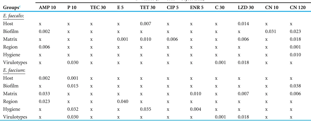
Table 3 Significance of correlation between the antibiotic resistances of different samples classification based of Chi-squared test.
P-values (P<0.05=reject null)a,b
Groupsc AMP 10 P 10 TEC 30 E 5 TET 30 CIP 5 ENR 5 C 30 LZD 30 CN 10 CN 120
E. faecalis:
Host x x x x 0.007 x x x 0.014 x x
Biofilm 0.002 x x x x x x x x 0.031 0.023
Matrix x x x 0.001 0.010 0.006 x x 0.006 x 0.018
Region 0.006 x x x x x x x x x 0.001
Hygiene x x x x x x x x x x 0.010
Virulotypes x 0.030 x x x x x 0.001 0.018 x x
E. faecium:
Host 0.002 0.001 x x x x x x x x x
Biofilm x 0.015 x x x x x x x x 0.038
Matrix 0.033 x x x x x 0.010 x 0.007 x 0.006
Region 0.023 x x 0.040 x x x x x x x
Hygiene x 0.032 x x 0.035 x 0.004 x x x x
Virulotypes x 0.030 x x x x x 0.001 0.018 x x
Notes.
aAMP, ampicillin; P, penicillin; TEC, teicoplanin; E, erythromycin; TET, tetracycline; ENR, enrofloxacin; CIP, ciprofloxacin; C, chloramphenicol; LZD, linezolid; CN,
gentam-icin.
bx, No significant correlation.
cSubject, human host and swine host; Biofilm refers to biofilm former and non-former; Matrix, oral, rectal, nasal, urine and fecal; Region, northern region and central region;
hy-giene, HP1, HP2, HP3.
were represented by Clusters C15, C16 and C22, comprised of strains isolated from both humans and pigs. However, regional- and host-specific clusters were also observed in the dendrogram (Files S4). Meanwhile, 35 pulsotypes ofE. faeciumstrains were grouped into 11 clusters and 6 unique patterns (Files S5). All the clusters were regional specific and Cluster C2 was made up of strains from both pigs and humans.
Antibiotic resistance profiles of E. faecalisandE. faeciumstrains
Correlations between the antibiotic resistance ofE. faecalis andE. faeciumstrains with different factors were determined by chi-squared test (Table 3). Ampicillin resistance of
E. faecaliswas correlated to the sampling region and biofilm-forming ability. High-level gentamicin resistance ofE. faecaliswas correlated with farm locality, sample matrix, farm hygiene practice and the biofilm-forming ability of the strains. In contrast, ampicillin resistance of E. faeciumwas correlated with all the environmental factors. Penicillin and high-level gentamicin resistance were correlated with the biofilm-forming ability of the strains. Strains isolated from different regions possessed different ampicillin and erythromycin resistance. The farm hygiene practice was found to correlate with the strains’ resistance towards ampicillin, penicillin, tetracycline and enrofloxacin (Table 3).
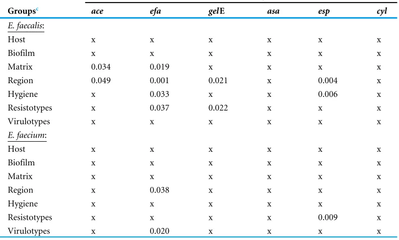
The presence ofacegene inE. faecalisstrains was dependent on the sample matrix and sampling region while theefagene was correlated with sampling region, sample matrix, farm hygiene practice and resistotypes (Table 4). On the other hand, the presence ofefa
Table 4 Correlation between the presence of virulence genes with environmental factors, virulotypes and resistotypes.
P-values (P<0.05=reject null)a,b
Groupsc ace efa gelE asa esp cyl
E. faecalis:
Host x x x x x x
Biofilm x x x x x x
Matrix 0.034 0.019 x x x x
Region 0.049 0.001 0.021 x 0.004 x
Hygiene x 0.033 x x 0.006 x
Resistotypes x 0.037 0.022 x x x
Virulotypes x x x x x x
E. faecium:
Host x x x x x x
Biofilm x x x x x x
Matrix x x x x x x
Region x 0.038 x x x x
Hygiene x x x x x x
Resistotypes x x x x 0.009 x
Virulotypes x 0.020 x x x x
Notes.
aace, collagen binding cell wall protein;efa, endocarditis specific antigen;gelE, gelatinase;asa, aggregation substance;esp,
ente-rococcal surface protein;cyl, cytolysin bx, No significant correlation.
cSubject: human host and swine host; Biofilm refers to biofilm former and non-former; Matrix, oral, rectal, nasal, urine and
fe-cal; Region, northern region and central region; Hygiene, HP1, HP2, HP3.
Distribution of the E. faecalisandE. faeciumstrains across locations, host and sample type, and its relationship to the biofilm-forming ability
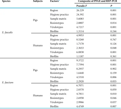
To determine the association between the genotypes, resistotypes, virulotypes and different environmental factors, a composite similarity matrix of the two typing methods was used to perform statistical analysis on the correlation between the genotype of the strains and different factors. Based on the DistLM results, the DNA fingerprints of strains isolated from both pigs and humans were significantly different among each other (Pseudo-F=2.5204,
P=0.020). We further evaluated the factors which might affect the genotypes of the strains such as sample matrix, sampling region, farm hygiene practice, biofilm-forming ability as well as the resistotypes and virulotypes of the strains (Table 5). For theE. faecalisisolated from pigs, a significant correlation was detected for all tested factors except biofilm-forming ability while for human strains, no association was found between the genotypic patterns in relation to farm hygiene practice, sample matrix and biofilm-forming ability. Conversely,
Table 5 Correlation between environmental factors, virulotypes and resistotypes with composition phylogenetic relationship inferred using REP-PCR and PFGE.
Species Subjects Factorsa Composite of PFGE and REP-PCR
PseudoF P
Region 26.229 0.001
Hygiene practice 24.392 0.001
Sample matrix 5.6083 0.001
Resistotypes 2.8807 0.014
Virulotypes 4.7217 0.001
Pigs
Biofilm 1.3114 0.244
Region 4.9953 0.001
Hygiene practice 0.5268 0.767
Sample matrix 1.7178 0.126
Resistotypes 2.3653 0.048
Virulotypes 6.0838 0.001
E. faecalis
Humans
Biofilm 1.1187 0.361
Region 9.3722 0.001
Hygiene practice 7.7394 0.001
Sample matrix 0.2937 0.902
Resistotypes 1.6440 0.159
Virulotypes 4.5310 0.006
Pigs
Biofilm 2.2920 0.055
Region 6.7071 0.001
Hygiene practice 2.8370 0.050
Sample matrix 4.7811 0.010
Resistotypes 2.6958 0.046
Virulotypes 2.9966 0.037
E. faecium
Humans
Biofilm 4.4749 0.007
Notes.
aSubject, human host and swine host; Biofilm refers to biofilm former and non-former; Matrix, oral, rectal, nasal, urine and
fe-cal; Region: northern region and central region; Hygiene, HP1, HP2, HP3.
ability and similar toE. faecalis, they were unaffected by the farm hygiene practice. Overall, the statistical analysis supported the finding that the strains isolated in this study are host and regional specific. Strong correlations were also found between the genotypes and the resistance and virulence profiles of the strains.
DISCUSSION
2017). According to the National Pharmaceutical Control Bureau of Ministry of Health, Malaysia, there are 97 antimicrobials registered for the use in livestock. A majority of these antimicrobials are used in poultry and swine farms despite the fact that some of these drugs are classified as Critically Important Antimicrobials by WHO (Health Action International Asia Pacific, 2013). In Malaysia, the Department of Veterinary Services (DVS) is responsible for certification, inspection, accreditation and implementation of legislation to ensure the production of quality livestock. They also ensure that the use of antibiotics is in accordance with the Feed Act 2009 of Malaysia. Taking into consideration the effect of antimicrobial resistance in public health and livestock industry, the Malaysian government has embarked multiple strategies against the antimicrobial resistance threat, which included increase public awareness and education on appropriate use of antibiotics, expedite surveillance and research, and improve infection prevention and control (MOH Malaysia, 2017). The unregulated use of antibiotics in swine husbandry, the co-transfer of resistance genes between enterococcal strains from different origins might have contributed to the high prevalence of resistant strains in this study (Silveira et al., 2013). The strains isolated in this study were mainly resistant to chloramphenicol, tetracycline, ciprofloxacin, gentamicin and erythromycin. Similar results were also observed in study on swine meat chain (Rizzotti, Rossi & Torriani, 2016).E. faeciumwas more resistant to penicillin and ampicillin whileE. faecaliswas more resistant to aminoglycosides.Larsen et al. (2011)reported that porcine-origin E. faecalis strains were genetically related to the strains isolated from infective endocarditis patients, suggesting that pigs can be a reservoir of human pathogens. Farm workers, veterinarians and those who are in close contact with the animals are at a higher risk of being colonized or infected by resistant bacteria harbored by the animal hosts. The resistance and virulence genes can also be transferred to other pathogenic bacteria such asStaphylococcus aureus,causing severe bacteremia (Kristich, Rice & Arias, 2014;Health Action International Asia Pacific, 2013). Strains with a particular resistance phenotype harbored the corresponding antibiotic resistance genes. Notably,E. faecalis
strains showing high-level gentamicin resistance harbored two resistance genes, which are theaac(60)-Ie-aph(200)-Iaandaph(30)-IIIa. Strains with only one aminoglycoside resistance gene were either susceptible or resistant to low-level gentamicin only. However, this trend was only observed in theE. faecalis strains. Vancomycin-resistant enterococcus (VRE) have been previously reported from swine farms located in Selangor, Perak, Johor and Penang (Getachew et al., 2010;Getachew et al., 2012;Getachew et al., 2013). However, these studies mainly focused on vancomycin resistance phenotype as VRE strains are important nosocomial pathogens and molecular evidence have also suggested that animals are reservoir of VRE (Courvalin, 2005). Although VRE was not found in our study, our findings showed that enterococcal strains found in the swine farms were multidrug-resistant. This indicates the importance of screening other antibiotics apart from vancomycin.
to antibiotics (Holmberg & Rasmussen, 2016). This probably explains the higher infection rate ofE. faecaliscompared toE. faecium,because the biofilm-formingE. faecalisstrains are more persistent thanE. faecium.Toledo-arana et al. (2001)reported thatespgene is involved in the biofilm formation of enterococcal strains.However, such correlation was not found in our study. In contrast,efaandgelE were found to have significant correlation with the resistotypes ofE. faecalisstrains.
The statistical analysis of the composite genotypic data showed thatE. faecalis and
E. faeciumwere significantly grouped according to their hosts (humans vs. pig), suggesting a host specific distribution. However, there are exceptions. CertainE. faecalisstrains isolated from different hosts with high percentage of similarity (>90%) were observed (Files S2 andS4). For instance, Enfs114 from nasal swab of a farmer and Enfs66 from a swine oral swab from PF4 possessed 100% and 91% similarity in REP and PFGE banding patterns, respectively. This result is consistent with the report byFreitas et al. (2011), which showed that certainE. faecalisandE. faeciumstrains in humans and pigs were indistinguishable by PFGE and MLST. It is also noteworthy that the strains isolated from swine oral and human nasal swabs were highly similar with the environmental strains, indicating a potential of inter-host dispersal of the enterococci strains, as strains could be transmitted from one host to another host through the environment. Detection of regional-specific strains could be explained by different swine farm management implemented in each farm as different disinfection and antibiotic exposure could act as a selective pressure.
CONCLUSION
This study showed a high occurrence of MDRE. faecalis andE. faeciumin the studied swine farms. MDR Enterococci strains from livestock industry harboring various antibiotic resistance and virulence genes pose a significant health risk to the public. Ubiquitous strains shared by different hosts were identified although majority of the strains were regional- and host-specific. A more stringent regulation is needed to ensure the proper use of antibiotics in swine husbandry to reduce the wide spread of MDR strains.
ACKNOWLEDGEMENTS
We thank the University of Malaya for facilities support.
ADDITIONAL INFORMATION AND DECLARATIONS
Funding
Grant Disclosures
The following grant information was disclosed by the authors:
High Impact Research (HIR): UM.C/625/1/HIR-MOHE/CHAN/11/02. UMRG: RP022-2012B.
University of Malaya Fellowship Scheme.
Competing Interests
The authors declare there are no competing interests.
Author Contributions
• Shiang Chiet Tan conceived and designed the experiments, performed the experiments, analyzed the data, prepared figures and/or tables, authored or reviewed drafts of the paper, approved the final draft.
• Chun Wie Chong, Cindy Shuan Ju Teh, Peck Toung Ooi and Kwai Lin Thong conceived and designed the experiments, analyzed the data, contributed reagents/materials/analysis tools, authored or reviewed drafts of the paper, approved the final draft.
Human Ethics
The following information was supplied relating to ethical approvals (i.e., approving body and any reference numbers):
The human samples collection was approved by Medical Ethics Committee, University Malaya Medical Centre (Ethic committee/IRB reference number: 1010.41).
Field Study Permissions
The following information was supplied relating to field study approvals (i.e., approving body and any reference numbers):
This study was approved by the Animal Care and Use Committee (ACUC), Faculty of Veterinary Medicine, UPM (UPM/IACUC/FYP-AUP-T006/2013).
Data Availability
The following information was supplied regarding data availability: The raw data are provided in theSupplemental Files.
Supplemental Information
Supplemental information for this article can be found online athttp://dx.doi.org/10.7717/ peerj.5353#supplemental-information.
REFERENCES
Baldassarri L, Cecchini R, Bertuccini L, Ammendolia MG, Iosi F, Arciola CR, Monta-naro L, Di Rosa R, Gherardi G, Dicuonzo G, Orefici G, Creti R. 2001.Enterococcus
spp. produces slime and survives in rat peritoneal macrophages.Medical Microbiol-ogy and ImmunolMicrobiol-ogy190:113–120DOI 10.1007/s00430-001-0096-8.
infections and infective endocarditis.Clinical Infectious Diseases67(2):303–309 DOI 10.1093/cid/ciy064.
Benjamin JK, Bruce AH, Lance BP. 2017.Food-animal production and the spread of antibiotic resistance: the role of ecology.Frontiers in Ecology and the Environment
15(6):309–318DOI 10.1002/fee.1505.
Clinical Laboratory Standards Institute (CLSI). 2015.Performance standards for antimicrobial susceptibility testing; twenty third informational supplement M100-S23 CLSI. Wayne: Clinical Laboratory Standards Institute.
Compassion in World Farming (CIWF). 2011. Antibiotics in animal farming: public health and animal welfare. Surrey, CIWF.Available athttps:// www.ciwf.org.uk/ media/ 3758863/ Antibiotics-in-Animal-Farming-Public-Health-and-Animal-Welfare. pdf (accessed on 15 January 2016).
Courvalin P. 2005.Antimicrobial drug resistance: ‘‘prediction is very difficult, especially about the future’’.Emerging Infectious Diseases11(10):1503–1506 DOI 10.3201/eid1110.051014.
Daniel DS, Lee SM, Dykes GA, Rahman S. 2015.Public health risks of multiple-drug-resistantEnterococcusspp. in Southeast Asia.Applied and Environmental Microbiology
81:6090–6097DOI 10.1128/AEM.01741-15.
Freitas AR, Coque TM, Novais C, Hammerum AM, Lester CH, Zervos MJ, Donabedian S, Jensen LB, Francia MV, Baquero F, Peixe L. 2011.Human and swine hosts share vancomycin-resistantEntercoccus faeciumCC17 and CC5 andEnterococcus faecium
CC2 clonal clusters harboring Tn1546 on indistinguishable plasmids.Journal of Clinical Microbiology49(3):925–931DOI 10.1128/JCM.01750-10.
Getachew Y, Hassan L, Zakaria Z, Aziz SA. 2013.Genetic variability of vancomycin-resistantEnterococcus faeciumandEnterococcus faecalisisolates from
hu-mans, chickens, and pigs in Malaysia.Applied and Environmental Microbiology
79(15):4528–4533DOI 10.1128/AEM.00650-13.
Getachew Y, Hassan L, Zakaria Z, Lokman N. 2010.Species distribution and resistance phenotypes of vancomycin-resistantEnterococcusisolated from pigs in Pulau Pinang, Malaysia.Pertanika Journal of Tropical Agricultural Science33(1):15–25.
Getachew Y, Hassan L, Zakaria Z, Zaid CZM, Yardi A, Shukor RA, Marawin LT, Embong F, Aziz SA. 2012.Characterization and risk factors of vancomycin-resistant
Enterococci(VRE) among animal-affiliated workers in Malaysia.Journal of Applied Microbiology113:1184–1195DOI 10.1111/j.1365-2672.2012.05406.x.
Health Action International Asia Pacific (HAIAP). 2013. Antibiotic use and antibiotic resistance in food animals in Malaysia: a threat to human and animal health. Third World Network (TWN) Penang in association with Consumers’ Association of Penang, Malaysia.Available athttp:// www.haiasiapacific.org/ wp-content/ uploads/ 2014/ 06/
Memo-on-Antibiotics-in-animal-feeds-the-case-for-Malaysia-21-Nov-2013-V1.pdf (accessed on 28 December 2016).
c7a49464268626d5231595734765457467359586c7a615746755830466a64476c76626c3 95162474675583039755830467564476c7461574e7962324a7059577866556d567a61584 e305957356a5a56386f54586c425543314254564970587a49774d5463744d6a41794d533
5775a47593d(accessed on 03 January 2018).
Holmberg A, Rasmussen M. 2016.Mature biofilms ofEnterococcus faecalisand En-terococcus faeciumare highly resistant to antibiotics.Diagnostic Microbiology and Infectious Disease84:19–21DOI 10.1016/j.diagmicrobio.2015.09.012.
Hwang IY, Ku HO, Lim SK, Park CK, Jung GS, Jung SC, Nam HM. 2009.Species distribution and resistance patterns to growth-promoting antimicrobials of ente-rococci isolated from pigs and chickens in Korea.Journal of Veterinary Diagnostic Investigation21:858–862DOI 10.1177/104063870902100616.
Jackson CR, Fedorka-Cray PJ, Barrett JB. 2004.Use of a genus- and species-specific multiplex PCR for identification of enterococci.Journal of Clinical Microbiology
42(8):3558–3565DOI 10.1186/s12866-015-0468-7.
Kristich CJ, Rice LB, Arias CA. 2014. Enterococcal infection—treatment and antibiotic resistance. In: Gilmore MS, Clewell DB, Ike Y, Shanker N, eds.Enterococci: from commensals to leading causes of drug resistant infection. Boston: Massachusetts Eye and Ear Infirmary, 123–184.
Larsen J, Schonheyder HC, Singh KV, Lester CH, Olsen SS, Porsbo LJ, Garcia-Migura L, Jensen LB, Bisgaard M, Murray BE, Hammerum AM. 2011.Porcine and human community reservoirs ofEnterococcus faecalis, Denmark.Emerging Infectious Diseases
17(12):2395–2397DOI 10.3201/eid17120101584.
Lebreton F, Willems R, Gilmore M. 2014. Enterococcusdiversity, origins in nature, and gut colonization. In: Gilmore MS, Clewell DB, Ike Y, Shanker N, eds.Enterococci: from commensals to leading causes of drug resistant infection. Boston: Massachusetts Eye and Ear Infirmary, 5–64.
Lim SY, Teh CSJ, Thong KL. 2017.Biofilm-related diseases and omics: global tran-scriptional profiling ofEnterococcus faeciumreveals different gene expression patterns in the biofilm and planktonic cells.OMICS A Journal of Integrative Biology
21(10):592–601DOI 10.1089/omi.2017.0119.
Novais C, Freitas AR, Silveira E, Antunes P, Silva R, Coque TM, Peixe L. 2013.Spread of multidrug-resistantEnterococcusto animals and humans: an underestimated role for the pig farm environment.Journal of Antimicrobial Chemotherapy68:2746–2754 DOI 10.1093/jac/dkt289.
Price VJ, McBride SW, Duerkop B, Palmer KL. 2018.CRISPR-Cas blocks antibiotic re-sistance plasmid transfer betweenEnterococcus faecalisstrains in the gastrointestinal tract.bioRxivDOI 10.1101/312751.
Rizzotti L, Rossi F, Torriani S. 2016.Biocide and antibiotic resistance ofEnterococcus fae-calisandEnterococcus faeciumisolated from the swine meat chain.Food Microbiology
60:160–164DOI 10.1016/j.fm.2016.07.009.
environment and foods) and clonal lineages.Journal of Antimicrobial Chemotherapy
69(4):899–906DOI 10.1093/jac/dkt479.
Stepanovic S, Vucovic D, Dakic I, Savic B, Svabic-Vlahovic M. 2000.A modified microtiter-plate test for quantification of staphylococcal biofilm formation.Journal of Microbiological Methods40(2):175–179DOI 10.1016/S0167-7012(00)00122-6. Toledo-arana A, Valle J, Solano C, Arrizubieta MJ, Cucarella C, Lamata M, Amorena B,
Leiva J, Penades JR, Lasa I. 2001.The enterococcal surface protein, esp, is involved inEnterococcus faecalisbiofilm formation.Applied and Environmental Microbiology
67(10):4538–4545DOI 10.1128/AEM.67.10.4538-4545.2001.
Turabelidze D, Kotetishvili M, Kreger A, Morris Jr JG, Sulakvelidze A. 2000.Improved pulsed-field gel electrophoresis for typing vancomycin-resistant enterococci.Journal of Clinical Microbiology38(11):4242–4245.
Versalovic J, Schneider M, De Bruijn F, Lupski JR. 1994.Genomic fingerprinting of bacteria using repetitive sequence-based polymerase chain reaction.Methods in Molecular and Cellular Biology 5:25–40.
Vidana R, Rashid MU, Ozenci V, Weintraub A, Lund B. 2015.The origin of endodontic
Enterococcus faecalisexplored by comparison of virulence factor patterns and antibiotic resistance to that of isolates from stool samples, blood cultures and food.
International Endodontic Journal 49(4):343–351DOI 10.1111/iej.12464. Woolhouse M, Ward M, Van Bunnik B, Farrar J. 2015.Antimicrobial resistance in
humans, livestock and the wider environment.Philosophical Transactions of the Royal Society B: Biological Sciences370(1670):20140083DOI 10.1098/rstb.2014.0083.
FURTHER READING
Aminov RI, Garrigues-JeanJean N, Mackie RI. 2001.Molecular ecology of tetracycline resistance: development and validation of primers for detection of tetracycline resistance genes encoding ribosomal protection proteins.Applied and Environmental Microbiology67(1):22–32DOI 10.1128/AEM.67.1.22-32.2001.
Clark NC, Cooksey RC, Hill BC, Swenson JM, Tenover FC. 1993.Characterization of glycopeptide-resistant enterococci from US hospitals.Antimicrobial Agents and Chemotherapy37:2311–2317DOI 10.1128/AAC.37.11.2311.
Dutka Malen S, Evers S, Courvalin P. 1995.Detection of glycopeptide resistance genotypes and identification to the species level of clinically relevant enterococci by PCR.Journal of Clinical Microbiology33:24–27.
Gever D, Danielsen M, Huys G, Swings J. 2003.Molecular characterization oftet(M) genes inLactobacillusisolates from different types of fermented dry sausages.Applied and Environmental Microbiology 69(2):1270–1275
DOI 10.1128/Aem.69.2.1270-1275.2003.
Mannu L, Pab A, Daga E, Comunian R, Zanetti S, Dupre I, Sechi LA. 2003.Comparison of the incidence of virulence determinants and antibiotic resistance between
Satake S, Clark N, Rimland D, Nolte FS, Tenover FC. 1997.Detection of vancomycin-resistant enterococci in fecal samples by PCR.Journal of Clinical Microbiology
35(9):2325–2330.
Shankar V, Baghdayan AS, Huycke MM, Lindahl G, Gilmore MS. 1999. Infection-derivedEnterococcus faecalisstrains are enriched inesp, a gene encoding a novel surface protein.Infection and Immunity67:193–200.
Vakulenko SB, Donabedian SM, Voskresenskiy AM, Zervos MJ, Lerner SA, Chow JW. 2003.Multiplex PCR for detection of aminoglycoside resistance genes in enterococci.Antimicrobial Agents and Chemotherapy 47(4):1423–1426 DOI 10.1128/AAC.47.4.1423-1426.2003.
Vankerckhoven V, Van Autgaerden T, Vael C, Lammens C, Chapelle S, Rossi R, Jabes D, Goossens H. 2004.Development of a multiplex PCR for the detection ofasaI,