www.impactjournals.com/oncotarget/ Oncotarget, November, Vol.3, No 11
Targeting HIF2α Translation with Tempol in VHL-Deficient Clear
Cell Renal Cell Carcinoma
Carole Sourbier1,*, Gaurav Srivastava1,*, Manik C. Ghosh2, Sanchari Ghosh1, Youfeng Yang1, Gopal Gupta1, William DeGraff3, Murali C. Krishna3, James B. Mitchell3, Tracey A. Rouault2, W. Marston Linehan1
1 Urologic Oncology Branch, Center for Cancer Research, National Cancer Institute 2 National Institute of Child Health and Human Development
3 Radiation Biology Branch, Center for Cancer Research, National Cancer Institute, National Institutes of Health, Bethesda, MD
* Authors contributed equally to this work.
Correspondence to: W. Marston Linehan, email: WML@nih.gov Keywords: HIF, Tempol, RCC, VHL, IRP1, iron metabolism
Received: July 19, 2012, Accepted: November 06, 2012, Published: November 08, 2012
Copyright: © Sourbier et al. This is an open-access article distributed under the terms of the Creative Commons Attribution License, which permits unrestricted use, distribution, and reproduction in any medium, provided the original author and source are credited.
ABSTRACT:
The tumor suppressor gene, Von Hippel-Lindau (VHL), is frequently mutated in
the most common form of kidney cancer, clear cell renal cell carcinoma (CCRCC). In hypoxic conditions, or when there is a VHL mutation, the hypoxia inducible factors, HIF1α and HIF2α, are stabilized and transcribe a panel of genes associated with cancer such as vascular endothelial growth factor receptor (VEGFR), platelet derived growth factor (PDGF), and glucose transporter 1 (GLUT1). Recent studies in clear cell kidney cancer have suggested that HIF2α, but not HIF1α, is the critical oncoprotein in the VHL pathway. Therefore, targeting HIF2α could provide a potential therapeutic approach for patients with advanced CCRCC. Since iron regulatory protein 1 (IRP1) is known to inhibit the translation of HIF2α, we investigated whether Tempol, a stable nitroxide that activates IRP1 towards IRE-binding, might have a therapeutic effect on a panel of human CCRCC cells expressing both HIF1α and HIF2α. We first evaluated the protein expression of HIF1α and HIF2α in 15 different clear cell renal carcinoma cell lines established from patient tumors in our laboratory. Tempol decreased the expression of HIF2α, and its downstream targets in all the cell lines of the panel. This effect was attributed to a dramatic increase of IRE-binding activity of IRP1. Several cell lines were found to have an increased IRP1 basal activity at 20% O2 compared to 5% O2, which may lower HIF2α expression in some of the cell lines in a VHL-independent manner. Taken together our data identify Tempol as an agent with potential therapeutic activity targeting expression of HIF2α in VHL-deficient clear cell kidney cancer and illustrate the importance of studying biochemical processes at relevant physiological O2 levels.
INTRODUCTION
In the United States, an estimated 64,770 new cases of kidney cancers will be diagnosed in 2012 and about 13,570 people will die of this disease [1]. The most common type of kidney cancers, clear cell renal cell carcinoma (CCRCC), is associated with mutations of the VHL gene [2,3,4]. In normal tissues, the product
of the VHL gene is associated with ubiquitination and degradation of the hypoxia inducible factor (HIF) through
an oxygen-sensing mechanism [5,6]. In normoxia, HIF1α and HIF2α are hydroxylated by prolyl-hydroxylases
in an iron-dependent manner. This post-translational
modification allows recognition of HIF by the VHL
presence of a mutated VHL gene, HIF1α and HIF2α
are stabilized and induce the expression of a panel of
transcriptional target genes such as VEGF, PDGF, and GLUT1, supporting the metabolic shift that underlies CCRCC tumorigenicity [9]. Even in presence of oxygen,
clear cell kidney cancer cells with VHL gene mutation display a “pseudo-hypoxic” phenotype. Although the
degradation of both HIF1α and HIF2α are regulated by VHL [10,11], HIF2α has been thought to be the dominant oncoprotein in VHL-deficient CCRCC cells [12,13,14]. It was also recently suggested that HIF1α may function as a tumor suppressor gene in VHL-deficient clear cell kidney
cancer [15]. However, despite the proposed critical role
of HIF2α in VHL-deficient CCRCC tumorigenesis, only a few HIF2α inhibitors have been described [16,17].
A molecular link between HIF2α expression and iron availability has recently been reported [18]. HIF2α, but not HIF1α, has an iron-responsive element (IRE) in the
5’ untranslated region (UTR) of its mRNA. Thus, when
the cells are deficient in iron, the iron regulatory proteins (IRPs) repress the translation of HIF2α protein by binding to its 5’IRE. In addition, Ghosh et al. have recently
shown that a stable nitroxide, Tempol (4-hydroxy-2,2,6,6-tetramethylpiperidine-N-oxyl) partially restored the
phenotypes of IRP2 knockout mice by activating the IRE binding activity of IRP1 and thus partially compensating for the loss of IRP2 [19].
In this report, using a panel of VHL-deficient CCRCC cell lines that express increased either HIF1α or HIF2α or both, we investigated whether Tempol inhibits the translation of HIF2α by inducing the IRE binding activity of IRP1 and whether Tempol might be a potential
targeted therapy for CCRCC.
RESULTS
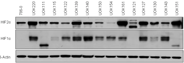
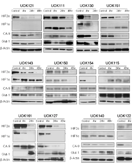
HIF1α and HIF2α expression in CCRCC cell lines.
We first characterized the HIFα status of 15 RCC
cell lines with mutated VHL gene (Figure 1 and Table 1).
All of the cells expressed HIF2α, but some, including
786-0 [7], UOK111, UOK121 and UOK151 did not express
HIF1α or expressed a shorter 75kDa splice isoform. Interestingly although expressed in UOK154, HIF2α
protein levels were low relative to other VHL-deficient
cells.
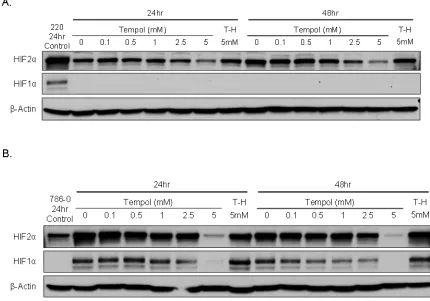
Tempol decreases HIF2α protein expression and inhibits its downstream targets.
We then assessed the effect of Tempol on HIF1α and HIF2α protein levels. Tempol decreased HIF1α / HIF2α levels in 786-0 cell line, which expresses only HIF2α [7], and in UOK220 cell line, which expresses both HIFα isoforms (Figure 2). The reduced form of Tempol
(Tempol-H), which was used as a negative control, had
no effect on either HIF1α or HIF2α expression (Figure 2). To further investigate the effect of Tempol on HIFα
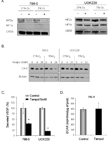
activity and pathway, we assessed the level of nuclear
HIF2α following Tempol treatment (5mM, 24h; Figure 3A, supplementary figure 1). Using 30μg of nuclear
extracts and similar to that observed in the previous
experiment, Tempol significantly decreased the level of nuclear HIF2α, suggesting that its transcriptional activity
was also compromised. The effect of Tempol on nuclear
HIF1α expression was, however, not consistent in all of the cell lines (Figure 3, supplementary figure 1). We then evaluated the expression of HIF2α downstream targets
following Tempol’s treatment. In 786-0 and UOK220
cells, both CA-9 expression and VEGF secretion, two known HIF2α targets and therapeutic targets for CCRCC
[20,21], were found to be decreased after 24h of treatment
with 5mM Tempol (Figure 3B-C). Also, since HIFα is
known to play a role in regulating tumor cells metabolism, we asked whether Tempol might have a metabolic effect
on 786-0 cells. As shown in Figure 3D and consistent with our observation that Tempol’s effect on HIFα is mainly on HIF2α, Tempol (24h, 5mM) did not affect the extra-cellular acidification rate of 786-0 cells (ECAR), a
surrogate for lactate secretion. This is consistent with other published works, such as the recent paper of Chiavarina et
[image:2.612.130.489.562.685.2]al which shows that HIF1α and not HIF2α is important for
glycolysis in a breast cancer model [22]. Further, the effect
of Tempol on HIF2α expression and on its downstream
targets was highly reproducible since it was also observed
in 12 other CCRCC cell lines displaying various HIF1α and HIF2α expression patterns (Figure 4 and Figure 1). Thus, our data suggest that Tempol is a potent HIF2α inhibitor, regardless of the HIF1α status of the CCRCC
cell line studied.
Tempol’s effect on IRP1 activity correlates with HIF2α.
To further investigate Tempol’s mechanism of action
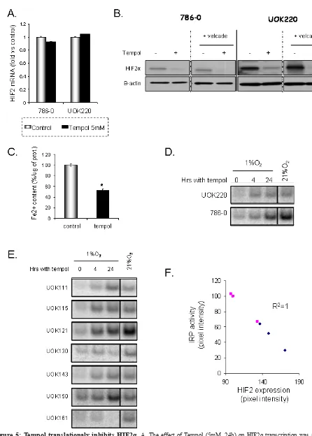
and how it decreases HIF2α expression, we first assessed whether Tempol might have an effect on HIF2α at a transcriptional level. We quantified HIF2α mRNA levels following Tempol treatment by semi-quantitative RT-PCR.
As shown in Figure 5A, 24h of Tempol treatment did not
significantly change the mRNA expression of HIF2α.
Then,in order to assess whether Tempol might have an
effect on HIF2α by inducing its degradation, we evaluated the effect of Tempol (5mM, 24h) on HIF2α expression
following inhibition of the proteasome with velcade (1µM, 24h). As shown in figure 5B, using 20μg of whole cell lysates, Tempol still decreased HIF2α protein expression, suggesting that Tempol’s effect on HIF2α happened at the translational level. Because HIF2α expression and
iron availability have recently been linked [18] and that
Ghosh et al. have shown that Tempol is able to activate
the IRE binding activity of IRP1[19], we thus investigated Tempol’s effect on iron availability and IRP1 activity. We
measured the amount of Fe2+ in cells following Tempol treatment as the nitroxide agent Tempol is able to lower Fe2+[23]. As shown in Figure 5C, Tempol significantly decreased the amount of available Fe2+. In addition,
Tempol increased IRP1 activity in a panel of CCRCC cell lines (Figure 5D-E), which inversely correlated with HIF2α expression (Figure 5F, R2=1). Taken together these
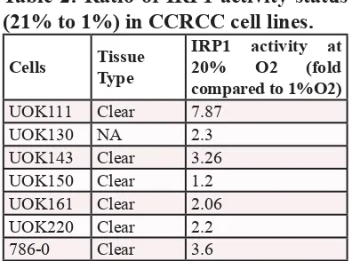
data suggest that Tempol regulates HIF2α expression at the translational level by increasing the IRE binding activity of IRP1. Interestingly, the basal level of IRP1 activation
[image:3.612.117.505.107.431.2]was highly increased in most of the cell lines at 21% O2 (Table 2) [24], suggesting that the O2 environment of the cell culture in vitro also affects HIFα expression in VHL-deficient cells.
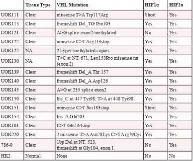
Table 1: CCRCC cell lines HIF status.
Summary of the CCRCC cell lines and
normal epithelial kidney cell line with their VHL mutation and their HIF status.
NA: not available. Short: a 75kD isoform of HIF1α is expressed but not the full
length.
Tissue Type VHL Mutation HIF1α HIF2α UOK111 Clear missense T>A Trp117Arg Short Yes UOK115 Clear frameshift Del_TG Pro103 Yes Yes UOK121 Clear A>G splice exon2/methylated No Yes UOK122 Clear nonsense C>T Arg113stop Yes Yes UOK127 NA 2 hyper-methylated copies Yes Yes UOK130 NA T>C at NT. 671, Leu153Pro missense mt (exon 2) Yes Yes
UOK139 Clear frameshift Del_A Thr 157 Yes Yes UOK140 Clear frameshift Del_A Asp126 Yes Yes UOK143 Clear A>G at 235 splice exon2 Yes Yes UOK150 Clear Ins_C at 447 Tyr98; T>A at 448 Tyr98 Yes Yes UOK151 Clear nonsense C>T Ser183stop Short Yes
UOK154 Clear Ins_A Gln203 Yes Yes
UOK161 Clear C>T Gln164stop Yes Yes
UOK220 Clear 2 missense T>A Asn78Lys C>T Arg79Cys Yes Yes 786-0 Clear 1bp Del at NT. 523, frameshift at Gly104, exon 1 No Yes
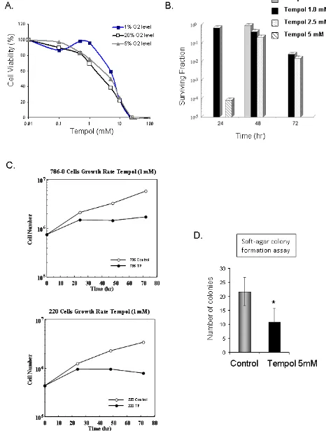
Tempol is cytotoxic to CCRCC cells.
Due to the observed effect of Tempol on HIF2α, CA9 and VEGF, all previously reported therapeutic targets
for CCRCC tumors, we investigated whether Tempol might have an anti-tumor effect in CCRCC cells. Cell cytotoxicity assays demonstrated that 48h treatment with Tempol is cytotoxic to CCRCC cells in vitro (Figure 6A), that Tempol inhibits the formation of colonies of 786-0
cells (Figure 6B), and inhibits the proliferation of CCRCC
cells 786-0 and UOK220 (Figure 6C). Also, Tempol
treatment (5mM) decreased by 50% the ability of 786-0
cells to form anchorage-independent colonies, a marker of tumor transformation assessed by soft-agar colony
formation assay (6D).
DISCUSSION
Targeting HIF2α is of particular interest in cancer therapy since HIF2α is the transcription factor of
numerous genes that have been involved not only in the development of CCRCC growth [20,21,25,26] but also in
the development of other type of tumors [27,28]. However,
to date, only few HIF2α inhibitors have been identified. In this report, we identified the nitroxide Tempol as a potent HIF2α inhibitor, with promising anti-tumor effects in a
panel of CCRCC tumor cell lines.
Mutations occurring on the VHL gene are a critical event leading to CCRCC development in both sporadic
[image:4.612.90.520.344.645.2]and hereditary forms of kidney tumors. This lack of VHL function leads to the stabilization of both HIF1α and HIF2α proteins as well as to an increase in DNA damage [10,11,29]. Although the expression of both HIF1α and HIF2α is similarly regulated by VHL, their roles in
tumor development, tumor metabolism, and response to therapy are sometimes contrasted [12,22,30,31]. Thus,
although our goal was to identify a new HIF2α inhibitor, we characterized the expression status of both HIF1α and HIF2α in 15 VHL-deficient clear cell kidney cancer cell lines and assessed the effect of Tempol on both HIFα isoforms. Regardless of the differences in HIF1α and HIF2α expression between CCRCC cell lines, Tempol decreased the expression and function of HIF2α in all the
cell lines. Surprisingly, Tempol also decreased the protein
Figure 3: Tempol decreases HIF2α nuclear activity. A. To assess the effect of Tempol on HIFα nuclear activities, expression of HIFα was assessed in 786-0 and UOK220 cells following Tempol treatment (5mM, 24h) and using 30μg nuclear lysates extracted at 21%
expression and activity of HIF1α in some of the CCRCC cell lines. As HIF1α does not have an IRE-binding domain, the reasons underlying Tempol’s effect on HIF1α remain
unknown, especially as it was inconsistent throughout the panel. Also, further work will be necessary to assess
whether this effect on HIF1α might be beneficial or not for
the survival of the tumors. Nevertheless, Tempol’s effect
on HIF2α was consistent in all the tested CCRCC cell lines regardless its effect on HIF1α.
[image:6.612.77.530.121.689.2]The presence of an iron-responsive
Figure 4: Tempol inhibits HIF2α in a panel of CCRCC cells. Twelve CCRCC cell lines were treated with 5mM Tempol for 4h, 24h or 48h before to be subjected to whole protein lysis, SDS-PAGE and immunoblotting for both HIFα isoforms and CA-9. Twenty
Figure 5: Tempol translationaly inhibits HIF2α. A. The effect of Tempol (5mM, 24h) on HIF2α transcription was assessed by semi-quantitative RT-PCR in 786-0 and UOK220 cells. B. The effect of Tempol (5mM, 24h) on HIF2α protein degradation was assessed by treating the cells with the proteasome inhibitor velcade (1µM, 24h). HIF2α expression was then assessed by immunoblotting using 20μg
of whole cell lysates. C. The amount of available ferrous iron (Fe2+) content was measured after Tempol treatment (5mM, 24h) in 786-0
element in the 5’-UTR of HIF2α mRNA regulating HIF2α translation (but not HIF1α) links HIF2α and iron
metabolism. This also opens new therapeutic strategies to
selectively target HIF2α. When Ghosh and collaborators demonstrate that Tempol is able to activate the IRE binding activity of IRP1[19], we thus hypothesized that Tempol might also be able to inhibit HIF2α translation.
In this report, we demonstrated that the decrease in
expression of HIF2α following Tempol treatment significantly correlated with an increased activation of IRP1, reinforcing the link between IRP1 activity and HIF2α expression. Moreover, Tempol treatment decreased
CCRCC cells viability, clonogenicity, proliferation, as well as anchorage-independent growth. Thus, by regulating the iron metabolism of CCRCC cells, Tempol may have a potential therapeutic value for cancers dependent on
HIF2α for growth and survival, such as CCRCC tumors.
Interestingly, at 21%O2, most of the CCRCC cell lines (with the exception of UOK150) were found to
have an artificially high level of IRP1 activity (Table2).
This suggests that culturing CCRCC cells at 21%O2 may
artificially lower HIF2α expression in a VHL-independent manner by oxidizing the [4Fe-4S] center in aconitase,
and therefore, activating the IRE-binding form of IRP1. These findings highlight the importance of working in an in vitro condition that properly recreates the in vivo tumor environment [24].
Although significant progress in understanding the
genetic basis of kidney cancer has been made over the past twenty years, there is still a need for the development of effective forms of therapy for patients with advanced
kidney cancer. Since the identification of the VHL gene,
seven novel agents that target the VHL pathway have been approved by the FDA. However, there are few
complete responses to these agents and most patients develop resistance to therapy and develop progressive
disease. Targeting HIF2α with an agent such as Tempol
has the potential to inhibit the oncoprotein critical to clear
cell kidney cancer carcinogenicity. The findings in this
work may provide the foundation for the development
of effective forms of therapy for this and other
HIF2α-dependent cancers.
METHODS
Cell lines and cell culture
The UOK cell lines (UOK-111, 115, 121, 122, 127, 130, 139, 140, 143, 150, 151, 154, 161, and UOK220)
were established in the Urologic Oncology Branch (National Cancer Institute, Bethesda, MD) [32]. 786-0 and
HK-2 were obtained from ATCC. Cells were cultured in
high glucose DMEM supplemented with 10% FBS. Cells
were harvested or treated when they reached 70-80%
confluence.
Chemical agents
Tempol and hydroxyl-Tempol were purchased from
Sigma-Aldrich (St Louis, MO).
Immunoblotting
Ten to thirty micrograms of protein was loaded
in 4-20% polyacrylamide gels (Biorad, Hercules, CA).
After elelectropheresis, the proteins were transferred on
PVDF membranes before being blocked with 5% fat-free milk for at least 1h. Primary antibodies were incubated over-night at 4°C. After serial washes with TBS-Tween,
Horseradish peroxidase-linked secondary antibodies (Sigma-Aldrich) were incubated 1-2h before development
with the ECL protein detection system (Pierce, Rockford, IL). Goat anti-HIF2α antibody was obtained from R&D Systems (Minneapolis, MN). Rabbit antibodies against β-actin, and carbonic anhydrase 9 (CA9) were from Cell Signaling Technology, Inc. (Danvers, MA). Mouse HIF1 antibody was from BD Transduction (San Jose, CA). All
the antibodies were used at 1:1000 dilution.
Cell cytotoxicity
Cell cytotoxicity was measured using the tetrazolium salt
3-(4,5-dimethylthiazol-2-yl)-2,5-diphenyltetrazolium bromide (MTT), as previously described [33]. Briefly, 5,000 cells were seeded into 96-well plate and treated as described in the results or figure legends. After 48 hrs, the MTT salt was added into the media (1:10) and incubated for 2 hrs at 37°C. During that
time, metabolically active cells transformed the yellow
MTT salt into purple formazan crystals. The media was then removed and the crystals were solubilized in 100 µl of DMSO. Data were read at 570nm. Cell survival was assessed by the clonogenic assay. Exponentially growing cells were exposed to Tempol (0.1 -5.0 mM) for 24-72
hr. Following treatment, cells were trypsinized, counted,
[image:8.612.73.271.57.204.2]plated, and incubated for 10-14 days. Colonies were fixed
Table 2: Ratio of IRP1 activity status
(21% to 1%) in CCRCC cell lines.
Cells Tissue Type IRP1 activity at 20% O2 (fold compared to 1%O2) UOK111 Clear 7.87
Figure 6: Anti-tumor effect of Tempol in CCRCC. A. The cytotoxic effect of Tempol in 786-0 cells was assessed by MTT after 48h
treatment at 1%, 5% and 20% O2. B. Cell viability following Tempol treatment was assessed by clonogenic assay in 786-0 cells. C. Effect of Tempol (1mM) on cell proliferation in 786-0 and UOK220 cells. D. The effect of Tempol (5mM) on the anchorage-independent growth
with methanol/acetic acid (3:1), stained with crystal violet,
counted (colonies >50 cells), and survival was corrected
for the plating efficiency of untreated controls.
RT-PCR.
Total RNA was extracted using RNeasy minin kit
(Qiagen, Valencia, CA) and cDNA was prepared with High Capacity cDNA Reverse Transcription kit (Applied Biosystem). RT-PCR was performed using Platinum SYBR Green qPCR SuperMix-UDG (Invitrogen).
RNA Mobility Shift Assays
Gel retardation assays were performed as follows:
cell lysates were prepared in an anaerobic chamber in
oxygen-depleted lysis buffer containing 10 mM Hepes (pH 7.2), 3 mM MgCl2, 40 mM KCl, 5% glycerol, 0.2% Nonidet P-40, 5 mM DTT, 1 mM the protease inhibitor AEBSF, 10 µg/ml Leupeptin and Complete EDTA-free
protease inhibitor mixture (Roche Applied Science).
Lysate (x µl) containing 10 µg of total protein was added
to (12.5-x) µl of bandshift buffer containing 25 mM Tris-HCl (pH 7.5) and 40 mM KCl. The samples were incubated for 5 min at room temperature (RT) with 12.5 µl of a reaction mixture containing 20% glycerol, 0.2 units/µl Super RNAsine (Ambion), 0.6 µg/µl yeast t-RNA, 5 mM DTT, and 20 nM 32P-labeled IRE from human ferritin H chain gene in 25 mM Tris-HCl (pH 7.5) and 40 mM KCl. A measure of 20 µl of this reaction mixture was loaded into a 10% acrylamide/TBE gel, which was run at 200 V for 4 h, and then the gel was fixed, dried, and exposed for
autoradiography.
Determination of Fe2+
The content of ferrous ion was assessed using
Iron Assay Kit (Biovision, Milpitas, CA) following the
manufacturer instructions and without iron reducers. Fe2+ contents were measured at 593 nm using a microplate reader.
Anchorage-independant colony formation assay
The ability of 786-0 cell line to form anchorage-independent colonies was assessed by soft-agar colony formation assay. Two layered agarose gels of 0.5% (bottom) or 0.4% (top) low melting agarase gels were pored into 60mm dishes. Two hundred thousands 786-0 cells were added to the top layer and cultured until colonies were formed (about 3 weeks). Colonies with a diameter of > 0.1 mm were then counted in 5 random
high-power fields using a phase contrast microscope.
Statistics
All values are expressed as mean ± Standard Error. All experiments were performed three times. Values were compared using the Student-Newman-Keul’s test. P < 0.05 was considered significant.
REFERENCE
1. Siegel R, Naishadham D, Jemal A. CA Cancer J Clin. 2012;
62:
2. Latif F, Tory K, Gnarra J, Yao M, Duh FM, Orcutt ML, Stackhouse T, Kuzmin I, Modi W, Geil L, . Science. 1993;
260: 1317-1320.
3. Gnarra JR, Lerman MI, Zbar B, Linehan WM. Sem Oncol. 1995; 22: 3-8.
4. Nickerson ML, Jaeger E, Shi Y, Durocher JA, Mahurkar S, Zaridze D, Matveev V, Janout V, Kollarova H, Bencko V, Navratilova M, Szeszenia-Dabrowska N, Mates D, Mukeria A, Holcatova I, Schmidt LS, Toro JR, Karami S, Hung R, Gerard GF, Linehan WM, Merino M, Zbar B, Boffetta P, Brennan P, Rothman N, Chow WH, Waldman FM, Moore LE. Clin Cancer Res. 2008; 14: 4726-4734.
5. Jaakkola P, Mole DR, Tian YM, Wilson MI, Gielbert J, Gaskell SJ, Kriegsheim A, Hebestreit HF, Mukherji M, Schofield CJ, Maxwell PH, Pugh CW, Ratcliffe PJ. Science. 2001; 292: 468-472.
6. Kaelin WG, Jr. Nature Reviews Cancer. 2002; 2: 673-682. 7. Maxwell PH, Wiesener MS, Chang GW, Clifford SC, Vaux
EC, Cockman ME, Wykoff CC, Pugh CW, Maher ER, Ratcliffe PJ. Nature. 1999; 399: 271-275.
8. Ohh M, Park CW, Ivan M, Hoffman MA, Kim TY, Huang LE, Pavletich N, Chau V, Kaelin WG. Nat.Cell Biol. 2000;
2: 423-427.
9. Smaldone MC, Maranchie JK. Urol.Oncol. 2009; 27:
238-245.
10. Krieg M, Haas R, Brauch H, Acker T, Flamme I, Plate KH. Oncogene. 2000; 19: 5435-5443.
11. Sufan RI, Jewett MA, Ohh M. Am.J.Physiol Renal Physiol. 2004; 287: F1-F6.
12. Raval RR, Lau KW, Tran MG, Sowter HM, Mandriota SJ, Li JL, Pugh CW, Maxwell PH, Harris AL, Ratcliffe PJ. Mol.Cell Biol. 2005; 25: 5675-5686.
13. Maranchie JK, Vasselli JR, Riss J, Bonifacino JS, Linehan WM, Klausner RD. Cancer Cell. 2002; 1: 247-255. 14. Kondo K, Klco J, Nakamura E, Lechpammer M, Kaelin
WG, Jr. Cancer Cell. 2002; 1: 237-246.
15. Shen C, Beroukhim R, Schumacher SE, Zhou J, Chang M, Signoretti S, Kaelin WG, Jr. Cancer Discov. 2011; 1:
222-235.
17. Kong HS, Lee S, Beebe K, Scroggins B, Gupta G, Lee MJ, Jung YJ, Trepel J, Neckers L. Mol.Pharmacol. 2010; 78:
1072-1078.
18. Sanchez M, Galy B, Muckenthaler MU, Hentze MW. Nat. Struct.Mol.Biol. 2007; 14: 420-426.
19. Ghosh MC, Tong WH, Zhang D, Ollivierre-Wilson H, Singh A, Krishna MC, Mitchell JB, Rouault TA. Proc.Natl. Acad.Sci.U.S.A. 2008; 105: 12028-12033.
20. Rini BI. Cancer. 2009; 115: 2306-2312.
21. Stillebroer AB, Mulders PF, Boerman OC, Oyen WJ, Oosterwijk E. Eur.Urol. 2010; 58: 75-83.
22. Chiavarina B, Martinez-Outschoorn UE, Whitaker-Menezes D, Howell A, Tanowitz HB, Pestell RG, Sotgia F, Lisanti MP. Cell Cycle. 2012; 11: 3280-3289.
23. Charloux C, Paul M, Loisance D, Astier A. Free Radic.Biol. Med. 1995; 19: 699-704.
24. Meyron-Holtz EG, Ghosh MC, Rouault TA. Science. 2004;
306: 2087-2090.
25. Sourbier C, Massfelder T. Cancer Lett. 2006; 240: 170-182. 26. Manisterski M, Golan M, Amir S, Weisman Y, Mabjeesh
NJ. Cell Cycle. 2010; 9: 3723-3729.
27. McDonald PC, Winum JY, Supuran CT, Dedhar S. Oncotarget. 2012; 3: 84-97.
28. Ellis LM, Hicklin DJ. Nat.Rev.Cancer. 2008; 8: 579-591. 29. Roe JS, Kim HR, Hwang IY, Ha NC, Kim ST, Cho EJ,
Youn HD. Cell Cycle. 2011; 10: 3920-3928.
30. Demidenko ZN, Blagosklonny MV. Cell Cycle. 2011; 10:
1557-1562.
31. Culver C, Melvin A, Mudie S, Rocha S. Cell Cycle. 2011;
10: 1249-1260.
32. Anglard P, Trahan E, Liu S, Latif F, Merino MJ, Lerman MI, Zbar B, Linehan WM. Cancer Res. 1992; 52: 348-356. 33. Sourbier C, Lindner V, Lang H, Agouni A, Schordan E,