http://dx.doi.org/10.4236/jbm.2016.47009
How to cite this paper: Slomiany, B.L. and Slomiany, A. (2016) Role of α-Tubulin Acetylation and Protein Kinase D2 Ser/Tyr
Role of
α
-Tubulin Acetylation and Protein
Kinase D2 Ser/Tyr Phosphorylation in
Modulation by Ghrelin of
Porphyromonas
gingivalis
-Induced Enhancement in Matrix
Metalloproteinase-9 (MMP-9) Secretion by
Salivary Gland Cells
Bronislaw L. Slomiany, Amalia Slomiany
Research Center, Rutgers School of Dental Medicine, Rutgers, The State University of New Jersey, Newark, NJ, USA
Received 1 June 2016; accepted 8 July 2016; published 11 July 2016
Copyright © 2016 by authors and Scientific Research Publishing Inc.
This work is licensed under the Creative Commons Attribution International License (CC BY). http://creativecommons.org/licenses/by/4.0/
Abstract
Keywords
Porphyromonas gingivalis, Oral Mucosa, Ghrelin, MMP-9, α-Tubulin Acetylation, Arf1, PKD2 Ser/Tyr Phosphorylation
1. Introduction
Porphyromonas gingivalis is recognized as a potent periodontopathic pathogen implicated in the etiology of pe-riodontitis, a chronic inflammatory disease that leads to progressive destruction of teeth-supporting tissue and is the major cause of adult tooth loss [1]-[3]. The oral mucosal reaction to P. gingivalis triggers a pattern of in-flammatory responses characterized by the disturbances in nitric oxide synthase and cyclooxygenase systems, up-regulation in EGFR and MAPK activation, and the induction in secretion of the glycosylated metalloprotei-nase-9 (MMP-9) [4]-[8]. Like the secretion of other salivary proteins and glycoproteins, the processing of MMP- 9 along the secretory pathway relies heavily on the co- and posttranslational modifications acquired during the protein transit between the endoplasmic reticulum (ER), Golgi, and trans-Golgi network (TGN) [9][10]. More-over, the trafficking of MMP-9 cargo occurs in concert with the changes in cytoskeleton microtubule (MT) stabi-lization, and remains under a strict control of factors that affect the membrane recruitment and activation of vari-ous coat and cargo proteins, including ADP-ribosylation factors (Arfs) and protein kinase D (PKD), [11]-[15].
Indeed, studies show that cytoskeleton plays an important role not only in cell division and motility, but also is involved in the intracellular transport and positioning of organelles and vesicles, and its largest component, MTs, have been directly implicated in the regulation of MMP-9 secretion in several different cell systems [11] [15]-[18]. These diverse aspects of MT specialization arise from the accumulation of a variety of posttransla-tional modifications on the tubulin subunits, one of which is α-tubulin acetylation, which is known to affect the binding of MT-associated proteins and cause the enhanced MT stabilization associated with the increased in MMP-9 secretion [11][15].
Equally important in controlling the secretory cargo trafficking and sorting through Golgi-TNG is the role of small GTP-binding proteins of the ADP-ribosylation factor (Arf) family [14] [19]. The Arfs are active when bound to GTP and inactive when bound to GDP, and the activation status is controlled by the guanine nucleotide exchange factors (GEFs) [19][20]. All six members of the Arf family are characterized by presence the N-ter- minal myristoylate tail, which allows their association with membranes, and based on their amino acid sequence homology are grouped into three classes. Of these, class I and class II Arfs are associated with the Golgi appa-ratus and implicated in the regulation of cell secretory function, while class III Arf, Arf6, functions in the endo-cytic system [13][21]. While inactive Arfs are cytosolic, the stimulus activated GTP-bound class I Arfs (Arf1, Arf2, and Arf3) rapidly translocate to the Golgi membrane compartments and assume the principal role in the recruitment of various cytosolic coat and cargo adaptor proteins, exchange factors, and lipid modifying enzymes, including phospholipase D and phosphatidylinositol 4-kinase, that are essential for regulation of ER-to-Golgi traffic [12][14].
Of particular significance to the secretory cargo processing is the recently established involvement of Arf1 in the recruitment from the cytoplasm to the TGN of PKD [21]-[23]. The PKD family includes three closely re-lated serine/threonine kinases (PKD1, PKD2, and PKD3), that play a crucial role in the regulation of Golgi structure and function by phosphorylation of the TGN-localized substrates required for subsequent shedding of cargo-containing vesicles [12][23]. Upon cell stimulation, the members of PKD family are first recruited from the cytosol to the diacylglycerol-rich membrane environment, including the Golgi complex, via interaction with Arf-GTP and then undergo activation by protein kinase C (PKC)-mediated transphosphorylation of their activa-tion loop on Ser744, followed by autophosphorylation on Ser748[12][24]. There are also indications that in addi-tion to PKC-mediated activaaddi-tion loop phosphorylaaddi-tion on Ser, the PKD activaaddi-tion may also involve Src-med- iated phosphorylation on Tyr [12] [25]. Thus, PKD along with PKC form a key convergence and integration node for signals triggered by Toll-like receptor (TLR) activation as well as those arising through G protein- coupled receptor (GPCR) stimulation.
ghrelin [26]-[28]. This endogenous ligand for the growth hormone secretagogue receptor type 1a (GHS-R1a), initially isolated from the stomach, and later identified in saliva and the acinar cells of salivary glands, has emerged recently as a principal modulator of the local inflammatory responses to bacterial infection [29]-[32].
As there are indications that P. gingivalis LPS along with the elevated levels of MMP-9, routinely detected in the circulation of periodontal disease patients, may be responsible for a decrease in cardiac function and cardi-ovascular disease [3][33], while the anti-inflammatory effect of ghrelin observed during progressive heart fail-ure has been linked to the suppression in MMP-9 expression [34], in this study we investigated the factors im-plicated in the regulation of MMP-9 secretion by salivary gland acinar cells in response to P. gingivalis LPS and ghrelin.
2. Materials and Methods
2.1. Salivary Gland Cell Incubation
The acinar cells of rat sublingual salivary gland were suspended in five volumes of ice-cold Dulbecco’s mod-ified (Gibco) Eagle’s minimal essential medium (DMEM), supplemented with fungizone (50 µg/ml), penicillin (50 U/ml), streptomycin (50 µg/ml), and 10% fetal calf serum, and gently dispersed by trituration with a syringe and settled by centrifugation [35]. The cells were then resuspended in the medium to a concentration of 2 × 10⁷ cell/ml, and transferred in 1 ml aliquots to DMEM in culture dishes and incubated under 95% O2 and 5% CO2 at 37˚C for up to 16 h in the presence of 0 - 100 ng/ml P. gingivalis LPS [35]. P. gingivalis used for LPS prepara-tion was cultured from clinical isolates obtained from ATCC No. 33277 [27]. In the experiments evaluating the effect of ghrelin (rat) and wide spectrum PKC inhibitor, GF109203X (Sigma), Src family protein tyrosine kinase (SFK-PTK) selective inhibitor, PP2, ER-to-Golgi transport inhibitor, Brefeldin A (BFA), microtubule stabilizing agent, tubacin (Tb) and microtubule destabilization agent, nocodazole (Noc), (Calbiochem), the cells were first preincubated for 30 min with the indicated dose of the agent or vehicle before the addition of the LPS.
2.2. Gelatin Zymography and Western Blot Analysis of MMP-9
The measurement of P. gingivalis LPS effect on the acinar cell MMP-9 activation was carried out by gelatin zymography [8][15]. The spent acinar cell media, collected by centrifugation, were mixed with Laemmli buffer, lacking 2-mercaptoethanol, and subjected to electrophoresis using 8% SDS-PGE containing 0.2% gelatin. Fol-lowing, electrophoresis, the gels were washed three times for 20 min in zymogram wash buffer (2.5% Triton X-100, 50 mM Tris-HCl, ph 7.5), and incubated for 24 h at 37˚C in a developing buffer containing 50 mM Tris- HCl (pH 7.5), 10 mM CaCl2, 5 µM ZnCl2, and 150 mM NaCl. The Gels were then stained with 0.25% Coomas-sie Brilliant Blue solution [15], and the gelatinolytic activities were detected as transparent bands against the dark background. For Western blot analysis, the spent culture media and total cell lysates were boiled in SDS sample buffer for 5 min, separated on 8% SDS-PAGE, transferred to nitrocellulose membranes, and following blocking (5% skim milk), the membranes were incubated overnight at 4˚C with the specific MMP-9 anti-body (Calbiochem).
2.3. Arf1-GTP Activity Assay
The measurement of Arf1 activation was carried out with Arf1 Pull-Down and Detection Kit (EMD Millipore). The salivary gland acinar cells from the control and experimental treatments were lysed in the lysis/binding/ wash buffer (25 mM Tris-HCl, pH 7.2, 0.15 M NaCl, 5 mM MgCl2, 1% Nonidet P-40, and 5% glycerol), con-taining protease inhibitor cocktail (10 µg/ml leupeptin, 10 µg/ml aprotinin, 1 mM sodium orthovanadate, 1 mM PAF, and 1 mM NaF), at 4˚C for 30 min and centrifuged at 12,000 × g for 10 min. The supernatants were prec-leared with GST beads and the beads were incubated with a 100 µg of GST-GGA3-PBD proteins PAK1 PBD- agarose for 1 h at 4˚C. The beads were washed three times in the lysis buffer, resuspended in Laemmli reducing sample buffer, resolved on 12% SDS-PAGE, and immunoblotted for GTP-bound Arf1 using anti-Arf1 antibody.
2.4. Golgi Membranes
mM Tris-HCl buffer, pH 7.4, containing 0.25 M sucrose, 5 mM EDTA, 25 mM KCl, 1 mM PMSF, 10 mM aprotinin, 10 mM leupeptin, and 10 mM chymostatin. The homogenate was filtered through a layer of gauze and centrifuged at 10,000 rpm for 20 min. The supernatant was collected, layered onto 1.25 M sucrose, buffered with 10 mM Tris-HCl, pH 7.4, and centrifuged at 25,000 rpm for 90 min in SW28 rotor. The crude membranes sedimented above the interface with the sucrose layer were collected by aspiration, adjusted to 1.2 M sucrose, and overlaid with 1.1, 1.0, and 0.5 M buffered (10 mM Tris-HCl, pH 7.4) sucrose. Following centrifugation for 2.5 h at 25,000 rpm in the SW28 rotor, the Golgi-enriched membranes were collected from the 0.5/1.0 sucrose interface.
2.5. Immunoprecipitation and Immunoblotting
The acinar cells from various experimental treatments were collected by centrifugation and resuspended for 30 min in ice-cold lysis buffer (20 mM Tris-HCl, pH 7.4, 150 mM NaCl, 10% glycerol, 1% Triton X-100, 2 mM EDTA, 1 mM sodium orthovanadate, 4 mM sodium pyrophosphate, 1 mM PMSF, and 1 mM NaF), containing 1 µg/ml leupeptin and 1 µg/ml pepstatin [5]. Following brief sonication, the lysates were centrifuged at 10,000 g for 10 min, and the supernatants were subjected to protein determination using BCA protein assay kit (Pierce). The cell lysates as well as those of Golgi membrane preparations were then used either for immunoblots analysis, or proteins of interest were subjected to immunoprecipitation by incubation with the respective primary antibo-dies for 2 h at 4˚C, followed by overnight incubation with protein G-Sepharose beads. The immune complexes were precipitated by centrifugation, washed with lysis buffer, boiled in SDS sample buffer for 5 min, and sub-jected to SDS-PAGE using 40 µg protein/lane. The separated proteins were transferred onto nitrocellulose membranes, blocked for 1 h with 5% skim milk in Tris-buffered Tween (20 mM Tris-HCl, pH 7.4, 150 mM NaCl, 0.1% Tween-20), and probed with specific antibodies directed against PKD2, phospho-PKD2 (Ser876), Arf1, and phosphotyrosine (4G10) (EMD Millipore), PKD1, α-tubulin and acetylated α-tubulin (Sigma).
2.6. Data Analysis
All experiments were carried out using duplicate sampling, and the results are expressed as means ± SD. Analy-sis of variance (ANOVA) and nonparametric Kruskal-Wallis tests were used to determine significance. Any difference detected was evaluated by means of post hoc Bonferroni test, and the significance level was set at P < 0.05.
3. Results
Although salivary glands are recognized as the principal contributor of proteins, glycoproteins and mucins found in saliva, their acinar cells are also known to respond rapidly to microbial challenge by the increased in nitric oxide and proinflammatory cytokine production, and the enhancement in secretion of the glycosylated metallo-proteinase, MMP-9 [3][37][38]. As the elaboration of glycosylated proteins relies on their processing along the ER-to-Golgi secretory pathway, in this study we investigated the nature of factors implicated in the regulation of salivary gland acinar cell MMP-9 secretion in response to P. gingivalis stimulation, as well as the influence of peptide hormone, ghrelin, on these processes. Employing zymography and Western blot analyses of the extra-cellular levels of MMP-9 released into the incubation medium by the acinar cells exposed to incubation with LPS of periodontopathic bacterium, P. gingivalis, we showed that the effect of the LPS was manifested in a marked up-regulation in MMP-9 secretion, while preincubation with ghrelin elicited a significant reduction in the LPS effect (Figure 1). Furthermore, we found that the stimulatory effect of the LPS on MMP-9 secretion was susceptible to suppression by a well-known inhibitor of protein glycosylation, Brefeldin A, which also pro-duced an additive result on the inhibitory effect of ghrelin. These data, thus suggest that the LPS as well as ghre-lin exert their effect on MMP-9 elaboration at the level of ER-to-Golgi processing.
Z: MMP-9 WB:MMP-9 α-tubulin
____
(a)
Medium Cell lysateC + - - - -LPS - + + + + Gh - - + - + BFA - - - + +
[image:5.595.84.536.80.233.2]0 1 2 3 4 5 0 1 2 3 4 5 Z WB MMP -9 ac tiv ity (f old o f c on tr ol) MMP -9 p rot ei n ( fol d of c on tr ol ) * * ** ** ** ** *** **** (b)
Figure 1. Effect of Brefeldin A (BFA) on the changes induced by P. gingivalis LPS and ghrelin (Gh) in MMP-9 secretion by
sublingual salivary gland acinar cells. The cells, preincubated with BFA at 5 µg/ml, were treated with 0.5 µg/ml Gh, and incubated for 16 h in the presence of 100 ng/ml LPS. Following centrifugation, the cell culture supernatants were collected and subjected to gelatin zymography (Z) or immunoblotting (WB) for MMP-9, while the total cell lysate was analyzed for
α-tubulin, used as a lane loading control (a). The relative level of MMP-9 protein and its gelatinolytic activity are expressed as fold of control (b). The data represent the mean ± SD of four experiments. *P < 0.05 compared with that of control. **P < 0.05 compared with that of LPS. ***P < 0.05 compared with that of Gh + LPS.
MMP-9 secretion (Figure 2). Tubacin, furthermore, exerted countering effect on the suppression in MMP-9 se-cretion observed in the presence of ghrelin. Moreover, we have also assessed the relationship between the de-gree of MT stabilization and MMP-9 secretion by monitoring the level of acetylated α-tubulin, a marker of MT stability. As shown in Figure 2, a significant increase in the acetylated α-tubulin level was attained following the acinar cell stimulation with the LPS and tubacin, while the effect of ghrelin was manifested in a marked re-duction in the LPS-induced α-tubulin acetylation. Together, these results strongly infer that the enhancement in MT stabilization plays an important role in the salivary gland acinar cell secretion of MMP-9 in response to P. gingivalis LPS challenge, and that the effect of ghrelin is associated with MT destabilization.
As the regulation of ER-to-Golgi and TGN trafficking relays on the sequential activation and inactivation of a small GTPase of the ADP-ribosylation factor family, Arf1 [14], in further assessment of the mechanism of MMP-9 secretion, we have examined the influence of P. gingivalis LPS and ghrelin on the acinar cell Arf1 acti-vation. The results of Arf1 GTPase activation assay revealed that effect of the LPS was manifested in the eleva-tion in Arf1-GTP, which paralleled that of the rise in MMP-9 secreeleva-tion, while the effect of ghrelin was reflected in the reduction in the LPS-induced Arf1-GTP formation as well as MMP-9 secretion (Figure 3). Further, we found that preincubation of the acinar cells with Brefeldin A led to a significant suppression in the LPS-induced MMP-9 secretion as well as Arf-1-GTP formation, thus implicating P. gingivalis LPS as well as ghrelin in mod-ulation of MMP-9 secretion and Arf1-GTP activation at the level of the ER-to-Golgi trafficking. Moreover, as Arf1 activation occurs through the exchange of GDP for GTP that is catalyzed by guanine nucleotide exchange factors (GEFs), we have followed the leads as to role of Src/PKC in Arf-GEF activation. The results of Arf-GEF activity assays revealed that the effect of the LPS was manifested by a significant increase in the acinar cell GEF activation, whereas preincubation with ghrelin elicited reduction in the LPS effect (Figure 4). Moreover, the ac-tivation of Arf-GEF by the LPS was susceptible to suppression not only by Brefeldin A, but also to a wide spec-trum PKC inhibitor, GF109203X, as well as the inhibitor of SFK-PTKs, PP2, thus affirming the involvement of SFK-PTKs and PKC, identified earlier as PKCδ[39], in the processes of BFA-susceptible salivary gland Arf- GEF activation.
(a) Medium Cell lysate MMP-9 Ac-tubulin α-tubulin C + - - -
-LPS - + + + + + + Gh - - + - + - + Noc - - - + + - -Tb - - - + +
[image:6.595.84.538.84.239.2]0 2 4 6 8 0 2 4 6 8 MMP-9 Ac-tubulin MMP -9 (f ol d of c on tr ol ) Ac et yl -α -tubul in (fol d of c on tr ol ) * * ** ** ** ** *** *** (b) *** *** ** **
Figure 2. Effect of P. gingivalis LPS and ghrelin (Gh) on MMP-9 secretion and a-tubulin acetylation in the salivary gland
acinar cells. The cells, preincubated with 10 µM of microtubule destabilizing agent, nocodazole (Noc) or 5 µM of micro- tubule stabilizing agent, tubacin (Tb), were treated with 0.5 µg/ml Gh, and incubated for 16 h in the presence of 100 ng/ml LPS. The cell culture supernatants were collected and subjected to Immunoblotting for MMP-9, while the total cell lysate was analyzed for acetylated α-tubulin and α-tubulin, used as a lane loading control (a). The relative levels of MMP-9 protein and acetyl-α-tubulin are expressed as fold of control (b). The data represent the mean ± SD of four experiments. *P < 0.05 compared with that of control. **P < 0.05 compared with that of LPS. ***P < 0.05 compared with that of Gh + LPS.
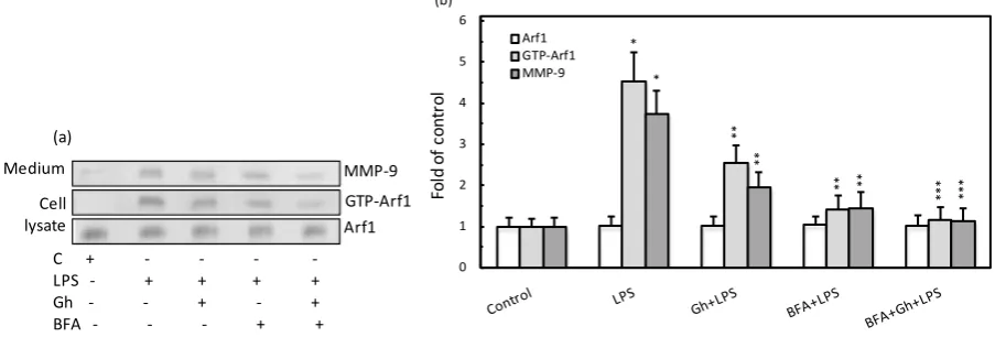
(a) Medium Cell lysate MMP-9 GTP-Arf1 Arf1
C + - - -
-LPS - + + + +
Gh - - + - +
BFA - - - + +
0 1 2 3 4 5 6 Arf1 GTP-Arf1 MMP-9 Fol d of c on tr ol * * ** ** ** ** *** *** (b)
Figure 3. Influence of ER-to-Golgi transport inhibitor, Brefeldin A (BFA), on the changes induced by P. gingivalis LPS and
ghrelin (Gh) in Arf1 activation in the salivary gland acinar cells. The cells, preincubated with 5 µg/ml BFA, were treated with 0.5 µg/ml Gh, and incubated with the LPS at 100 ng/ml. After 2 h of incubation the cell lysates were analyzed for Arf1 and Arf1-GTP, whereas the conditioned media following16 h of incubation were subjected to immunoblotting for MMP-9 (a). The relative levels of protein are expressed as fold of control (b). The data represent the mean ± SD of four experiments. *
P < 0.05 compared with that of control. *P < 0.05 compared with that of control. **P < 0.05 compared with that of LPS. ***P
< 0.05 compared with that of Gh + LPS.
the PKD2 was subject to interference by BFA.
[image:6.595.87.539.334.487.2]0 1 2 3 4 5
Ar
f-G
EF
ac
tiv
ity
(f
old
o
f c
on
tr
[image:7.595.143.537.79.230.2]ol)
Figure 4. Effect of ghrelin (Gh) on P. gingivalis LPS-induced changes in sublingual salivary gland acinar cell expression of
Arf-GEF activity (GTP-Arf1). The cells, preincubated with 5 µg/ml of Brefeldin A (BFA), 30 µM of SFK-PTKs inhibitor, PP2 or 5 µM of wide spectrum PKC inhibitor, GF109203X (GF), were treated with 0.5 µg/ml Gh, and incubated for 2 h in the presence of 100 ng/ml LPS. The data represent the mean ± SD of four experiments. *P < 0.05 compared with that of control. *P < 0.05 compared with that of control. **P < 0.05 compared with that of LPS. ***P < 0.05 compared with that of Gh + LPS.
(a) IP:Arf1
WB:PKD1 WB:PKD2 WB:Arf1
C + - - -
-LPS - + + + +
Gh - - + - +
BFA - - - + +
0 0.4 0.8 1.2
1.6 Arf1PKD2 PKD1
Fol
d of
c
on
tr
ol
(b)
*
*
Figure 5. Impact of Brefeldin A (BFA) on the association of Arf1 with PKD1 and PKD2 induced in the acinar cells by P.
gingivalis LPS and ghrelin (Gh). The cells, preincubated with 5 µg/ml BFA, were treated with 0.5 µg/ml Gh, and incubated
for 2 h in the presence of 100 ng/ml LPS. Cell lysates were immunoprecipitated (IP) with anti-Arf1 antibody and imm- unoblotted (WB) with anti-PKD1, and anti-PKD2 antibody (a). The relative densities of proteins are expressed as fold of Arf1 control (b). The data represent the mean ± SD of four experiments. *P < 0.05 compared with that of control.
association of PKD2 with Src, on the other hand was dependent on PKD2 phosphorylation on Tyr, and dis-played susceptibility to SFK-PTKs inhibitor, PP2. Together, these data suggest that PKCδ-mediated phosphory-lation of PKD2 on Ser plays a major role in the LPS-induced up-reguphosphory-lation in MMP-9 secretion, while the SFK- PTKs-mediated phosphorylation of PKD2 on Tyr reflects the modulatory influence on the acinar cell MMP-9 secretion by ghrelin.
4. Discussion
[image:7.595.90.539.313.490.2](a)
_____
Golgi Cell lysate pTyr pPKD2 PKD2 PKD2 C LPS Gh+LPS0 1 2 3 4 5
Control LPS Gh+LPS
[image:8.595.83.539.83.245.2]PKD2 pSer pTyr Fol d of c on tr ol * * * ** ** (b)
Figure 6. Effect P. gingivalis LPS and ghrelin (Gh) on PKD2 phosphorylation in the salivary gland acinar cells. The cells
were preincubated with 0 or 5 µg/ml Gh and incubated for 2h in the presence of 100 ng/ml LPS. The lysates of whole cells and the corresponding Golgi membrane fractions were analyzed for PKD2, phospho-PKD2 (Ser), and phosphotyrosine (pTyr) with specific antibodies (a). The relative densities of proteins are expressed as fold of control (b). The data represent the means ± SD of four experiments. *P < 0.05 compared with that of control. **P < 0.05 compared with that of LPS.
(a)
IP:PKD2
WB:PKCδ
WB:PKD2
C + - - -
-LPS - + + + +
Gh - - + - +
GF - - - + +
0 0.6 1.2 1.8 0 0.6 1.2 1.8 PKD2 PKCd PK D 2 (f ol d of c on tr ol ) PK C δ (f ol d of c on tr ol ) * ** ** ** (b) (c) IP:PKD2 WB:Src WB:PKD2
C + - - -
-LPS - + + + +
Gh - - + - +
PP2 - - - + +
0 0.6 1.2 1.8 0 0.6 1.2 1.8 PKD2 Src PK D 2 (f ol d of c on tr ol ) Sr c ( fol d of c on tr ol ) ** * *** (d) ***
Figure 7. Effect of P. gingivalis LPS and ghrelin (Gh) on the association of PKD2 with PKCδ in the salivary gland acinar
[image:8.595.89.538.316.641.2]LPS along with the elevated content of MMP-9, routinely detected in the circulation of patients with periodonti-tis, may be responsible for a decrease in cardiac function and cardiovascular disease [3][33]. Moreover, the li-terature data on the course of inflammatory responses associated with progressive hearth failure as well as the spinal cord injury suggest that up-regulation in MMP-9 secretion is subject to suppression by a peptide hormone, ghrelin [34][40]. Hence, in the present study we investigated the nature of factors implicated in the modulatory influence of ghrelin on P. gingivalis LPS-induced up-regulation in salivary gland acinar cell MMP-9 secretion.
Employing zymography and Western blot analyses; our results revealed that incubation of sublingual salivary gland acinar cells with the LPS elicited a marked enhancement in MMP-9 secretion, while the effect of ghrelin was reflected in a significant reduction in the LPS effect. Furthermore, we found that the stimulatory effect of the LPS on MMP-9 secretion was susceptible to suppression by a well-known ER-to-Golgi secretory cargo inhi-bitor, Brefeldin A [41], which also produced an additive result on the inhibitory effect of ghrelin. Taking into consideration that MMP-9 is a highly glycosylated protein comprising mainly of O-glycans, and the fact that N-glycosylation is a co-translational event taking place in the ER, while the O-glycosylation occurs later in the Golgi and TGN [9][10], the above findings attest to the modulatory influence of ghrelin on the rate of MMP-9 elaboration at the level of ER-to-Golgi processing.
Moreover, relying on the literature evidence as to the role of cytoskeleton MT stabilization in the secretory vesicle trafficking [11][15][16], we assessed the impact of the LPS and ghrelin on the acinar cell secretion of MMP-9 in the presence of pharmacological agents affecting MT stability. We observed that the LPS elicited in-duction in MMP-9 secretion was susceptible to suppression by MT destabilizing agent, nocodazole, which also produced further amplification in the countering effect of ghrelin, while preincubation with MT stabilizing agent, tubacin, led to the enhancement of the LPS-induced MMP-9 secretion and the inhibition in the countering effect of ghrelin. Our findings are thus in keeping with the data indicating that microtubule stabilization is not only of paramount importance to cell shape, motility and division, but also plays a major role in MMP-9 secretion
[15]-[18]. Furthermore, as the extent of MT stabilization has been directly linked to the degree of its tubulin subunit acetylation on Lys40[11][15], we have monitored the impact of P. gingivalis LPS and ghrelin on the level of acetylated α-tubulin in the acinar cells. A significant increase in the acetylated α-tubulin level was ob-served following the acinar cell stimulation with the LPS and MT stabilizing agent, tubacin, while the effect of ghrelin and MT destabilizing agent, nocodazole, was reflected in a marked reduction in the LPS-induced α-tu- bulin acetylation. Accordingly, we concluded that P. gingivalis LPS as well as ghrelin exert their influence on the acinar cell secretion of MMP-9 by affecting MT stabilization through α-tubulin acetylation. In this connec-tion, it is pertinent to note that MT α-tubulin acetylation is reversible [11][42], and hence presents a tempting target for therapeutic intervention in the secretory processes of MMP-9.
In further assessment of the role of ghrelin in modulation of pathways utilized by P. gingivalis LPS to en-hance the acinar cell secretion of MMP-9, we have turned our attention to factors that affect trafficking and sorting of secretory cargo through the Golgi-TGN, namely Arf and PKD [12]-[14]. Following the literature leads as to the involvement of Arf1 in controlling the Golgi-TGN cargo trafficking through GDP to GTP-dependent activation [14], we have identified Arf1 in the acinar cells and demonstrated that the elevation in Arf1-GTP formation upon the LPS-stimulation paralleled that of the rise in MMP-9 secretion, while the effect of ghrelin was reflected in the reduction of the LPS-induced Arf1-GTP formation as well as MMP-9 secretion. Further-more, as the activation status of Arfs is controlled by GEFs [19][20], we have assessed the requirements for the acinar cell Arf-GEF activity and found that the effect of the LPS was manifested by a significant increase in the Arf-GEF activation, whereas preincubation with ghrelin led to the reduction in the LPS effect. Moreover, the ac-tivation of Arf-GEF by the LPS was susceptible to suppression not only by Brefeldin A, but also to the inhibi-tors of PKCd and SFK-PTKs. The sensitivity to Brefeldin A indicates that the acinar cell Arf1-GEF belongs to the class of high (>100 kDa) molecular weight GEFs [14], while the susceptibility to the inhibition by PP2 and GF109203X suggests that the process of the acinar cell Arf1-GEF activation, like that of Rac1-GEF, Dock180
[35], occurs with the involvement of Src/PKCδ-dependent phosphorylation.
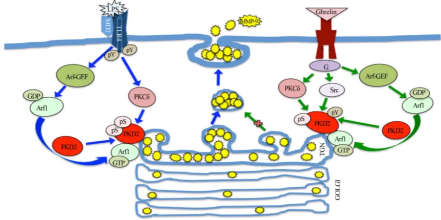
Figure 8. Schematic diagram of the regulatory role of Arf1/PKD2 in mediation of salivary gland acinar cell MMP-9 secretion in response to P. gingivalis LPS stimulation, and the modulatory influence of ghrelin. Engagement of TLR4 by the LPS triggers Arf-GEF-mediated Arf activation and its translocation to the Golgi. This promotes the localization of PKD2 with Arf1-GTP at the TGN, and its activation by phosphorylation on Ser by the PKCδ. The activated PKD2, by acting on the TGN-localized substrates, affects the vesicle fission events and thus enhances the secretion of MMP-9. Ligation by ghrelin ofGHS-R1a, on the other hand, leads to Arf-GEF-mediated Arf1 activation and PKD2-to-TGN localization, followed by the SFK-PTKs-mediated phosphorylation of PKD2 on Tyr that maintains the PKD2 activity at its normal regulatory level. G heterotrimeric G protein, pS phosphoserine, pY phosphotyrosine.
MMP-9 secretion, we examined the effect of the LPS and ghrelin on the recruitment and phosphorylation of PKD2 in the Golgi. Taking into account evidence as to the PKDs activation through phosphorylation on Ser as well as Tyr residues [12][25], we demonstrated that the effect of the LPS was manifested by a marked elevation in the Golgi membrane content of PKD2, and its phosphorylation on Ser and some Tyr, while preincubation with ghrelin evoked to a significant increase in PKD2 phosphorylation on Tyr and was accompanied by a marked decrease in the phosphorylation on Ser. Moreover, employing co-immunoprecipitation analysis in con-junction with pharmacological inhibitors we have established that the LPS-induced up-regulation in the acinar cell MMP-9 secretion is associated with PKCδ-mediated PKD2 phosphorylation on Ser, while the modulatory influence of ghrelin is reflected in the SFK-PTKs-dependent phosphorylation of PKD2 on Tyr.
Thus it is becoming increasingly apparent that PKC along with PKD form a key convergence and integration node for signals triggered by TLR activation as well as those arising through GPRC stimulation. Indeed, PKD/ PKC axis is not only the signaling target of LPS-induced TLR4 activation that defines the extent of inflam-matory involvement, but also plays a major role in modulation of inflammation by ghrelin, an endogenous li-gand of GPCR. The schematic diagram of the regulatory role of Arf1/PKD2 in mediation of salivary gland acinar cell MMP-9 secretion in response to P. gingivalis LPS and the modulatory influence of ghrelin is de-picted in Figure 8.
5. Conclusion
[image:10.595.86.537.88.313.2]References
[1] Nonnenmacher, C., Mutters, R. and de Jacoby, L.F. (2001) Microbiological Characteristics of Subgingival Microbiota in Adult Periodontitis, Localized Juvenile Periodontitis and Rapidly Progressive Periodontitis Subjects. Clinical
Mi-crobiology and Infection, 7, 213-221. http://dx.doi.org/10.1046/j.1469-0691.2001.00210.x
[2] Colombo, A.P., Boches, S.K. and Cotton, S.L. (2009) Comparisons of Subgingival Microbial Profiles of Refractory Periodontitis, Severe Periodontitis, and Periodontal Health Using the Human Oral Microbe Identification Microarray.
Journal of Periodontology, 80, 1421-1432. http://dx.doi.org/10.1902/jop.2009.090185
[3] Mysak, J., Podzimek, S., Sommerova, P., et al. (2014) Porphyromonas gingivalis: Major Periodontopathic Pathogen Overview. Journal of Immunology Research, 2014, Article ID: 476068. http://dx.doi.org/10.1155/2014/476068
[4] Slomiany, B.L. and Slomiany, A. (2011) Ghrelin-Induced cSrc Activation through Constitutive Nitric Oxide Synthase- Dependent S-Nitrosylation in Modulation of Salivary Gland Acinar Cell Inflammatory Responses to Porphyromonas
gingivalis. American Journal of Molecular Biology, 2, 43-51. http://dx.doi.org/10.4236/ajmb.2011.12006
[5] Slomiany, B.L. and Slomiany, A. (2015) Porphyromonas gingivalis-Stimulated TACE Activation for TGF-α Ectodo-mian Shedding and EGFR Transactivation in Salivary Gland Cells Requires Rac1-Dependent p38 MAPK Membrane Localization. Journal of Biosciences and Medicines, 3, 42-53. http://dx.doi.org/10.4236/jbm.2015.311005
[6] Ejeil, A.L., Igondio-Tchen, S., Ghomrasseni, S., Pellat, B., Godeau, G. and Gogly, B. (2003) Expression of Matrix Metalloproteinases (MMPs) and Tissue Inhibitors of Metalloproteinases (TIMPs) in Healthy and Diseased Human Gingiva. Journal of Periodontology, 74, 188-195. http://dx.doi.org/10.1902/jop.2003.74.2.188
[7] Jotwani, R., Eswaran, S.V.K., Moonga, S. and Cutler, C.W. (2010) MMP-9/TIMP-1 Imballance Induced in Human Dendritic Cells by Porphyromonas gingivalis. FEMS Immunology and Medical Microbiology, 50, 314-321.
http://dx.doi.org/10.1111/j.1574-695X.2009.00637.x
[8] Slomiany, B.L. and Slomiany, A. (2016) Role of Rac1/p38 and ERK-Dependent Cytosolic Phospholipase A2 Activa-tion in Porphyromonas gingivalis-Evoked Induction in Matrix Metalloproteinase-9 (MMP-9) Release by Salivary Gland Cells. Journal of Biosciences and Medicines, 4, 68-79. http://dx.doi.org/10.4236/jbm.2016.44010
[9] Van den Steen, P.E., Van Aelst, I., Hvidberg, V., et al. (2006) The Hemopexin and O-Glycosylated Domains Tune Gelatinase B/MMP-9 Bioavailability via Inhibition and Binding to Cargo Receptors. Journal of Biological Chemistry, 281,18626-18637. http://dx.doi.org/10.1074/jbc.M512308200
[10] Vandooren, J., Van den Steen, P.E. and Opdenakker, G. (2013) Biochemistry and Molecular Biology of Gelatinase B or Matrix Metalloproteinase-9 (MMP-9): The Next Decade. Critical Reviews in Biochemistry and Molecular Biology, 48, 222-272. http://dx.doi.org/10.3109/10409238.2013.770819
[11] Howes, S.C., Alushin, G.M., Shida, T., Nachury, M.V. and Nogales, E. (2014) Effects of Tubulin Acetylation and Tu-bulin Acetyltransferase Binding on Microtubule Structure. Molecular Biology of the Cell, 25, 257-266.
http://dx.doi.org/10.1091/mbc.E13-07-0387
[12] Rozengurt, E. (2011) Protein Kinase D Signaling: Multiple Biological Functions in Health and Disease. Physiology, 26, 23-33. http://dx.doi.org/10.1152/physiol.00037.2010
[13] Bonnemaison, M.L., Eipper, B.A. and Mains, R.E. (2013) Role of Adaptor Proteins in Secretory Granule Biogenesis and Maturation. Frontiers in Endocrinology, 4, Article 101. http://dx.doi.org/10.3389/fendo.2013.00101
[14] Bourgoin, S.G. and El Azreq, M.A. (2012) Small Inhibitors of ADP-ribosylation Factor Activation and Function in Mammalian Cells. World Journal of Pharmacology, 1, 55-64. http://dx.doi.org/10.5497/wjp.v1.i4.55
[15] Hanania, R., Sun, H.S., Xu, K., et al. (2012) Classically Activated Macrophages Use Stable Microtubules for Matrix Metalloproteinase-9 (MMP-9) Secretion. Journal of Biological Chemistry, 287, 8468-8483.
http://dx.doi.org/10.1074/jbc.M111.290676
[16] Goode, B.L., Drubin, D.G. and Barnes, G. (2000) Functional Cooperation between the Microtubule and Actin in Cy-toskeletons. Current Opinions in Cell Biology, 12, 63-71. http://dx.doi.org/10.1016/S0955-0674(99)00058-7
[17] Gu, S., Liu, Y., Zhu, B., et al. (2016) Loss of α-Tubulin Acetylation Is Associated with TGF-Beta-Induced Epithelial- Mesenchymal Transition. Journal of Biological Chemistry, 291, 5396-5405.
http://dx.doi.org/10.1074/jbc.M115.713123
[18] Sbai, O., Ould-Yahoui, A., Ferhart, I., et al. (2010) Differential Vesicular Distribution and Trafficking of MMP-2, MMP-9, and Their Inhibitors in Astrocytes. Glia, 58, 344-366.
[19] Donaldson, J.G. and Jackson, C.L. (2011) ARF Family G Proteins and Their Regulators: Roles in Membrane Transport, Development and Disease. Nature Reviews Molecular Cell Biology, 12, 362-375. http://dx.doi.org/10.1038/nrm3117
[20] Cherfils, J. and Zeghouf, M. (2013) Regulation of Small GTPases by GEFs, GAPs, and GDIs. Physiological Reviews, 93, 269-309. http://dx.doi.org/10.1152/physrev.00003.2012
Pro-tein Kinase D2 Interaction with ADP-Ribosylation Factor 1, Transgolgi-Golgi Network Recruitment, and ProPro-tein Transport. Molecular Biology of the Cell, 21, 1011-1022. http://dx.doi.org/10.1091/mbc.E09-09-0814
[22] Wille, C. Kohler, C., Armacki, M., et al. (2014) Protein Kinase D2 Induces Invasion of Pancreatic Cancer Cells by Regulating Matrix Metalloproteinases. Molecular Biology of the Cell, 25, 324-336.
http://dx.doi.org/10.1091/mbc.E13-06-0334
[23] Eiseler, T., Wille, C., Koehler, C., Illing, A. and Seufferlein, T. (2016) Protein Kinase D2 Assembles a Multiprotein Complex at the Trans-Gologi Network to Regulate Matrix Metalloproteinase Secretion. Journal of Biological Chemi-stry, 291, 462-477. http://dx.doi.org/10.1074/jbc.M115.673582
[24] Waldron, R.T. and Rosengurt, E. (2003) Protein Kinase C Phosphorylates Protein Kinase D Activation Loop Ser744 and Ser748 and Releases Autoinhibition by the Pleckstrin Homology Domain. Journal of Biological Chemistry, 278, 154-163. http://dx.doi.org/10.1074/jbc.M208075200
[25] Doppler, H. and Storz, P. (2007) A Novel Tyrosine Phosphorylation Site in Protein Kinase D Contributes to Oxidative Stress-Mediated Activation. Journal of Biological Chemistry, 282,31873-31881.
http://dx.doi.org/10.1074/jbc.M703584200
[26] Ivison, S.M., Graham, N.R., Bernales, C.Q., et al. (2007) Protein Kinase D Interaction with TLR5 Is Required for In-flammatory Signal in Response to Bacterial Flagellin. Journal of Immunology, 178, 5735-5743.
http://dx.doi.org/10.4049/jimmunol.178.9.5735
[27] Slomiany, B.L. and Slomiany, A. (2011) Role of Ghrelin-Induced cSrc Activation in Modulation of Gastric Mucosal Inflammatory Responses to Helicobacter pylori. Inflammopharmacology, 19, 197-204.
[28] Lodeiro, M., Alen, B.O., Mosteiro, C.S., et al. (2011) The SHP-1 Protein Tyrosine Phosphatase Negatively Modulates Akt Signaling in the Ghrelin/GHSR1a System. Molecular Biology of the Cell, 22, 4182-4191.
http://dx.doi.org/10.1091/mbc.E11-04-0373
[29] Kojima, M., Hosoda, H., Date, Y., Nakazato,M., Matsuo, H. and Kangawa, K. (1999) Ghrelin Is a Growth-Hormone- Releasing Acetylated Peptide from Stomach. Nature, 402, 656-660. http://dx.doi.org/10.1038/45230
[30] Groschl, M., Topf, H.G., Bohlender, J., et al. (2005) Identification of Ghrelin in Human Saliva: Production by Salivary Glands and Potential Role in Proliferation of Oral Keratinocytes. Clinical Chemistry, 51, 997-1006.
http://dx.doi.org/10.1373/clinchem.2004.040667
[31] Slomiany, B.L. and Slomiany, A. (2010) Suppression by Ghrelin of Porphyromonas gingivalis-Induced Constitutive Nitric Oxide Synthase S-Nitrosylation and Apoptosis in Salivary Gland Acinar Cells. Journal of Signal Transduction, 2010, Article ID: 643642. http://dx.doi.org/10.1155/2010/643642
[32] Slomiany, B.L. and Slomiany, A. (2011) Ghrelin Protects against the Detrimental Consequences of Porphyromonas
gingivalis-Induced Akt Inactivation through S-Nitrosylation on Salivary Mucin Synthesis. International Journal of
In-flammation, 2011, Article ID: 807279. http://dx.doi.org/10.1007/s10787-014-0206-z
[33] Beck, J., Garcia, R., Heiss, G., Vokonas, P.J. and Offenbacher, S. (1996) Periodontal Disease and Cardiovascular Dis-ease. Journal of Periodontology, 67, 1123-1137. http://dx.doi.org/10.1902/jop.1996.67.10s.1123
[34] Huang, C.X., Yuan, M.J., Huang, H., et al.(2009) Ghrelin Inhibits Post-Infarct Myocardial Remodeling and Improves Cardiac Function through Anti-Inflammatory Effect. Peptides, 30, 2286-2291.
http://dx.doi.org/10.1016/j.peptides.2009.09.004
[35] Slomiany, B.L. and Slomiany, A. (2015) Porphyromonas gingivalis-Induced GEF Dock180 Activation by Src/PKCδ- Dependent Phosphorylation Mediates PLCγ2 Amplification in Salivary Gland Acinar Cells: Effect of Ghrelin. Journal
of Biosciences and Medicines, 3, 66-77. http://dx.doi.org/10.4236/jbm.2015.37008
[36] Slomiany, A., Grabska, M., Slomiany, B.A., Grzelinska, E., Morita, M. and Slomiany, B.L. (1993) Intracellular Tran- sport, Organelle Biogenesis and Establishment of Golgi Identity: Impact of Brefeldin a on the Activity of Lipid Syn-thesizing Enzymes. International Journal of Biochemistry, 25, 891-901.
http://dx.doi.org/10.1016/0020-711X(93)90245-A
[37] Slomiany, B.L., Murty, V.L.N., Piotrowski, J. and Slomiany, A. (1996) Salivary Mucins in Oral Mucosal Defense.
General Pharmacology, 27, 761-771. http://dx.doi.org/10.1016/0306-3623(95)02050-0
[38] Slomiany, B.L. and Slomiany, A. (2004) Platelet-Activating Factor Mediates Porphyromonas gingivalis Lipopolysac-charide Interference with Salivary Mucin Synthesis via Phosphatidylinositol 3-Kinase-Dependent Constitutive Nitric Oxide Synthase Activation. Journal of Physiology and Pharmacology, 55, 85-98.
[39] Slomiany, B.L. and Slomiany, A. (2014) Modulation of Gastric Mucosal Inflammatory Responses to Helicobacter
py-lori via Ghrelin-Induced Protein Kinase Cδ Tyrosine Phosphorylation. Inflammopharmacology, 22, 251-262.
http://dx.doi.org/10.1007/s10787-014-0206-z
Acta, 1842, 2403-2412. http://dx.doi.org/10.1016/j.bbadis.2014.09.006
[41] Ivessa, N.E., De Lemos-Chiarandini, C., Gravotta, D., Sabatini, D.D. and Kreibich, G. (1995) The Brefeldin A-induced Retrograde Transport from the Golgi Apparatus to the Endoplasmic Reticulum Depends on Calcium Sequestered to Intracellular Stores. Journal of Biological Chemistry, 270, 25960-25967. http://dx.doi.org/10.1074/jbc.270.43.25960
[42] Hubert, C., Guardiola, A., Shao, R., et al.(2002) HDAC6 Is a Microtubule-Associated Deacylase. Nature, 417, 455- 458. http://dx.doi.org/10.1038/417455a
[43] Hauser, A., Storz, P., Martens, S., Link, G., Toker, A. and Pfizenmaier, K. (2005) Protein Kinase D Regulates Vesicu-lar Transport by Phosphorylating and Activating Phosphatidylinositol-4-Kinase IIIBeta at the Golgi Complex. Nature
Cell Biology, 7, 880-886. http://dx.doi.org/10.1038/ncb1289