R E S E A R C H A R T I C L E
Open Access
Renal expression and serum levels of high
mobility group box 1 protein in lupus nephritis
Agneta Zickert
1*, Karin Palmblad
2, Birgitta Sundelin
3, Sangeeta Chavan
4, Kevin J Tracey
4, Annette Bruchfeld
5and
Iva Gunnarsson
1Abstract
Introduction:High mobility group box 1 protein (HMGB1) is a nuclear DNA binding protein acting as a pro-inflammatory mediator following extracellular release. HMGB1 has been increasingly recognized as a pathogenic mediator in several inflammatory diseases. Elevated serum levels of HMGB1 have been detected in autoimmune diseases including Systemic lupus erythematosus (SLE). However, the local expression of HMGB1 in active lupus nephritis (LN) is not known. Here we aimed to study both tissue expression and serum levels of HMGB1 in LN patients with active disease and after induction therapy.
Methods:Thirty-five patients with active LN were included. Renal biopsies were performed at baseline and after standard induction therapy; corticosteroids combined with immunosuppressive drugs. The biopsies were evaluated according to the World Health Organization (WHO) classification and renal disease activity was estimated using the British Isles lupus assessment group (BILAG) index. Serum levels of HMGB1 were analysed by western blot. HMGB1 expression in renal tissue was assessed by immunohistochemistry at baseline and follow-up biopsies in 25 patients. Results:Baseline biopsies showed WHO class III, IV or V and all patients had high renal disease activity (BILAG A/B). Follow-up biopsies showed WHO I to II (n= 14), III (n= 6), IV (n= 3) or V (n= 12), and 15/35 patients were regarded as renal responders (BILAG C/D).
At baseline HMGB1 was significantly elevated in serum compared to healthy controls (P < 0.0001). In all patients, serum levels decreased only slightly; however, in patients with baseline WHO class IV a significant decrease was observed (P = 0.03). Immunostaining revealed a pronounced extranuclear HMGB1 expression predominantly outlining the glomerular endothelium and in the mesangium. There was no clear difference in HMGB1 expression comparing baseline and follow-up biopsies or any apparent association to histopathological classification or clinical outcome.
Conclusions:Renal tissue expression and serum levels of HMGB1 were increased in LN. The lack of decrease of HMGB1 in serum and tissue after immunosuppressive therapy in the current study may reflect persistent inflammatory activity. This study clearly indicates a role for HMGB1 in LN.
Introduction
Systemic lupus erythematosus (SLE) is a chronic inflam-matory autoimmune disease characterized by multiple organ involvement, production of autoantibodies to nuclear components, and immune complex deposition [1]. Lupus nephritis (LN) is regarded as one of the most severe organ manifestations of SLE, affecting approxi-mately 35% to 50% of patients with lupus. Despite
increased knowledge of pathogenesis and improved treatment regimens, LN remains a main cause of mor-bidity among patients with SLE [2].
High-mobility group box 1 protein (HMGB1), a nuclear protein found in all mammalian cells, is known as a DNA-binding protein participating in chromatin structure and transcriptional regulation [3,4]. Extracellu-lar HMGB1 has been identified as a proinflammatory mediator and, owing to its proinflammatory and immu-nostimulatory properties, has been proposed to contri-bute to the pathogenesis of multiple chronic inflammatory and autoimmune diseases [5-8]. HMGB1
* Correspondence: [email protected]
1
Department of Medicine, Unit of Rheumatology, D2:00 Solna, Karolinska University Hospital, Karolinska Institute, S-171 76 Stockholm, Sweden Full list of author information is available at the end of the article
is actively secreted from activated immune cells such as macrophages and monocytes and is passively released from injured or necrotic cells. When translocated from the nucleus to the extracellular milieu, HMGB1 can act as an ‘alarmin’, a danger signal that can activate the immune system and has been demonstrated as a key factor in necrosis-induced inflammation [9,10]. More-over, HMGB1 induces other cytokines such as tumor necrosis factor and interleukin-1 (IL-1), IL-6, and IL-8 and is an activator of endothelial cells leading to the upregulation of adhesion molecules [11,12].
Elevated serum levels of HMGB1 have been found in different inflammatory conditions such as sepsis [13], rheumatoid arthritis [14,15], anti-neutrophilic cytoplas-matic antibody (ANCA)-associated vasculitis [16], and chronic kidney disease [17] as well as in SLE [18-20]. Previous studies have shown increased HMGB1 expres-sion in skin leexpres-sions of patients with SLE [21], thus indi-cating that HMGB1 may be an important mediator of inflammation in target organs in lupus.
Interestingly, increased levels of HMGB1 were recently demonstrated in patients with active LN in comparison with patients with active non-renal disease [22]. How-ever, specified data on the severity of renal disease, his-topathological findings, or tissue expression were not included in that study. The aim of this study was to investigate renal tissue expression and serum levels of HMGB1 in correlation not only with renal histopatholo-gical and clinical activity but also with response to ther-apy in order to further investigate its role in patients with LN.
Materials and methods
Patients
Thirty-five patients with SLE and biopsy-proven active LN during the period of 1996 to 2008 were included in this study. All patients fulfilled the 1982 American Col-lege of Rheumatology classification criteria for SLE [23] and participated in a prospective control program for LN at the rheumatology clinic of Karolinska University Hospital in Stockholm, Sweden. Thirty of the 35 patients were female (86%) and five were men (14%), and the mean age was 34 years (range of 18 to 50). The patients were treated, in accordance with standard ther-apy for LN, with corticosteroids combined with
cyclo-phosphamide (n= 21), mycophenolate mofetil (n= 8),
or rituximab (n= 5), and one patient was treated with
azathioprin. After induction therapy for a mean duration of 8 months (range of 6 to 14), all patients underwent a second renal biopsy. At both biopsies, clinical data and blood and urinary samples were collected. Serum sam-ples were frozen at -70°C for future analyses. As a con-trol group for serum levels of HMGB1, blood samples from 48 healthy controls were used [17]. The clinical
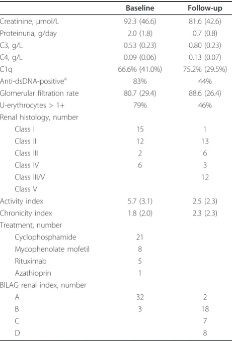
characteristics of patients and nephritis data at baseline and follow-up are presented in Table 1.
Evaluation of renal function, histopathology, and renal activity
Renal evaluation at the time point of the first and sec-ond biopsies included urine analyses (dipslide proce-dure), urinary sediment assessment, and investigation of 24-hour urine albumin excretion. Renal function was determined by serum creatinine levels (expressed as micromoles per liter) and estimated glomerular filtration rate (GFR) by using the modification in diet in renal dis-ease (MDRD) formula [24].
[image:2.595.305.536.365.705.2]Renal biopsies were performed by percutaneous ultra-sonography-guided puncture. The renal tissue obtained was evaluated by light microscopy, immunofluorescence, and electron microscopy. The biopsies were graded according to the World Health Organization (WHO) classification of nephritis [25] and additionally scored for activity and chronicity indices [26]. Renal tissue
Table 1 Clinical characteristics of all patients at baseline and follow-up
Baseline Follow-up
Creatinine,μmol/L 92.3 (46.6) 81.6 (42.6) Proteinuria, g/day 2.0 (1.8) 0.7 (0.8)
C3, g/L 0.53 (0.23) 0.80 (0.23)
C4, g/L 0.09 (0.06) 0.13 (0.07)
C1q 66.6% (41.0%) 75.2% (29.5%)
Anti-dsDNA-positivea 83% 44%
Glomerular filtration rate 80.7 (29.4) 88.6 (26.4)
U-erythrocytes > 1+ 79% 46%
Renal histology, number
Class I 15 1
Class II 12 13
Class III 2 6
Class IV 6 3
Class III/V 12
Class V
Activity index 5.7 (3.1) 2.5 (2.3) Chronicity index 1.8 (2.0) 2.3 (2.3) Treatment, number
Cyclophosphamide 21
Mycophenolate mofetil 8
Rituximab 5
Azathioprin 1
BILAG renal index, number
A 32 2
B 3 18
C 7
D 8
Values are presented as mean (standard deviation) unless otherwise indicated.
a
from an unaffected part of a kidney that was nephrecto-mized owing to carcinoma was used as control. Evalua-tion of renal activity was estimated by using the British Isles lupus assessment group (BILAG) index [27,28]. Patients with renal BILAG C or D at follow-up were regarded as responders (as previously suggested [29]).
Serology and complement measures
Assessments of serum IgG anti-double-stranded DNA (anti-dsDNA) antibodies were carried out by
immuno-fluorescence microscopy by usingCrithidiae luciliaeas
a source of antigen. Analyses of complement component C1q were performed by rocket electrophoresis by using rabbit anti-C1q as the antibody. Levels of C1q were expressed as the percentage of the levels of healthy blood donors (normal range of 76% to 136%). C3 (nor-mal range of 0.67 to 1.43 g/L) and C4 (nor(nor-mal range of 0.12 to 0.32 g/L) levels were determined by nephelometry.
HMGB1 serum determination
HMGB1 was analyzed by Western blot in sera from 20 of the patients at baseline and at repeat biopsy by a method previously described [17].
Immunohistochemical staining of renal biopsies
Immunohistochemical stainings of HMGB1 expression were performed on formaldehyde-fixed paraffin-embedded serial 4-μm sections of renal biopsies. Slides were deparaffinized in xylene and rehydrated with etha-nol. Antigen retrieval treatment prior to staining was omitted to allow clearer visualization of extranuclear HMGB1. To block endogenous peroxidase activity,
sec-tions were treated with 3% H2O2 followed by a serum
block with 2% human AB sera for 30 minutes and an avidin/biotin-blocking step (Vector Laboratories Inc., Burlingame, CA, USA). The slides were thereafter incu-bated overnight with an affinity-purified monoclonal mouse IgG2b anti-HMGB1 antibody (concentration of
10 μg/mL, 2G7; Critical Therapeutics Inc., Lexington,
MA, USA). A biotin-labeled horse anti-mouse antibody (Vector Laboratories Inc.) containing 2% normal horse serum was used for detection. Stainings were developed by using a DAB kit (Vector Laboratories Inc.) in accor-dance with the instructions of the manufacturer.
Sec-tions were counterstained with Mayer’s hematoxylin.
Phosphate-buffered saline supplemented with 0.1% sapo-nin was used in all subsequent washes and incubation steps in order to permeabilize the cells. In each assay, controls for specificity of the HMGB1 staining were included on the basis of parallel staining studies omit-ting the primary antibody, and a primary isotype-matched immunoglobulin of irrelevant antigen specifi-city (negative mouse IgG2b control; Dako Cytomatation,
Glostrup, Denmark) was used. The specificities of extra-cellular and intraextra-cellular HMGB1 immunoreactivities were further verified by blocking experiments with pre-absorption of the HMGB1-specific antibody with recom-binant HMGB1 prior to staining.
Statistics
For comparisons of variables at baseline and at repeat biopsy, the non-parametric Wilcoxon matched pair test was used. For comparisons of variables between groups, the non-parametric Mann-Whitney test was used. Cor-relations were calculated by using Spearman’s rank
cor-relation. P values of less than 0.05 were considered
statistically significant. Statistical evaluation was made with statistical software (STATISTICA 9; StatSoft, Inc., Tulsa, OK, USA).
Ethics
Informed consent was obtained from all subjects, and the study protocol was approved by the regional ethics committee in Stockholm.
Results
Histopathology and renal activity
All patients had an active nephritis at baseline; biopsies showed WHO class III (n= 15), III/V (n= 2), IV (n=
12), or V (n = 6). All patients had high renal disease
activity; 32 out of 35 had renal BILAG A and three out
of 35 a BILAG B. Follow-up biopsies showed WHO I (n
= 1), II (n= 13), III (n= 6), IV (n= 3), or V (n= 12). Two out of 35 patients had renal BILAG A, 18 out of 35 BILAG B, seven out of 35 BILAG C, and eight out of 35 BILAG D. Fifteen patients were thus regarded as responders and 20 out of 35 were regarded as non-responders according to BILAG. The mean renal activity index decreased significantly from 5.7 to 2.5 (P< 0.001), whereas no significant findings regarding chronicity index were observed (data presented in Table 1).
HMGB1 serum determination and evaluation of renal activity
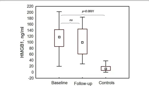
At baseline, HMGB1 was significantly elevated in the patient group (mean of 108.4 ± 48.0 ng/mL, median of 117.1, range of 19.8 to 202.4) in comparison with healthy controls (mean of 13 ± 10 ng/mL, median of 12.0, range of 0 to 38.0) (P< 0.0001) (Figure 1). Serum HMGB1 levels were significantly higher in the patients with WHO class III (mean of 133.8 ± 47.2 ng/mL, med-ian of 130.0, range of 29.6 to 202.0) in comparison with those with class IV (mean of 87.7 ± 39.1 ng/mL, median
of 87.7, range of 19.8 to 155.9) (P = 0.01). When all
183.9) (P value not significant), although the levels remained significantly high in comparison with those of the control group (P< 0.0001) (Figure 1). In the patients with WHO class III at baseline biopsy, no difference in HMGB1 levels was observed at follow-up (mean of 136.9 ± 42.9 ng/mL, median of 142.7, range of 40.3 to 183.9), whereas in class IV, a significant decrease was found (mean of 71.0 ± 35.6 ng/mL, median of 71.4, range of 27.9 to 145.9) (P= 0.03) (Figure 2).
The responder patients (BILAG C/D) had lower serum levels of HMGB1 at follow-up (mean of 93.7 ± 54.2 ng/ mL, median of 94.0, range of 27.9 to 181.5) versus those with persisting renal BILAG A or B (mean of 107.4 ± 49.0 ng/mL, median of 108.8, range of 40.3 to 183.9), but the difference was not statistically significant (Figure 3). Though not statistically significant, the serum levels
were higher at follow-up in the group of patients (n=
4) who developed membranous nephritis (WHO V) in comparison with other WHO classes at follow-up biopsy. There was an inverse correlation between activ-ity index at baseline biopsies and HMGB1 levels both at baseline (r = -0.61,P< 0.05) and at follow-up (r = -0.58,
P < 0.05). HMGB1 showed no significant correlation
with complement levels, creatinine, GFR, proteinuria, or anti-dsDNA antibody positivity (data not shown). Data
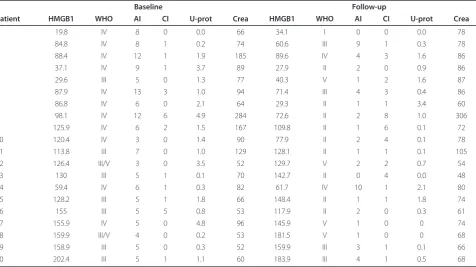
on serum levels of HMGB1, WHO classification, protei-nuria, and renal function are presented in Table 2.
Immunohistochemistry
Immunostaining revealed an extranuclear HMGB1 expression found in all of the examined biopsies from the patients with LN. The staining was predominantly found outlining the glomerular endothelium and was also documented in the mesangium. A less pronounced HMGB1 expression was documented in vessels and tub-ular cells (Figure 4). To investigate the amount of HMGB1 expression, an arbitrary scale was used for comparisons between biopsies. Five patients had unchanged amounts, eight had a decrease, and 12 had more abundant amounts of HMGB1 staining. Overall, no difference in the comparison of first and second biopsies was demonstrated (data not shown), and no clear correlation with histopathological classification or clinical outcome could be documented.
[image:4.595.58.537.88.365.2]HMGB1 is abundantly present in all cell nuclei, thus generating a strong expression that might interfere with visualization of a cytoplasmatic and extranuclear stain-ing, which most often is weaker. By omission of antigen retrieval treatment prior to staining, a clearer visualiza-tion of extranuclear HMGB1 expression is allowed. In
kidney biopsies from control renal tissue, HMGB1 expression was predominantly negative or, if present, was restricted to the cell nuclei (Figure 4).
Discussion
To the best of our knowledge, we are the first to demonstrate HMGB1 expression in affected renal tissue
[image:5.595.61.539.87.291.2]from LN patients, in whom we clearly show an increased expression in active disease as well as after immunosuppressive therapy. HMGB1 tissue stainings were predominantly found outlining the glomerular endothelium but also were expressed in the mesangium. In this study, we could also confirm the findings of ele-vated serum levels of HMGB1 in LN measured by
Figure 2Serum HMGB1 levels in different World Health Organization (WHO) classes. Serum levels at baseline and follow-up in lupus nephritis patients with WHO III (n= 9) versus IV (n= 11) are shown. After induction therapy, serum levels of HMGB1 were significantly
decreased in patients with WHO class IV. However, HMGB1 levels remained unchanged in patients with WHO class III. Data are expressed as means, and vertical bars denote 95% confidence intervals. HMGB1, high-mobility group box 1 protein.
[image:5.595.59.539.463.682.2]Western blot, as recently shown by Abdulahad and col-leagues [22].
We found a clear tissue staining for HMGB1 in LN and this staining was absent in non-lupus control renal tissue. There was no distinct difference in expression of HMGB1 either in the proliferative glomerular lesions or in sites with infiltrates of inflammatory cells in compari-son with less affected glomeruli, and the exact origin of the increased renal expression of HMGB1 is not fully clear. The staining outlining the glomerular endothelium could emanate from local extracellular release but also may reflect capture from circulating HMGB1. Thus, one may speculate that the findings of increased serum levels as well as tissue expression of HMGB1 reflect both systemic and local inflammation within the kidney. In glomeruli, the pronounced endothelial staining and the increased expression in the mesangium suggest a co-localization for HMGB1 and immune depositions in LN. However, further studies with other methodologies are required to address this issue.
Elevated HMGB1 expression has previously been demonstrated in lupus patients with cutaneous photoin-duced inflammation, in whom cytoplasmatic and extra-cellular HMGB1 were detected in biopsies from the most clinically active skin lesions in comparison with inactive or non-lesional skin [21]. That study also demonstrated that HMGB1 was still expressed in
persisting dermal infiltrates in a proportion of healing lesions, thus suggesting that HMGB1 might be a media-tor of both early and late inflammation and may partici-pate in the inflammatory process over longer periods of time.
[image:6.595.61.538.100.367.2]In a recent study by Bruchfeld and colleagues [16], increased serum levels and renal tissue expression of HMGB1 were demonstrated in ANCA-associated vascu-litis with renal involvement. In that study, high baseline serum levels of HMGB1 decreased significantly after treatment, and in contrast to our findings in LN, a more distinct histopathological response and a more distinct clinical response were demonstrated. These findings are consistent with other studies showing that patients with LN, even patients with quiescent clinical pictures, still may have histopathological inflammatory activity after induction treatment [30,31]. Of note, less than half of the patients in the present study were regarded as renal responders according to the definition used. Our obser-vation that serum HMGB1 remained significantly ele-vated combined with persistent expression in renal tissue at follow-up may reflect an ongoing inflammatory activity in patients with LN despite immunosuppressive therapy. The findings in patients with vasculitis are of particular interest since LN with a focal segmental glo-merular pattern (WHO class III), which had the highest HMGB1 levels in our study, has been considered to Table 2 Serum levels of HMGB1 in 20 patients
Baseline Follow-up
Patient HMGB1 WHO AI CI U-prot Crea HMGB1 WHO AI CI U-prot Crea
1 19.8 IV 8 0 0.0 66 34.1 I 0 0 0.0 78
2 84.8 IV 8 1 0.2 74 60.6 III 9 1 0.3 78
3 88.4 IV 12 1 1.9 185 89.6 IV 4 3 1.6 86
4 37.1 IV 9 1 3.7 89 27.9 II 2 0 0.9 86
5 29.6 III 5 0 1.3 77 40.3 V 1 2 1.6 87
6 87.9 IV 13 3 1.0 94 71.4 III 4 3 0.4 86
7 86.8 IV 6 0 2.1 64 29.3 II 1 1 3.4 60
8 98.1 IV 12 6 4.9 284 72.6 II 2 8 1.0 306
9 125.9 IV 6 2 1.5 167 109.8 II 1 6 0.1 72
10 120.4 IV 3 0 1.4 90 77.9 II 2 4 0.1 78
11 113.8 III 7 0 1.0 129 128.1 II 1 1 0.1 105
12 126.4 III/V 3 0 3.5 52 129.7 V 2 2 0.7 54
13 130 III 5 1 0.1 70 142.7 II 0 4 0.0 48
14 59.4 IV 6 1 0.3 82 61.7 IV 10 1 2.1 80
15 128.2 III 5 1 1.8 66 148.4 II 1 1 1.8 74
16 155 III 5 5 0.8 53 117.9 II 2 0 0.3 61
17 155.9 IV 5 0 4.8 96 145.9 V 1 0 0 74
18 159.9 III/V 4 0 0.2 53 181.5 V 1 0 0 68
19 158.9 III 5 0 0.3 52 159.9 III 3 1 0.1 66
20 202.4 III 5 1 1.1 60 183.9 III 4 1 0.5 68
B
A
C D
G H
E F
N
M
J
L
I
[image:7.595.52.545.88.629.2]K
have features and possibly immune mechanisms similar to those of lesions of systemic vasculitis [32] in compari-son with the more widespread global pattern character-ized by more pronounced immune depositions seen in patients with class IV. The difference observed in HMGB1 levels when focal and diffuse proliferative nephritis were compared may suggest differences in pathogenic mechanisms and deserves further studies.
In the study by Abdulahad and colleagues [22], a cor-relation between serum HMGB1 and proteinuria was shown in active SLE patients having some degree of proteinuria but was not confirmed in our study on patients with active biopsy-proven LN only. The two study populations may not be fully comparable, how-ever. Although we could confirm the finding of elevated HMGB1 levels in patients with LN as determined by Western blot, the methods used in the two studies may not be fully comparable (whole serum in the study by Abdulahad and colleagues [22] in comparison with low molecular serum components in the present study). This methodological issue may have contributed to the discrepancies regarding circulating HMGB1 and disease activity between the two studies. Further studies would be of interest to determine the optimal method for HMGB1 determination in patients with LN.
Although HMGB1 levels have been shown to correlate inversely with renal function in patients with chronic kidney disease [17], no such correlation was found in our study or in the previous study of patients with SLE [22]. HMGB1 has been proposed to be involved in the pathogenesis of SLE, and several biological properties of HMGB1 are currently of major interest in SLE research [8]. Inappropriate apoptosis is regarded as a key event in the pathogenesis of SLE. Nuclear autoantigens from not properly cleared apoptotic cells may be excessively pre-sented to the immune system, resulting in loss of self-tolerance and generation of nuclear autoantibodies [33]. HMGB1 released from apoptotic cells may therefore be of special interest in SLE. In primary apoptosis, there is almost no release of HMGB1; however, in conditions with inappropriate clearance of apoptotic material, apoptotic cells may undergo secondary necrosis leading to HMGB1 release [34,35]. HMGB1 has been demon-strated to bind to the chromatin of cells during apopto-sis and to remain bound to nucleosomes when released from apoptotic cells. Furthermore, HMGB1-nuclesome complexes have recently been demonstrated to induce the production of proinflammatory cytokines from macrophages and dendritic cells and to induce anti-dsDNA and anti-histone IgG responses and therefore may play a central role in breaking tolerance against nuclear antigens [36]. In that study, circulating HMGB1-nucleosome complexes were detected in serum from patients with SLE but not in healthy controls.
Previous studies have demonstrated high levels of anti-HMGB1 antibodies in patients with SLE [22,37]. This finding raises the possibility that the monoclonal anti-HMGB1 antibody used for tissue staining could have difficulties in binding to HMGB1 if the epitope was already occupied by deposited anti-HMGB1 antibo-dies. This could possibly contribute to underestimating the local renal expression of HMGB1 and, if so, could explain why there were no clear differences in HMGB1 expression in the most proliferative inflammatory lesions in comparison with less affected glomeruli.
Biomarkers available for assessing renal activity and classification, response to therapy, remission, and flares are insufficient in SLE and do not always reflect the actual inflammatory activity in the renal tissue [30,38]. Although renal biopsy is regarded as the‘gold standard’ for assessing renal activity [39], there is a need for new biomarkers for evaluation of disease activity in LN. In the present study, we were not able to determine whether serum levels of HMGB1 could be used for
monitoring of renal disease activity per se but found
persistently elevated levels of serum as well as tissue expression, suggesting that HMGB1 is an important inflammatory mediator in LN. To determine whether a more pronounced reduction of HMBG1 levels can be achieved in a less active phase of LN, more long-term studies with sequential serum HMGB1 determination and repeat biopsies will be required, however.
As therapeutic targeting of HMGB1 has been found to be protective against tissue injury in various preclinical inflammatory disease models (reviewed in [40]), HMGB1 blocking may be of future interest in the devel-opment of new treatment strategies in autoimmune dis-ease and also in SLE. As our study was of limited size, additional extended studies will be required to study the role of HMGB1 as a biomarker for renal disease activity in patients with lupus.
Conclusions
The increased tissue staining for HMGB1, most pro-nounced outlining the endothelium and in the mesan-gium, clearly indicates a role for HMGB1 in the inflammatory process of LN. Serum levels of HMGB1 were significantly increased in patients with LN and remained elevated after induction treatment, possibly reflecting persistent inflammatory activity despite immu-nosuppressive therapy.
Abbreviations
Acknowledgements
Financial support was provided by a grant from King Gustaf V’s 80th Birthday Fund, the Karolinska Institute Foundation, the Swedish Association against Rheumatism, the Swedish Medical Research Council, Åke Wiberg’s Foundation, and the Swedish Kidney Association and through the regional agreement on medical training and clinical research (ALF) between Stockholm County Council and Karolinska Institute.
Author details
1
Department of Medicine, Unit of Rheumatology, D2:00 Solna, Karolinska University Hospital, Karolinska Institute, S-171 76 Stockholm, Sweden.
2
Women’s and Children’s Health, Astrid Lindgren Children’s Hospital, Solna, Karolinska University Hospital, Karolinska Institute, S-171 76 Stockholm, Sweden.3Department of Pathology and Cytology, Solna, Karolinska
University Hospital, Karolinska Institute, S-171 76 Stockholm, Sweden.
4Laboratory of Biomedical Sciences, Feinstein Institute for Medical Research,
350 Community Drive, Manhasset, NY 11030, USA.5Department of Renal Medicine, K56 Huddinge, Karolinska University Hospital, Karolinska Institute, S-141 86 Stockholm, Sweden.
Authors’contributions
AZ participated in patient characterization, acquisition of data, statistical analyses, interpretation of results, and manuscript writing. KP participated in immunohistochemistry staining, interpretation of results, and manuscript writing. BS participated in evaluation of renal biopsies and interpretation of results of HMGB1 tissue staining. SC and KJT were responsible for serum HMGB1 measurement and interpretation of data. AB participated in interpretation of results and manuscript writing. IG participated in study design, interpretation of results, and manuscript writing. All authors were involved in revising the manuscript critically for content and read and approved the final manuscript.
Competing interests
The authors declare that they have no competing interests.
Received: 17 May 2011 Revised: 4 January 2012 Accepted: 20 February 2012 Published: 20 February 2012
References
1. Rahman A, Isenberg DA:Systemic lupus erythematosus.N Engl J Med
2008,358:929-939.
2. Cervera R, Khamashta MA, Font J, Sebastiani GD, Gil A, Lavilla P, Mejia JC, Aydintug AO, Chwalinska-Sadowska H, de Ramon E, Fernández-Nebro A, Galeazzi M, Valen M, Mathieu A, Houssiau F, Caro N, Alba P, Ramos-Casals M, Ingelmo M, Hughes GR, European Working Party on Systemic Lupus Erythematosus:Morbidity and mortality in systemic lupus erythematosus during a 10-year period: a comparison of early and late manifestations in a cohort of 1,000 patients.Medicine (Baltimore)2003,
82:299-308.
3. Bianchi ME, Beltrame M, Paonessa G:Specific recognition of cruciform DNA by nuclear protein HMG1.Science1989,243:1056-1059. 4. Muller S, Ronfani L, Bianchi ME:Regulated expression and subcellular
localization of HMGB1, a chromatin protein with a cytokine function.J Intern Med2004,255:332-343.
5. Voll RE, Urbonaviciute V, Furnrohr B, Herrmann M, Kalden JR:The role of high-mobility group box 1 protein in the pathogenesis of autoimmune diseases.Curr Rheumatol Rep2008,10:341-342.
6. Andersson U, Erlandsson-Harris H, Yang H, Tracey KJ:HMGB1 as a DNA-binding cytokine.J Leukoc Biol2002,72:1084-1091.
7. Pisetsky DS, Erlandsson-Harris H, Andersson U:High-mobility group box protein 1 (HMGB1): an alarmin mediating the pathogenesis of rheumatic disease.Arthritis Res Ther2008,10:209.
8. Andersson U, Harris HE:The role of HMGB1 in the pathogenesis of rheumatic disease.Biochim Biophys Acta2010,1799:141-148.
9. Scaffidi P, Misteli T, Bianchi ME:Release of chromatin protein HMGB1 by necrotic cells triggers inflammation.Nature2002,418:191-195. 10. Wang H, Yang H, Tracey KJ:Extracellular role of HMGB1 in inflammation
and sepsis.J Intern Med2004,255:320-331.
11. Andersson U, Wang H, Palmblad K, Aveberger AC, Bloom O, Erlandsson-Harris H, Janson A, Kokkola R, Zhang M, Yang H, Tracey KJ:High mobility
group 1 protein (HMG-1) stimulates proinflammatory cytokine synthesis in human monocytes.J Exp Med2000,192:565-570.
12. Treutiger CJ, Mullins GE, Johansson AS, Rouhiainen A, Rauvala HM, Erlandsson-Harris H, Andersson U, Yang H, Tracey KJ, Andersson J, Palmblad JE:High mobility group 1 B-box mediates activation of human endothelium.J Intern Med2003,254:375-385.
13. Sunden-Cullberg J, Norrby-Teglund A, Rouhiainen A, Rauvala H, Herman G, Tracey KJ, Lee ML, Andersson J, Tokics L, Treutiger CJ:Persistent elevation of high mobility group box-1 protein (HMGB1) in patients with severe sepsis and septic shock.Crit Care Med2005,33:564-573.
14. Kokkola R, Sundberg E, Ulfgren AK, Palmblad K, Li J, Wang H, Ulloa L, Yang H, Yan XJ, Furie R, Chiorazzi N, Tracey KJ, Andersson U, Harris HE:
High mobility group box chromosomal protein 1: a novel
proinflammatory mediator in synovitis.Arthritis Rheum2002,46:2598-2603. 15. Goldstein RS, Bruchfeld A, Yang L, Qureshi AR, Gallowitsch-Puerta M,
Patel NB, Huston BJ, Chavan S, Rosas-Ballina M, Gregersen PK, Czura CJ, Sloan RP, Sama AE, Tracey KJ:Cholinergic anti-inflammatory pathway activity and High Mobility Group Box-1 (HMGB1) serum levels in patients with rheumatoid arthritis.Mol Med2007,13:210-215.
16. Bruchfeld A, Wendt M, Bratt J, Qureshi AR, Chavan S, Tracey KJ, Palmblad K, Gunnarsson I:High-mobility group box-1 protein (HMGB1) is increased in antineutrophilic cytoplasmatic antibody (ANCA)-associated vasculitis with renal manifestations.Mol Med2011,17:29-35.
17. Bruchfeld A, Qureshi AR, Lindholm B, Barany P, Yang L, Stenvinkel P, Tracey KJ:High Mobility Group Box Protein-1 correlates with renal function in chronic kidney disease (CKD).Mol Med2008,14:109-115. 18. Urbonaviciute V, Furnrohr BG, Weber C, Haslbeck M, Wilhelm S,
Herrmann M, Voll RE:Factors masking HMGB1 in human serum and plasma.J Leukoc Biol2007,81:67-74.
19. Jiang W, Pisetsky DS:Expression of high mobility group protein 1 in the sera of patients and mice with systemic lupus erythematosus.Ann Rheum Dis2008,67:727-728.
20. Li J, Xie H, Wen T, Liu H, Zhu W, Chen X:Expression of high mobility group box chromosomal protein 1 and its modulating effects on downstream cytokines in systemic lupus erythematosus.J Rheumatol
2010,37:766-775.
21. Barkauskaite V, Ek M, Popovic K, Harris HE, Wahren-Herlenius M, Nyberg F:
Translocation of the novel cytokine HMGB1 to the cytoplasm and extracellular space coincides with the peak of clinical activity in experimentally UV-induced lesions of cutaneous lupus erythematosus.
Lupus2007,16:794-802.
22. Abdulahad DA, Westra J, Bijzet J, Limburg PC, Kallenberg CG, Bijl M:High mobility group box 1 (HMGB1) and anti-HMGB1 antibodies and their relation to disease characteristics in systemic lupus erythematosus.
Arthritis Res Ther2011,13:R71.
23. Tan EM, Cohen AS, Fries JF, Masi AT, McShane DJ, Rothfield NF, Schaller JG, Talal N, Winchester RJ:The 1982 revised criteria for the classification of systemic lupus erythematosus.Arthritis Rheum1982,25:1271-1277. 24. Levey AS, Bosch JP, Lewis JB, Greene T, Rogers N, Roth D:A more accurate
method to estimate glomerular filtration rate from serum creatinine: a new prediction equation. Modification of Diet in Renal Disease Study Group.Ann Intern Med1999,130:461-470.
25. Churg J, Bernstein J, Glassock RJ:Renal Disease: Classification and Atlas of Glomerular Diseases.2 edition. New York, Tokyo: Igakuso Shoin; 1995. 26. Austin HA, Muenz LR, Joyce KM, Antonovych TA, Kullick ME, Klippel JH,
Decker JL, Balow JE:Prognostic factors in lupus nephritis. Contribution of renal histologic data.Am J Med1983,75:382-391.
27. Hay EM, Bacon PA, Gordon C, Isenberg DA, Maddison P, Snaith ML, Symmons DP, Viner N, Zoma A:The BILAG index: a reliable and valid instrument for measuring clinical disease activity in systemic lupus erythematosus.Q J Med1993,86:447-458.
28. Isenberg DA, Rahman A, Allen E, Farewell V, Akil M, Bruce IN, D’Cruz D, Griffiths B, Khamashta M, Maddison P McHugh N, Snaith M, The LS, Yee CS, Zoma A, Gordon C:BILAG 2004. Development and initial validation of an updated version of the British Isles Lupus Assessment Group’s disease activity index for patients with systemic lupus erythematosus.
Rheumatology (Oxford)2005,44:902-906.
29. Yee CS, Farewell V, Isenberg DA, Griffiths B, Teh LS, Bruce IN, Ahmad Y, Rahman A, Prabu A, Akil M, McHugh N, Edwards C, D’Cruz D,
to change for assessment of SLE disease activity.Rheumatology (Oxford)
2009,48:691-695.
30. Gunnarsson I, Sundelin B, Heimburger M, Forslid J, van Vollenhoven R, Lundberg I, Jacobson SH:Repeated renal biopsy in proliferative lupus nephritis–predictive role of serum C1q and albuminuria.J Rheumatol
2002,29:693-699.
31. Hill GS, Delahousse M, Nochy D, Remy P, Mignon F, Mery JP, Bariety J:
Predictive power of the second renal biopsy in lupus nephritis: significance of macrophages.Kidney Int2001,59:304-316. 32. Najafi CC, Korbet SM, Lewis EJ, Schwartz MM, Reichlin M, Evans J:
Significance of histologic patterns of glomerular injury upon long-term prognosis in severe lupus glomerulonephritis.Kidney Int2001,
59:2156-2163.
33. Janko C, Schorn C, Grossmayer GE, Frey B, Herrmann M, Gaipl US, Munoz LE:Inflammatory clearance of apoptotic remnants in systemic lupus erythematosus (SLE).Autoimmun Rev2008,8:9-12.
34. Bell CW, Jiang W, Reich CF, Pisetsky DS:The extracellular release of HMGB1 during apoptotic cell death.Am J Physiol Cell Physiol2006,291: C1318-1325.
35. Kruse K, Janko C, Urbonaviciute V, Mierke CT, Winkler TH, Voll RE, Schett G, Munoz LE, Herrmann M:Inefficient clearance of dying cells in patients with SLE: anti-dsDNA autoantibodies, MFG-E8, HMGB-1 and other players.Apoptosis2010,15:1098-1113.
36. Urbonaviciute V, Furnrohr BG, Meister S, Munoz L, Heyder P, De Marchis F, Bianchi ME, Kirschning C, Wagner H, Manfredi AA, Kalden JR, Schett G, Rovere-Querini P, Herrmann M, Voll RE:Induction of inflammatory and immune responses by HMGB1-nucleosome complexes: implications for the pathogenesis of SLE.J Exp Med2008,205:3007-3018.
37. Hayashi A, Nagafuchi H, Ito I, Hirota K, Yoshida M, Ozaki S:Lupus antibodies to the HMGB1 chromosomal protein: epitope mapping and association with disease activity.Mod Rheumatol2009,19:283-292. 38. Christopher-Stine L, Siedner M, Lin J, Haas M, Parekh H, Petri M, Fine DM:
Renal biopsy in lupus patients with low levels of proteinuria.J Rheumatol
2007,34:332-335.
39. Gordon C, Jayne D, Pusey C, Adu D, Amoura Z, Aringer M, Ballerin J, Cervera R, Calvo-Alén J, Chizzolini C, Dayer J, Doria A, Ferrario F, Floege J, Guillevin L, Haubitz M, Hiepe F, Houssiau F, Lesavre P, Lightstone L, Meroni P, Meyer O, Moulin B, O’Reilly K, Praga M, Schulze-Koops H, Sinico R, Smith K, Tincani A, Vasconcelos C,et al:European consensus statement on the terminology used in the management of lupus
glomerulonephritis.Lupus2009,18:257-263.
40. Yang H, Tracey KJ:Targeting HMGB1 in inflammation.Biochim Biophys Acta2010,1799:149-156.
doi:10.1186/ar3747
Cite this article as:Zickertet al.:Renal expression and serum levels of high mobility group box 1 protein in lupus nephritis.Arthritis Research & Therapy201214:R36.
Submit your next manuscript to BioMed Central and take full advantage of:
• Convenient online submission
• Thorough peer review
• No space constraints or color figure charges
• Immediate publication on acceptance
• Inclusion in PubMed, CAS, Scopus and Google Scholar
• Research which is freely available for redistribution