Bagneris, C and Rogala, K and Baratchian, M. and Zamfir, V.S. and Kunze,
M.B.A. and Dagles, S and Pirker, K.F. and Collins, M. and Hall, B.A. and
Barrett, Tracey E. and Kay, C.W.M. (2015) Probing the solution structure
of IB Kinase (IKK) subunit
and its interaction with Kaposi’s Sarcoma
Associated Herpes Virus Flice Interacting Protein and IKK Subunit by EPR
Spectroscopy. Journal of Biological Chemistry 290 , pp. 16539-16549. ISSN
0021-9258.
Downloaded from:
Usage Guidelines:
Please refer to usage guidelines at or alternatively
! "!
Probing the Solution Structure of I!B Kinase (IKK) Subunit " and its Interaction with Kaposi’s Sarcoma Associated Herpes Virus Flice Interacting Protein and IKK Subunit # by EPR Spectroscopy*
Claire Bagnéris1, Kacper B. Rogala1,2,§, Mehdi Baratchian3, Vlad Zamfir1,2, Micha B. A. Kunze2,#, Selina Dagles2, Katharina F. Pirker2,$, Mary K. Collins3, Benjamin A. Hall4 , Tracey E. Barrett,1
and Christopher W. M. Kay2,5
1Department of Biological Sciences, Institute of Structural and Molecular Biology, Birkbeck College, University of London, London WC1E 7HX, UK
2Institute of Structural and Molecular Biology, Darwin Building, University College London, Gower Street, London WC1E 6BT, UK
3MRC Centre for Medical Molecular Virology, UCL Cancer Institute, University College London, London WC1E 6BT, U.K. and National Institute for Biological Standards and Control, Blanche Lane,
South Mimms, Potters Bar, Herts EN6 3QG, UK
4MRC Cancer Unit, University of Cambridge, Hutchison/MRC Research Centre, Box 197, Cambridge Biomedical Campus, Cambridge, United Kingdom CB2 0XZ, UK
5London Centre for Nanotechnology, University College London, 17-19 Gordon Street, London WC1H 0AH, UK
*Running title: Solution Structure of IKK!
To whom correspondence should be addressed: Ben Hall, MRC Cancer Unit, University of Cambridge, Hutchison/MRC Research Centre, Box 197, Cambridge Biomedical Campus, Cambridge, United Kingdom CB2 0XZ, Tel.: +44 (0) 1223 763268; Fax: +44(0) 1223 763241. Tracey Barrett,Department of Biological Sciences, Institute of Structural and Molecular Biology, Birkbeck College, University of London, London WC1E 7HX, UK, Tel.: +44 (0) 207 631 6822; fax: +44(0) 207 631 6803; E-mail: [email protected]. Chris Kay, Institute of Structural and Molecular Biology, Darwin Building, University College London, Gower Street, London WC1E 6BT, UK, Tel.: +44 (0) 207 679 7312; fax: +44 (0) 207 679 7096; E-mail: [email protected]
Keywords: NF-!B; IKK"; NEMO; KSHV; vFLIP; EPR; nitroxide spin labelling; coarse-grained molecular dynamics
CAPSULE:
Background: The I!B kinase subunit " (IKK") is an essential modulator of the IKK signalosome whose solution structure is unsolved
Results: IKK" is a parallel coiled coil comprising two registers accommodated by a twist
Conclusion: The response of IKK" to binding of signaling partners is twisting and stiffening, rather than major rearrangement.
Significance: Viral activation of IKK" exploits the structural motif of the twist
ABSTRACT
Viral flice interacting protein (vFLIP), encoded by the oncogenic Kaposi’s sarcoma associated herpes virus (KSHV), constitutively activates the canonical nuclear factor kappa-light-chain-enhancer of activated B cells (NF-!B) pathway. This is achieved through
subversion of the I!B kinase (IKK) complex (or signalosome) which involves a physical interaction between vFLIP and the modulatory subunit IKK". Although this interaction has been examined both in vivo and in vitro, the mechanism by which vFLIP activates the kinase remains to be determined. Since IKK" functions as a scaffold, recruiting both vFLIP and the IKK#/$ subunits, it has been proposed that binding of vFLIP could trigger a structural rearrangement in IKK" conducive to activation. To investigate this hypothesis we engineered a series of mutants along the length of the IKK" molecule that could be individually modified with nitroxide spin labels. Subsequent distance measurements using electron paramagnetic resonance (EPR) spectroscopy combined with molecular modelling and
! #!
molecular dynamics (MD) simulations revealed that IKK" is a parallel coiled coil whose response to binding of vFLIP or IKK$ is localised twisting/stiffening and not large-scale rearrangements. The coiled coil comprises N- and C-terminal regions with distinct registers accommodated by a twist: this structural motif is exploited by vFLIP, allowing it to bind and subsequently activate the NF-!B Pathway. In vivo assays confirm that NF-!B activation by vFLIP only requires the N-terminal region up to the transition between the registers which is located directly C-terminal of the vFLIP binding site.
Activation of the canonical nuclear factor kappa-light-chain-enhancer of activated B cells (NF-!B) transcriptional pathway occurs in response to a wide variety of cellular stimuli, including cell differentiation, infection and stress responses (1). It is normally tightly regulated since constitutive activation can lead to prolonged cellular survival and the production of inflammatory cytokines both of which have been directly linked to cancer and inflammatory diseases. The pathway converges on a set of NF-!B transcription factors that in resting cells are localised to the cytoplasm owing to their association with inhibitory I!B proteins (2-4). Liberation of the NF-!B transcription factors and their transition to the nucleus requires degradation of the I!Bs that are first phosphorylated and subsequently targeted for proteolysis by the 26S proteasome following Lys48 ubiquitination. Phosphorylation is facilitated by I!B kinase (IKK) or signalosome that minimally comprises the kinase subunits IKK$ and/or IKK# together with a modulatory element IKK"!(also known as NEMO: NF-!B essential modulator). This assembly is the target for several viruses since its constitutive activation results in the downstream overproduction of proteins that indirectly promote viral propagation and proliferation through their capacity to prolong cellular survival (5,6). One such virus is Kaposi’s sarcoma associated herpes virus (KSHV) that during its latent phase encodes vFLIP (7). The pathogenicity of vFLIP appears to derive from its capacity to render IKK constitutively active by associating with its modulatory element IKK" (8,9). This prolonged activation has been directly linked to Kaposi’s
sarcoma (KS) and other KSHV associated malignancies that includes primary effusion lymphoma (PELs) and multicentric Castleman’s disease where knockdown of vFLIP alone is sufficient to arrest the growth of KS tumours and kill PELs cells (10,11). The crystal structure of the vFLIP-IKK" complex (12) has revealed the nature of this interaction at the atomic level, which was later verified in vivo (13). However, the mechanism by which vFLIP is able to activate the IKK$ and IKK# kinases remained unclear, especially because the kinases associate with the N-terminus of IKK" which is more than 200 residues from the vFLIP binding site. Furthermore, recent studies have shown that the ability of vFLIP to activate the canonical NF-!B pathway appears to be independent of up and downstream effectors such as: tumour necrosis factor (TNF) receptor associated factors (TRAF)-2, TRAF-3, TRAF-6; linear ubiquitin chain assembly complex (LUBAC); and transforming growth factor (TGF)-# activated kinase 1 (TAK1) that are all essential for the mechanisms utilised by pro-inflammatory cytokines (14).
It has been proposed that vFLIP activation of IKK involves conformational changes within the IKK" molecule that effectively switches the assembly from an inactive to an active state that would favourably juxtapose IKK$/# for either trans or autophosphorylation (12). Since there is currently neither a crystal structure of full length IKK" nor a fragment that encompasses both the kinase and vFLIP binding sites (either alone or in the relevant complexes) to allow direct testing of this hypothesis in terms of both local and global transitions, we used electron paramagnetic resonance (EPR) spectroscopy to determine distances between spin-labelled cysteine residues introduced at intervals along IKK". These allowed in silico models of IKK" to be validated, enabling a solution structure of IKK" to be obtained for the first time. Measurements were also performed in the presence of vFLIP and separately an IKK# fragment comprising the IKK" binding site to provide readout of any induced conformational changes.
EXPERIMENTAL PROCEDURES
! $! residues 40-354. Human IKK" (amino acids 40 to 354) was amplified by polymerase chain reaction (PCR) using the plasmid pGEX-KT IKK" (8). This was performed using the primers 5’-TTTTGGATCCCACCTGCCTTCAG-3’ and 5’ TTTTGAATTCCTAGTCCTCGATCCTGGC 3’ containing the BamHI and EcoRI (underlined) restriction sites respectively. The resulting PCR product was cloned into pETM442 vector (details can be found in the supplemental data of (12) that had previously been digested using the same enzymes. The resulting construct included a tobacco etch virus (TEV) protease cleavable N-terminal 6His-NusA tag directly 5’ to the IKK" insert as an aid to protein solubilisation and purification. To prevent further proteolysis, Gln83 was mutated to alanine. All IKK" mutants were produced using the QuikChange kit (Agilent). A list of primers is provided (Supplemental Table 1). The IKK" interaction domain of IKK! (amino acids 644 to 756) was PCR amplified from the pRC-actin IKK! plasmid that incorporated the full human construct using the primers 5’-CACCGTCCGGCTGCAGGAG-3’ and 5’-TCATGAGGCCTGCTCCAGGC-3’. It was cloned into pET151/D-TOPO" (Invitrogen) to
provide an N-terminal 6His-tag cleavable by TEV protease. pETM6T1 vFLIP (1-178) from KSHV, also encompassing a TEV protease cleavable N-terminal 6His-NusA-tag, is described elsewhere (12).
Expression– Wild type and mutant pETM442 IKK" (40-354) and pETM6T1-vFLIP (1-178) were transformed into BL21(DE3)Star (Invitrogen) that harboured the pRARE2 plasmid encoding rare tRNAs (Novagen). pET151 IKK! (644-756) was transformed into Rosetta-gami B (DE3) (Novagen). The cells were grown in 100 mL 2YT media (1.6% (w/v) bacto-tryptone (Invitrogen), 1% bacto-yeast extract (Invitrogen) and 0.5% NaCl, adjusted to pH = 7.2) containing chloramphenicol (35 %g mL-1) and supplemented with either ampicillin (100 µg mL-1) for IKK",
kanamycin (24 %g mL-1) for vFLIP, or kanamycin (24 %g mL-1), ampicillin (100 %g mL-1) and tetracyclin (12.5 %g mL-1) for IKK!, and grown at 37°C overnight. 1 in 100 dilutions of overnight culture were inoculated into 2YT medium and grown to an OD600 of 1.0 at 37°C before induction with 1 mM isopropyl 1-thio-!
-D-galactopyranoside (IPTG) for IKK" and vFLIP or 0.1 mM IPTG for IKK!, and allowed to grow at: 30°C for 3 h (IKK"); 16°C overnight (vFLIP); or 20°C overnight (IKK!). The cells were then harvested by centrifugation, washed with buffer A (200 mM NaCl, 25 mM Tris pH 8.5) and stored at -70°C.
Protein Purification– Cell extracts were re-suspended in buffer A supplemented with DNase I (10 µg mL-1 final concentration) and
EDTA-free protease inhibitor cocktail tablets (Roche). After sonication on ice, lysates were clarified by centrifugation (46,000 g for 1 h at 40C) and supernatants filtered through a 0.45 µm
filter prior to loading onto a 5 mL HisTrapTM FF column (GE-Healthcare). The column was washed with 20 column volumes (CVs) of buffer A containing 50 mM imidazole and the protein eluted with 5 CVs of buffer A containing 500 mM imidazole. The tags were cleaved with TEV protease and overnight dialysis against buffer A supplemented with 1 mM DTT and 0.5 mM EDTA for vFLIP and IKK!. For IKK", the buffer pH was adjusted to 7.5 and maintained at this pH for the following anion exchange step. Cleaved protein solutions were diluted to 50 mM NaCl before loading onto a 5 mL HiTrap Q FF column (GE-Healthcare) and the proteins eluted with a 50 to 500 mM NaCl linear gradient (20 CVs). IKK" was then spin labelled as described in the following section. IKK! and vFLIP were dialysed against 25 mM Tris, pH = 7.5, 200 mM NaCl, 10% glycerol and concentrated to 2 mg mL-1 prior to addition of spin-labelled IKK".
! %! size exclusion chromatography as described for IKK" alone SDS-PAGE coomassie-stained of final size-exclusion profiles following purification for the 56-95 double mutant are depicted in Fig. 1. All other constructs gave similar results. The complexes were subsequently buffer exchanged into D2O buffer before analysis. The stability of
each labelled mutant was assessed by comparing its elution volume on a size exclusion chromatography to that of wild type IKK": all had similar profiles. The final concentration of samples for spectroscopic analysis was in the 25-100 µM range. These gave labelling efficiencies of above 70% as determined by continuous-wave EPR.
EPR spectroscopy– EPR-based distance measurements were performed at 50 K after flash freezing the samples in liquid nitrogen on an ELEXSYS E580 spectrometer (Bruker) operating at 9.6 GHz equipped with an ER 4118 X-MD5 resonator, a CF935 continuous flow cryostat (Oxford Instruments) and ITC503 temperature controller. The 4-pulse Double Electron-Electron Resonance sequence (15) used was #/2($obs)-%l
-#($obs)-t-#($pump)-(%l+%2-t)- #($obs)-%2-echo, where
the observer pulse length was 16 ns for #/2 and 32 ns for # pulses. The pump pulse length was 12 ns. The long interpulse delay (%2) was 3500-4500 ns
for the single mutants and 4000-5500 ns for the double mutants. All other parameters were according to (15) with %1= 400 ns and &%1 = 56 ns.
Data points were collected in 8 ns time steps. The total measurement time for each sample was in the range of 8 to 36 h. The spectra were analysed using the programme DeerAnalysis2011 (16). The background was corrected by a homogeneous three-dimensional fit and the distance distributions evaluated by Tikhonov regularization.
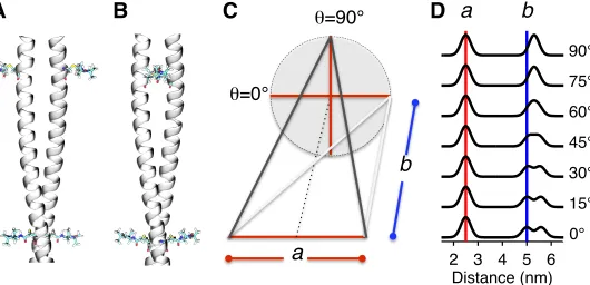
Pairs of residues were chosen so that the intra-pair distance was 2-3 nm, and the inter-pair distance was 4-6 nm. This choice ensured that the r-3 dependency of the dipolar interaction, whereby doubling the distance results in 8-fold lower dipolar frequency, separated the inter-pair from the intra-pair frequency while keeping the longer distance accessible. As illustrated in Fig. 2, in a coiled coil, the geometry is constrained by the intra-pair distance (designated a), and the inter-pair distance (designated b) and dihedral angle (designated ') between the vectors joining the
pairs. There are two long distances between the labels which are given by the expression:
!!
! !! !!"#!! ! !!. The two limiting cases are when the vectors joining the pairs are parallel (Fig. 2A) or when the vectors joining the pairs are perpendicular (Fig. 2B). In the former ' = 0° and the labels form a rectangle with two distinct long distances: corresponding to the sides, b, and the diagonals!! !!! !! (Fig. 2C, gray lines). In the latter ' = 90° and there is a single long distance:
!!
! ! !! (Fig. 2C, black lines). As illustrated in Fig. 2D, the shape of the distance distribution is strongly dependent on ', and hence the geometry of the coiled coil and changes thereto may be evaluated by EPR spectroscopy.
Note that for several cases, fewer spin pairs were observed for the IKK# or IKK"-vFLIP complexes; this is apparent from the relative modulation depth of the dipolar evolution. However, such differences can arise from the labelling efficiency varying between the samples, making interpretation ambiguous so we do not pursue it here.
MD simulations– Coarse-grained MD simulations were performed using the MARTINI force field over 1%s (17,18). In brief, approximately 4 non-hydrogen atoms are modelled as a single coarse grained particle. Non-bonded Lennard-Jones interactions are based on 4 classes of particles (polar, apolar, non-polar and charged), which are further divided into up to 5 subtypes. Interactions between different classes and sub-classes of particle are calculated based on an interaction table with 9 distinct interaction types. Furthermore, Lennard-Jones interactions were shifted to zero between 0.9 and 1.2 nm. Electrostatics were treated coulombically with a cut off at 1.2 nm (interactions are shifted to zero between 0 and 1.2 nm). Alpha helical secondary structure was maintained through dihedral angle restraints. Temperature and pressure were coupled at 323 K and 1 bar respectively using the Berendsen weak coupling algorithm (TT = 1 ps and
TP = 10 ps). Simulations were performed with
! &!
of these structures represents a distinct point in the dynamic ensemble, each of the structures has similar limitations to the original model. To include all of the conformational variability observed across the simulation, distance predictions using a rotamer library approach (20) were made for the labels in each frame and combined to generate a single distribution representing all label positions for all observed backbone bending, across the complete dynamic ensemble. This approach is a variation on serial multiscale modelling approaches (21), which explicitly samples across the entire trajectory rather than the end point alone.
NF-"B luciferase reporter assays–
Reporter assays were performed on 70Z/3 or Jurkat IKK" null cells stably reconstituted with IKK" mutants as previously described (13). Reconstituted cells were transduced with a lentiviral vector (LV) encoding an NF-!B responsive luciferase reporter. 5x10⁴ cells were
seeded in optical bottom 96-well plate and transduced with a vFLIP encoding LV. 48 h later, 50 %l BrightGlo luciferase substrate (Promega) was added to each well and the NF-!B induced luminescence was detected using Varioskan Flash multimode reader (Thermo Scientific). Where indicated cells were treated for 6 h with lipopolysaccharide (10 µg mL-1) prior to substrate
addition.
RESULTS
Analysis of IKK! using single pairs of spin
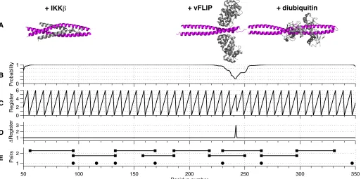
labels– The existing crystal structures of IKK"
fragments (12,22-25) all exhibit coiled-coil structures (Fig. 3A). Furthermore, 4 different coils prediction algorithms (26-29) suggest that the entire molecule (excluding the C-terminal zinc finger) adopts a parallel coiled-coil conformation concomitant with a switch in helical register close to Val250. The results from Multicoil2 (28) are depicted in Fig. 3B-D. In order to validate the prediction we employed EPR distance measurements; nitroxide spin-labelling of a single cysteine in a homo-dimeric protein such as IKK"
automatically gives rise to a pair of labels, whose separation may be measured by EPR spectroscopy.
IKK", however, contains 6 native cysteine residues distributed throughout the sequence. To establish whether their removal (in order to produce mutants containing a single cysteine for
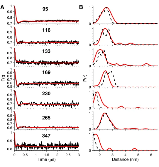
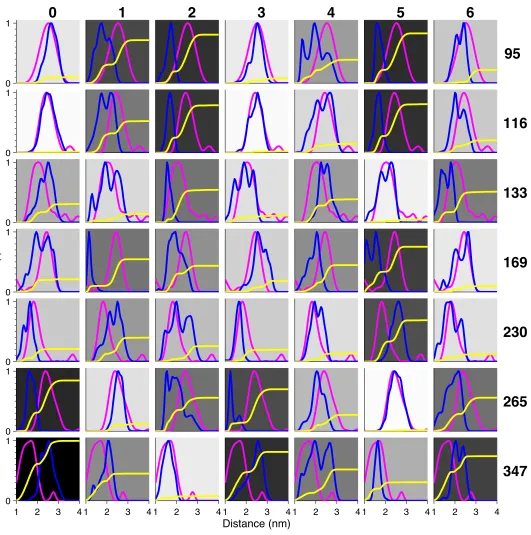
spin-labelling studies) would have a deleterious effect on the activity of IKK", a mutant involving the full-length protein in which 5 cysteines were mutated to serines (leaving only Cys167) was constructed and tested in vivo using a NF-!B luciferase reporter assay. Although the levels of NF-!B activation were reduced relative to wild type IKK", the mutant was still able to activate the pathway when stimulated by vFLIP (Fig. 4). In contrast, significant attenuation for this mutant was observed following stimulation using lipopolysaccharide (LPS) indicating that at least some of these residues have a role in cytokine induced activation. Occasionally, it was found that certain mutants containing a single native cysteine (and 5 serines) were unsuitable for EPR studies or were poorly expressed. In these cases, alternative mutants were constructed in which the cysteine was shifted to a neighbouring site. Thus, mutants containing a single cysteine at 7 different positions (95; 116; 133; 169; 230; 265 and 347) along the length of the 40-354 IKK" fragment were constructed, expressed and spin labelled. The positions of the labels in the sequence are indicated in Fig. 3E (solid circles).
The background-corrected dipolar evolution and derived fits (Fig. 5A, black and red lines, respectively) obtained by Tikonhov regularization all show a damped oscillation that has reached a plateau after several hundred nanoseconds. The corresponding distance distributions (Fig. 5B, red lines) show a single major peak centred below 3 nm. This confirms that IKK" is a parallel coiled coil along its entire length consistent with the crystal structures of fragments and prediction algorithms.
A static model of IKK!– An initial model
! '!
metric of the (dis-)agreement between them (Fig. 6, yellow lines). To aid visual comparison, the background of each plot has been gray-shaded according to the difference: a white implies perfect agreement; whereas black implies the largest deviation.
Register-3 and, to a slightly less extent, Register-0 gave the best agreement for positions 95, 116, 133, 169 and 230; while Register-5 gave the best agreement for positions 265 and 347. We therefore constructed two hybrid models combining the 0 or 3 and 5 registers for the N- and C-termini respectively, with the junction of the two registers in the region between amino acid residues Gln231 and Lys264. The key difference between the models is the handedness of the twist between different registers; the twist of the 0/5 hybrid is right handed, whilst the twist of the 3/5 hybrid is left handed. Both maintain the hydrophobic interface, whilst changing the specific angle the helices adopt relative to one another. The predicted distance distributions derived from the 3/5 hybrid gave best agreement with the EPR data (black dashed lines, Figure 5B).
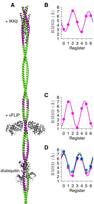
An overlay of the 3/5 hybrid with three crystal structures is presented in Fig. 7A. To obtain a metric of the deviation of the 7 models with crystal structures of IKK" fragments, the root mean square deviations (RMSDs) of the positions of the non-hydrogenic atoms in these regions were calculated (Fig. 7B-D). As expected the RMSDs follow a sinusoidal pattern covering two complete oscillations over 7 residues. The IKK" + IKK#
crystal structure (PDB entry = 3BRT, (22), residues: 50-110) has the closest match with Register-4 followed by Register-0 (Fig. 7B) whilst the crystal structure of IKK" + vFLIP (PDB entry = 3CLC, (12), residues: 195-250) has closest match with Register-3 followed by Register-6 (Fig. 7C). By contrast, crystal structures of the ubiquitin binding region both alone (PDB entry = 2ZVN, (24), residues: 270-330) and in complex with diubiquitin (PDB entry = 3FX0, (23)) and Hiop (PDB entry =!4OWF, (25), residues: 255-335) have closest matches with Register-2 followed by Register-5 (Fig. 7D). Thus both 0/5 and 3/5 hybrid models that we constructed from the EPR data are consistent with crystal structures giving confidence that we have derived a realistic overall view of the structure of IKK" in solution.
Analysis of IKK! alone and in complex
with vFLIP and IKK# using two pairs of spin
labels– The results presented above do not answer
the further question as to whether the coiled coil does or does not deviate from an extended structure. Hence, in order to provide a more detailed survey of the IKK" architecture and investigate any large-scale reorganisation of IKK"
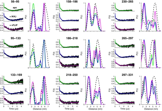
involving N- and C-terminal movements following association with vFLIP or IKK#, a series of 9 cysteine double mutants (56-95; 95-133; 133-169; 158-186; 186-218; 218-250; 230-265; 265-297; and 297-331) were constructed and spin labelled. The positions of the pairs in the sequence are given in Fig. 2E (solid squares).
The dipolar evolution of the 9 double mutants was recorded for IKK" alone (green lines) and in complex with IKK# (blue lines) and vFLIP (magenta). Inspection of the background-corrected dipolar evolution (Fig. 8) reveals that they contain both a high frequency component comparable to those seen in the spectra for pairs of spin labels (Fig. 5A) and a lower frequency component that extends out to several microseconds, indicative of longer distances.
Many of the data sets show small changes in profile upon binding of IKK# or vFLIP, especially at the inflection around 200 ns, whereas the lower frequency component is largely unchanged. These observations are reflected in the corresponding distance distributions (Fig. 8). These exhibit a rich variety of shapes and all show a major peak with a maximum in the range 2-3 nm as expected for the intra-pair distance and additional peaks in the range 4-6 nm, which arise from the inter-pair distances.
56-95, 95-133, and 133-169, give single sharp long distance peaks at just below 6 nm. This indicates that the pairs of vectors joining the spin labels are close to being perpendicular and that the coiled coil is well defined in this region. The distributions are only subtly affected by binding of IKK# or vFLIP, although we note that vFLIP induced denaturation of the 95-133 construct for reasons that are not understood.
! (! binding induces a broadening of the long distance distribution that may be indicative of twisting.
186-218, 218-250 and 230-265 all show a much broader distribution for the long distances, indicating that not only is the dihedral angle between the pairs of vectors joining spin labels far from 90°, but that the coiled coil is reasonably flexible in this region. Neither IKK# nor vFLIP have a significant effect on the distance distributions for 186-218 and 218-250, whereas for 230-265 vFLIP binding results in a remarkable sharpening of the intensity at 6 nm (magenta line), indicating that the angle between the pairs of vectors joining the spin labels tends towards 90° and that the coiled coil has become more rigid in this complex.
265-297 and 297-331 both display a pair of long distance peaks indicating that the dihedral angle between the pairs of vectors joining the spin labels deviates significantly from 90° and that the coiled coil is more rigid. Only minor broadening effects are observed when IKK# or vFLIP bind.
To summarise, the double mutants provide a picture of IKK" as an extended coiled coil that remains largely unchanged upon binding of either IKK# or vFLIP, although the binding of vFLIP stiffens the region spanning 230-265 (where it binds) and twists the region spanning 158-186.
Towards a dynamic model of IKK!– The 3/5 hybrid model derived above gives reasonable agreement with the distance distributions observed with both one and two pairs of spin labels, although the predictions exhibit fine structure absent from the experimental data that is likely to have arisen because the model is static whereas IKK" is potentially a dynamic molecule that can flex and bend. Hence, we performed a 1%s MARTINI (17,18) coarse-grained MD simulation of the 3/5 hybrid structure in water, with counter-ions that allowed for bending of the structure (Supplemental Movie). This dynamic model results in a better fit to the experimental distance distributions of the doubly-labelled IKK" (Fig. 8, black dashed lines) supporting our conclusion that the structure is an extended coiled coil. Additionally, visual inspection of the simulation suggests that the transition between the 3- and 5-registers that we identified as occurring in the region between Gln231 and Lys264 gives rise to a twist centred on residues Lys246 to Ser248, and
that the whole structure appears to hinge about the twist.
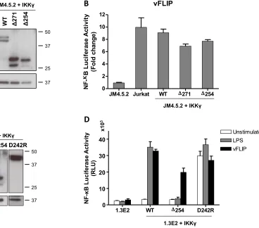
Activation of IKK! by vFLIP– We have shown that IKK" is essentially composed of two regions with different helical registers. The C-terminal region containing the ubiquitin binding region ends just before the vFLIP binding site. To investigate whether its absence would have any impact on the capacity of vFLIP to activate the canonical NF-!B pathway, IKK" truncation mutants were generated that terminated at Arg254 ((254-419) within the discontinuity and at Glu271 ((271-419) C-terminal to this region. Both mutants induced near wild type levels of activation in a NF-!B luciferase reporter assay (Fig. 9). This is in contrast to induction with LPS where they were found to be highly defective owing to deletion of crucial ubiquitin binding residues C-terminal to Glu271. Interestingly, a point mutation D242R at the vFLIP binding site within the transition bwtween the registers renders IKK" constitutively active in the absence of LPS (Fig. 9). Our findings thus confirm that the N-terminal region of IKK" is alone sufficient for vFLIP-induced activation and are consistent with previous results for an IKK"-estrogen receptor fusion protein in which IKK" was truncated at Gly251 (32), but also illustrate that the observed change in register is functionally important with respect to activation by endogenous cytokines.
DISCUSSION
! )!
To test this hypothesis given the absence of a full-length structure of IKK" (either in isolation or in complex with vFLIP and IKK$/#
simultaneously), we used EPR spectroscopy to obtain distances between pairs and quartets of spin-labelled cysteines positioned at intervals along the length of the IKK" molecule. The inter-label distance distributions observed for 56-95, 95-133, 133-169 and 158-186 towards the N-terminus and 265-297 and 297-331 towards the C-terminus are narrow, indicating a relatively rigid structure, while those observed for 186-218, 218-250 and 230-265 in the central region of the coiled coil are broad indicating a more flexible structure. The distance distributions were subsequently used to generate a model of the entire IKK" molecule as well as being analysed for changes indicative of
conformational rearrangements following
incubation with either vFLIP or IKK#. Apart from stiffening of the coiled coil observed in the vicinity of the vFLIP binding site, our results demonstrate that IKK" does not undergo gross structural reorganisation in response to binding. Furthermore, although the twisting observed for 133-169 (and the denaturation found for 95-133) indicates that subtle changes appear to be transmitted towards the N-terminus of the molecule, no effect is observed for 56-96 in the vicinity of the IKK# binding site.
The main structural insight gained is that to accommodate the change in register between the N- and C-termini, the region between them contains a twist located around residues Lys246 to Ser248. This builds tension into the structure as neither region can find a low energy conformation. As observed by X-ray crystallography, this region is essential for formation of the IKK"-vFLIP complex (12), which in turn leads to activation of the canonical NF-kB pathway. The D242R mutation in this region appears to mimic the effect of vFLIP binding: formation of a salt-bridge to Glu240 might induce a subtle change in structure that results in constitutive activation. The EPR data also demonstrate that binding of vFLIP stiffens this region of IKK".
Thus, it appears that although for the human cell, IKK" is simply designed with different registers so that the N- and C-termini can
perform their separate roles, namely kinase activity and regulation, respectively, the Kaposi’s sarcoma associated herpes virus has managed to exploit the structure of the transition between them in order to hijack the canonical NF-!B pathway.
We note that bacterial chemoreceptors, another class of coiled-coil signalling proteins have been proposed to signal through subtle modifications of a frustrated domain (34). Given the importance of this motif in signalling proteins, it is intriguing to speculate that this might be a common mechanism within the wider superfold family.
Since the induction of conformational changes can now be excluded as a potential mechanism for vFLIP-mediated NF-!B activation, alternatives need to be considered. Potentially, vFLIP may function by recruiting as yet unknown cofactors to IKK" or through blocking those that are known to downregulate the pathway in pro-inflammatory cytokine induced mechanisms, for example phosphatases (33). The former seems less likely considering recent reports where the absence of several factors essential to cytokine-induced activation failed to diminish vFLIP’s capacity to activate the pathway (14). In agreement with this, truncation of IKK" at Arg254 in the region of altered helical register directly C-terminal to the vFLIP binding site has little impact on NF-!B activation. Interestingly, a crystal structure of IKK" has recently been reported in complex with Hoip, a component of the linear ubiquitin chain assembly ligase essential for IKK"
polyubiquitination (25). The Hoip-IKK" interface encompasses residues in close proximity to this region which may therefore have an important role in NF-!B activation by pro-inflammatory cytokines in contrast to vFLIP. Although the mechanism by which vFLIP activates the IKK complex remains unclear – given its apparent
failure to induce anything but subtle
! *! REFERENCES
1. Scheidereit, C. (2006) I!B kinase complexes: gateways to NF-!B activation and transcription.
Oncog.25, 6685-6705
2. DiDonato, J. A., Hayakawa, M., Rothwarf, D. M., Zandi, E., and Karin, M. (1997) A cytokine-responsive I!B kinase that activates the transcription factor NF-!B. Nature388, 548-554 3. Mercurio, F., Zhu, H., Murray, B. W., Shevchenko, A., Bennett, B. L., Li, J., Young, D. B.,
Barbosa, M., Mann, M., Manning, A., and Rao, A. (1997) IKK-1 and IKK-2: cytokine-activated I!B kinases essential for NF-!B activation. Science278, 860-866
4. Israel, A. (2010) The IKK complex, a central regulator of NF-!B activation. Cold Spring Harb. Perspect. Biol.2, a000158
5. Hiscott, J., Nguyen, T. L., Arguello, M., Nakhaei, P., and Paz, S. (2006) Manipulation of the NF-!B pathway and the innate immune response by viruses. Oncog.25, 6844-6867
6. Keller, S. A., Hernandez-Hopkins, D., Vider, J., Ponomarev, V., Hyjek, E., Schattner, E. J., and Cesarman, E. (2006) NF-!B is essential for the progression of KSHV- and EBV-infected lymphomas in vivo. Blood107, 3295-3302
7. Jenner, R. G., Alba, M. M., Boshoff, C., and Kellam, P. (2001) Kaposi's sarcoma-associated herpesvirus latent and lytic gene expression as revealed by DNA arrays. J. Virol.75, 891-902 8. Field, N., Low, W., Daniels, M., Howell, S., Daviet, L., Boshoff, C., and Collins, M. (2003)
KSHV vFLIP binds to IKK" to activate IKK. J. Cell Sci.116, 3721-3728
9. Matta, H., Sun, Q., Moses, G., and Chaudhary, P. M. (2003) Molecular genetic analysis of human herpes virus 8-encoded viral FLICE inhibitory protein-induced NF-!B activation. J. Biol. Chem. 278, 52406-52411
10. Guasparri, I., Keller, S. A., and Cesarman, E. (2004) KSHV vFLIP is essential for the survival of infected lymphoma cells. J. Exp. Med.199, 993-1003
11. Grossmann, C., Podgrabinska, S., Skobe, M., and Ganem, D. (2006) Activation of NF-!B by the latent vFLIP gene of Kaposi's sarcoma-associated herpesvirus is required for the spindle shape of virus-infected endothelial cells and contributes to their proinflammatory phenotype. J. Virol.80, 7179-7185
12. Bagneris, C., Ageichik, A. V., Cronin, N., Wallace, B., Collins, M., Boshoff, C., Waksman, G., and Barrett, T. (2008) Crystal structure of a vFlip-IKK" complex: insights into viral activation of the IKK signalosome. Mol. Cell30, 620-631
13. Shimizu, A., Baratchian, M., Takeuchi, Y., Escors, D., Macdonald, D., Barrett, T., Bagneris, C., Collins, M., and Noursadeghi, M. (2011) Kaposi's sarcoma-associated herpesvirus vFLIP and human T cell lymphotropic virus type 1 Tax oncogenic proteins activate I!B kinase subunit gamma by different mechanisms independent of the physiological cytokine-induced pathways. J. Virol.85, 7444-7448
14. Matta, H., Gopalakrishnan, R., Graham, C., Tolani, B., Khanna, A., Yi, H., Suo, Y., and Chaudhary, P. M. (2012) Kaposi's sarcoma associated herpesvirus encoded viral FLICE inhibitory protein K13 activates NF-!B pathway independent of TRAF6, TAK1 and LUBAC.
PLoS One7, e36601
15. Pannier, M., Veit, S., Godt, A., Jeschke, G., and Spiess, H. W. (2000) Dead-Time Free
Measurement of Dipole–Dipole Interactions between Electron Spins. J. Magn. Reson.142, 331-340
16. Jeschke, G., Chechik, V., Ionita, P., Godt, A., Zimmermann, H., Banham, J., Timmel, C. R., Hilger, D., and Jung, H. (2006) DeerAnalysis2006—a comprehensive software package for analyzing pulsed ELDOR data. Appl. Magn. Reson.30, 473-498
17. Marrink, S.-J., de Vries, A. H., and Mark, A. E. (2004) Coarse grained model for semiquantitative lipid simulations. J. Phys. Chem. B108, 750-760
! "+!
19. Pronk, S., Pall, S., Schulz, R., Larsson, P., Bjelkmar, P., Apostolov, R., Shirts, M. R., Smith, J. C., Kasson, P. M., van der Spoel, D., Hess, B., and Lindahl, E. (2013) GROMACS 4.5: a high-throughput and highly parallel open source molecular simulation toolkit. Bioinformatics (Oxford, England) 29, 845-854
20. Polyhach, Y., Bordignon, E., and Jeschke, G. (2011) Rotamer libraries of spin labelled cysteines for protein studies. Phys. Chem. Chem. Phys. 13, 2356-2366
21. Stansfeld, P. J., and Sansom, M. S. P. (2011) From Coarse Grained to Atomistic: A Serial Multiscale Approach to Membrane Protein Simulations. J. Chem. Theo. Comp. 7, 1157-1166 22. Rushe, M., Silvian, L., Bixler, S., Chen, L. L., Cheung, A., Bowes, S., Cuervo, H., Berkowitz, S.,
Zheng, T., Guckian, K., Pellegrini, M., and Lugovskoy, A. (2008) Structure of a NEMO/IKK-Associating Domain Reveals Architecture of the Interaction Site. Structure 16, 798-808
23. Lo, Y.-C., Lin, S.-C., Rospigliosi, C. C., Conze, D. B., Wu, C.-J., Ashwell, J. D., Eliezer, D., and Wu, H. (2009) Structural Basis for Recognition of Diubiquitins by NEMO. Mol. Cell 33, 602-615 24. Rahighi, S., Ikeda, F., Kawasaki, M., Akutsu, M., Suzuki, N., Kato, R., Kensche, T., Uejima, T.,
Bloor, S., Komander, D., Randow, F., Wakatsuki, S., and Dikic, I. (2009) Specific Recognition of Linear Ubiquitin Chains by NEMO Is Important for NF-!B Activation. Cell 136, 1098-1109 25. Fujita, H., Rahighi, S., Akita, M., Kato, R., Sasaki, Y., Wakatsuki, S., and Iwai, K. (2014)
Mechanism underlying I!B kinase activation mediated by the linear ubiquitin chain assembly complex. Mol. Cell Biol. 34, 1322-1335
26. Lupas, A., Van Dyke, M., and Stock, J. (1991) Predicting coiled coils from protein sequences. Science 252, 1162-1164
27. McDonnell, A. V., Jiang, T., Keating, A. E., and Berger, B. (2006) Paircoil2: improved prediction of coiled coils from sequence. Bioinformatics (Oxford, England) 22, 356-358
28. Trigg, J., Gutwin, K., Keating, A. E., and Berger, B. (2011) Multicoil2: Predicting Coiled Coils and Their Oligomerization States from Sequence in the Twilight Zone. PLoS ONE 6, e23519 29. Delorenzi, M., and Speed, T. (2002) An HMM model for coiled-coil domains and a comparison
with PSSM-based predictions. Bioinformatics (Oxford, England) 18, 617-625
30. Stafford, R. L., Tang, M.-Y., Sawaya, M. R., Phillips, M. L., and Bowie, J. U. (2011) Crystal Structure of the Central Coiled-Coil Domain from Human Liprin-#2. Biochem. 50, 3807-3815 31. Eswar, N., Marti-Renom, M. A., Webb, B., Madhusudhan, M. S., Eramian, D., Shen, M., Pieper,
U., and Sali, A. (2006) Comparative protein structure modeling with MODELLER. Curr. Protoc. Bioinform. 15, 5.6.1-5.6.30
32. Tolani, B., Matta, H., Gopalakrishnan, R., Punj, V., and Chaudhary, P. M. (2014) NEMO is essential for Kaposi's sarcoma-associated herpesvirus-encoded vFLIP K13-induced gene expression and protection against death receptor-induced cell death, and its N-terminal 251 residues are sufficient for this process. J. Virol. 88, 6345-6354
33. Hong, S., Wang, L. C., Gao, X., Kuo, Y. L., Liu, B., Merling, R., Kung, H. J., Shih, H. M., and Giam, C. Z. (2007) Heptad repeats regulate protein phosphatase 2a recruitment to I-!B kinase gamma/NF-!B essential modulator and are targeted by human T-lymphotropic virus type 1 tax. J. Biol. Chem. 282, 12119-12126
34. Hall, B. A., Armitage, J. P., and Sansom, M. S. P. (2012) Mechanism of Bacterial Signal Transduction Revealed by Molecular Dynamics of Tsr Dimers and Trimers of Dimers in Lipid Vesicles. PLoS Comput Biol 8, e1002685
! ""!
Acknowledgements–We thank Professors Fabrice Agou and Alain Israël (Institut Pasteur, Paris, France) for their generous gift of the pRC-actin IKK# plasmid that incorporated the full human gene, Professor Chris Boshoff (UCL) for kindly providing the pGEX-KT IKK" plasmid, and Dr. Enrico Salvadori and Karen Fung (UCL) for helpful discussions.
FOOTNOTES
*This work was funded by a Medical Research Council grant (ID18218) to T.E.B., a Cancer Research UK grant (A12595) and a UCL Cancer Institute grant to M.K.C., a Wellcome Trust PhD Studentship to M.B.A.K. and a Royal Society Fellowship to B.A.H.. We declare that no conflict of interest arises from the work presented here.
§Current address: Department of Biochemistry, University of Oxford, South Parks Road, Oxford, UK. #Current address: Structural Biology and NMR Laboratory, Department of Biology, University of
Copenhagen, Copenhagen, Denmark
$Current address: Division of Biochemistry, Department of Chemistry, BOKU - University of Natural
Resources and Life Sciences, Muthgasse 18, Vienna, Austria.
The abbreviations used are:
vFLIP, Viral flice interacting protein;
KSHV, Kaposi’s sarcoma associated herpes virus;
NF-!B, nuclear factor kappa-light-chain-enhancer of activated B cells; IKK, I!B kinase;
NEMO, NF-!B essential modulator; EPR, electron paramagnetic resonance; MD, molecular dynamics;
TNF, tumour necrosis factor;
TRAF, TNF receptor associated factors; PCR, polymerase chain reaction; TEV, tobacco etch virus;
IPTG, isopropyl 1-thio-!-D-galactopyranoside;
CVs, column volumes; LV, lentiviral vector;
RMSDs, root mean square deviations.
AUTHOR CONTRIBUTIONS
! "#!
FIGURE LEGENDS
Figure 1. SDS-PAGE coomassie-stained gels of the final size-exclusion profiles obtained for: IKK"; IKK"–vFLIP complex; IKK"–IKK# complex; IKK"–vFLIP–IKK# ternary complex of the 56-95 double mutant.
Figure 2. Diagram illustrating how the register of the coiled coil translates into distance distributions between two pairs of nitroxide spin labels. Limiting cases in which (A) all 4 labels are in a plane or (B) the two vectors joining the pairs are perpendicular. (C) The geometry is constrained by the intra-pair distance (designated a), the inter-pair distance (designated b) and dihedral angle (designated ') between the vectors joining the pairs. (D) Illustration of the variation of the distance distribution for two pairs of labels as a function of ' for a = 2.5 nm and b = 5 nm with Gaussian linewidth of 0.2 nm.
Figure 3. (A) Crystal structures of fragments of IKK" (magenta) in complex with IKK#, vFLIP and diubiuitin (gray) aligned according to their position in the sequence. (B) The probability of a region adopting a coiled-coil arrangement based on a window of 28 residues predicted from the primary sequence of IKK". (C) Predicted position of residues within a coiled coil based on the heptameric repeat of the motif: each residue has a predicted position from 0-6 and a regular zigzag pattern implies a perfect coiled coil. (D) Increment of the register of a residue with respect to its predecessor. An increment of +1 implies a perfect coiled coil. (E) Positions of nitroxide spin labels for singly labelled IKK" (solid circles) and doubly labelled IKK" (solid squares joined by solid lines).
Figure 4. NF-!B reporter assays for the vFLIP mutant in which all 5 out of 6 cysteines were mutated to serine. (A) Western blot analysis of IKK" expression level in IKK" knockout mouse PreB cell line 1.3E2, respective parental cells 70Z/3 and IKK" wild type or mutant reconstituted 1.3E2 cells (B) NF-!B activation was measured in the above-mentioned cell lines using the BrightGlo Luciferase assay system, 6 h after stimulation with LPS (10 µg mL-1) or 48 h after transduction with KSHV vFLIP LV (MOI = 50). Bars represent mean fold induction values ± SD.
Figure 5. EPR distance measurement on singly-labelled IKK". (A) Background-corrected dipolar evolution (black lines) and their fits (red lines). (B) Distance distributions (red lines) derived from the data in (A) and predicted distance distributions (black dashed lines) derived from the static 3/5 hybrid model.
Figure 6. Validation of the model structures. Comparison of the distance distributions obtained from the EPR experiments for the singly-labelled IKK" (magenta lines) with the predicted distance distributions (blue lines) using a rotamer library approach for the 0-6 register models. The cumulative sum of the squares of the differences between the experimental and predicted distributions is used to provide a metric for goodness of fit (yellow lines). The background has been gray-shaded according to the relative final amplitude of the yellow lines: a white background implies perfect match, whereas a black background implies poor agreement.
! "$!
Figure 8. EPR distance measurements on doubly-labelled IKK", apo and complexed with IKK# and vFLIP. Fits to the baseline-corrected dipolar evolution and corresponding distance distributions for apo
IKK" (green lines) and IKK" complexed with IKK# (blue lines) and vFLIP (magenta lines). The distance
distributions for the doubly-labelled IKK" derived from the coarse-grained MD simulation of the 3/5 hybrid model are shown as a dashed black line.
Figure 9. NF-!B luciferase reporter assays for the IKK"(254-419 and (271-419 truncation mutants and D242R mutant. (A) Western blot analysis of IKK" expression level in IKK" knockout human T-cells JM4.5.2, parental Jurkat cells and IKK" wild type or mutant reconstituted JM4.5.2 cells (B) NF-!B activation was measured using the BrightGlo Luciferase assay system 48 h after transduction with KSHV vFLIP LV (MOI = 50). Bars represent mean fold induction values ± SD. (C) Western blot analysis of
IKK" expression levels in IKK" knockout mouse PreB cell line 1.3E2, parental cells 70Z/3 and 1.3E2
!"
#$
#"
%$
&$
"$
ladder
IKK
γ
- vFLIP
IKK
γ
[kDa]
MW
IKKγ(40-354)
IKK
γ
- IKK
β
IKK
γ
- vFLIP - IKK
β
[image:15.595.40.547.162.683.2]2 3 4 5 6
Distance (nm)
0°
15°
30°
45°
60°
75°
90°
a
b
e
=0°
e=90°
a
b
[image:16.595.38.568.289.545.2]0 1
Probability
0 2 4 6
Register
1 2 3
6
Register
50 100 150 200 250 300 350
1 2
Residue number
Pairs
A
B
C
D
E
[image:17.595.45.558.291.546.2]IKK
!
GAPDH
1.3E2 70Z/3
1.3E2 + IKK
!
WT
Mutant
1.3E2
70Z/3
WT
Mutant
1.3E2 + IKK
!
NF-"
B
Lu
ci
fe
ra
se
Act
ivi
ty
(F
ol
d
C
ha
ng
e)
A
!
Data 1
1.3E2
70Z/3
WT
mutant
0
10
20
30
LPS
vFLIP
NF
-!
B
L
u
ci
fe
ra
se
Ac
ti
vi
ty
(F
o
ld
c
h
an
g
e)
Data 1
1.3E2
70Z/3
WT
mutant
0
10
20
30
LPS
vFLIP
NF
-!
B
L
u
ci
fe
ra
se
Ac
ti
vi
ty
(F
o
ld
c
h
an
g
e)
B
!
[image:18.595.48.556.131.685.2]0
1
0.7
0.8
0.9
1
0
1
0.7
0.8
0.9
1
0
1
0.7
0.8
0.9
1
0
1
P(r)
0.5
0.6
0.7
0.8
0.9
1
F(t)
0
1
0.6
0.7
0.8
0.9
1
0
1
0.6
0.7
0.8
0.9
1
2
3
4
5
6
0
1
Distance (nm)
0
0.5
1
1.5
2
2.5
3
0.8
0.9
1
Time (µs)
95
116
133
169
230
265
347
[image:19.595.29.571.137.709.2]0 1
0 1
0 1
0 1
P(r)
0 1
0 1
1 2 3 4
0 1
1 2 3 4 1 2 3 4 1 2 3 4
Distance (nm)
1 2 3 4 1 2 3 4 1 2 3 4
0
1
2
3
4
5
6
95
116
133
169
230
265
[image:20.595.40.571.146.681.2]0 1 2 3 4 5 6
1
2
3
4
5
6
RM
S
D
(
˚
A)
Register
0 1 2 3 4 5 6
1
2
3
4
5
6
7
8
RM
S
D
(
˚
A)
Register
0 1 2 3 4 5 6
1
2
3
4
5
6
7
8
RM
S
D
(
˚
A)
Register
C
D
+ IKK
`
+ vFLIP
[image:21.595.142.462.60.757.2]0 1 P(r) 0.4 0.6 0.8 1 0.4 0.6 0.8 1 F(t) 0.4 0.6 0.8 1 0 1 P(r) 0.4 0.6 0.8 1 0.6 0.8 1 F(t)
2 3 4 5 6 7
0 1 P(r) Distance (nm) 0.2 0.4 0.6 0.8 1 0.4 0.6 0.8 1 F(t)
0 1 2 3 4
0.4 0.6 0.8 1
Time (!s)
0 1 P(r) 0.4 0.6 0.8 1 0.2 0.4 0.6 0.8 1 F(t) 0.4 0.6 0.8 1 0 1 P(r) 0.4 0.6 0.8 1 0.4 0.6 0.8 1 F(t) 0.4 0.6 0.8 1
2 3 4 5 6 7
0 1 P(r) Distance (nm) 0.4 0.6 0.8 1 0.4 0.6 0.8 1 F(t)
0 1 2 3 4
0.4 0.6 0.8 1
Time (!s)
0 1 P(r) 0.4 0.6 0.8 1 0.6 0.8 1 F(t) 0.4 0.6 0.8 1 0 1 P(r) 0.4 0.6 0.8 1 0.4 0.6 0.8 1 F(t) 0.2 0.4 0.6 0.81
2 3 4 5 6 7
0 1 P(r) Distance (nm) 0.4 0.6 0.8 1 0.4 0.6 0.8 1 F(t)
0 1 2 3 4
0.4 0.6 0.8 1
Time (!s)
95ï133 56ï95
133ï169
158ï186
186ï218
218ï250
230ï265
265ï297
297ï331
apo
+ IKK`
[image:22.595.33.572.237.606.2]vFLIP
JM4.5.2 Jurkat WT !271 !254 0
2 4 6 8 10 12
NF
-"B
L
u
ci
fe
ra
se
Ac
ti
vi
ty
(F
o
ld
c
h
an
g
e)
JM4.5.2 + IKK!
IKK!
GAPDH
WT "271 "254 JM4.5.2 + IKK!
37 50
25
37
IKK!
GAPDH
WT "254
1.3E2 + IKK!
37 50
25
37
!"
#"
$"
%"
D242R
1.3E2 + IKK!
1.3E2 WT !254 D242R
0 10000 20000 30000 40000
Unstimulated LPS vFLIP
0 10 20 30 40
NF-#
B
L
u
ci
fe
ra
se
A
cti
vi
ty
(R
L
U
)
x10
[image:23.595.171.539.258.582.2]![MWIKKβ - vFLIP - IKKβ - IKKγγγIKKIKK[kDa]Figure 1γIKK - vFLIPladder](https://thumb-us.123doks.com/thumbv2/123dok_us/8867818.940752/15.595.40.547.162.683/mwikkb-vflip-ikkb-ikkgggikkikk-kda-figure-gikk-vflipladder.webp)