N A N O E X P R E S S
Open Access
A turn-on fluorescent solid-sensor for Hg(II)
detection
Mayela De la Cruz-Guzman
1, Angelica Aguilar-Aguilar
1, Luis Hernandez-Adame
1, Alan Bañuelos-Frias
3,
Francisco J Medellín-Rodríguez
2and Gabriela Palestino
1*Abstract
A rhodamine organosilane derivative (Rh-UTES) has been obtained by one-pot synthesis. The chemical structure of Rh-UTES was confirmed by nuclear magnetic resonance (NMR) and infrared (FTIR) techniques. To obtain an inorganic-organic hybrid sensor, Rh-UTES was covalently immobilized on a porous silicon microcavity (PSiMc) via triethoxysilane groups. The attachment of the organic derivative into PSiMc was confirmed by FTIR, specular reflectance, and scanning electron microscopy (SEM). The optical performance of Rh-UTES receptor for Hg2+ detection was investigated by fluorescent spectroscopy and microscopy. Upon the addition of increasing amounts of Hg2+ions, a remarkable enhancement in emission intensity was produced in both systems. In the solid phase, an increase of integrated fluorescent emission of 0.12- and 0.15-fold after Hg2+receptor coordination was observed. The light harvesting capability of PSiMc devices allowed obtaining an enhanced fluorescent emission after Rh-UTES immobilization (277-fold). The fluorescence microscopy of hybrid PSiMc sensor provided an optical qualitative test for Hg2+detection.
Keywords:Chemosensor; Porous silicon; Rhodamine derivative; Fluorescence; Heavy metal
Background
The toxicity of mercury (Hg) and its complex forms on ecosystems and human health is well known. The need to create new sensitive and practical analytical methods to detect the mercury ions in different sources has in-creased. Recently, ion-selective sensors have attracted attention due to their diverse potential applications as tools for the quantitative and qualitative monitoring of metal ions in many biological and environmental pro-cesses [1-6]. Ion-selective sensors could find applicability in monitoring metal ion concentrations and can be prac-tical solutions to monitor industrial waste effluent streams and potable water. Emphasis has been placed on com-pound development that selectively responds to the pres-ence of specific metal ions through a change in one or more properties of the system, such as redox potentials [7], absorption [8], or fluorescence spectra [9]. Such sensors based on ion-induced changes in fluorescence appear to be
particularly attractive due to their simplicity, high sensitiv-ity, high selectivsensitiv-ity, and instantaneous response [10]. Fluorescent chemosensors based on xanthenes and related derivatives for the Hg2+ions detection have been increas-ing due to the low cost and high applicability in industrial and biological processes [11]. During recent years, novel inorganic-rhodamine hybrid sensors have been published. The rhodamine derivatives have been immobilized into the different inorganic receptors. Huang et al. reported fluores-cent gold nanoparticle sensors for detection of Hg2+ions [12]. Since gold nanoparticles (AuNPs) are highly efficient fluorescence quenchers, the rhodamine derivative had to be released from the AuNPs to restore the rhodamine fluorescence. Lee et al. and Zhou's group developed a covalently bonded mesoporous silica rhodamine derivative [13,14]. Childress and co-workers reported dye-doped polymer nanoparticles that are able to detect mercury ions. The nanoparticles were prepared by precipitation of highly fluorescent conjugated polymers and doped with rhoda-mine derivatives [15]. Recently, Wang and Gao designed a mercury sensor using β-NaYF4:Yb3+/Eu3+ nanorods as the excitation source and a rhodamine derivative as a probe [16]. In this proposal, our research group has
* Correspondence:[email protected]
1Biopolymers and Nanostructures Laboratory, Faculty of Chemical Sciences,
Universidad Autónoma de San Luis Potosí, Av. Manuel Nava No. 6, San Luis Potosí, San Luis Potosí 78210, México
Full list of author information is available at the end of the article
designed a new functional rhodamine derivative (Rh-UTES) that acts as a receptor of heavy metal ions. The Rh-UTES derivative was covalently bonded to porous silicon microcavity (PSiMc) to develop a hybrid sensor. The main advantage of the proposed method is the sim-plicity of the system and the fact that the hybrid sensor should be easy to carry for field applications. The PSiMc has proven to be a suitable material with unique optical properties for the development of this kind of fluores-cent sensor [17]. Our previous approaches in this field have shown that the detection of fluorescent molecules is possible using the optical properties of specific PSi structure (mirror or microcavity) [18]. Increased excitation and enhanced emission, both driven by the efficient reflec-tion of light and resonance effects within the PSi micro-cavities, allowed the enhancement of the fluorescent response of the Rh-UTES derivative even at low molecular concentration. Hence, the variation of this method was used here to produce detection of low concentrations of heavy metals by forming metallic complexes within the pores that turn on the luminescence emission.
Methods
Rhodamine base, ethylenediamine, m-xylenediisocyanate, 3-aminopropyltriethoxysilane (APTES), hydrochloric acid, hydrofluoric acid, nitric acid, sodium hydroxide, and mer-cury nitrate were purchased from Sigma-Aldrich (St. Louis, MO, USA). All solvents were analytical reagent grade and used as received.
Instruments and spectroscopy measurements
The reflectivity spectra were recorded in an Agilent Cary 60 UV-Vis spectrophotometer (Agilent Technologies, Sta. Clara, CA, USA) coupled with a 30° specular reflection unit. PSi samples were illuminated with the xenon source, and the reflected beam was detected with the silicon diode detector. The resulting spectra were captured in the range from 500 to 900 nm. The fluorescence images of PSiMc/ Rh-UTES sensor were recorded in a Nikon Optiphot-2 fluorescence microscope (super high pressure mercury lamp power supply; Nikon, Tokyo, Japan). The Fourier transform infrared spectra (FTIR) were recorded in a Bruker Tensor 27 spectrophotometer (Bruker Corporation, Billerica, MA, USA), with 128 scans and 4-cm−1 reso-lution, coupled with a diamond crystal attenuated total reflectance unit (ATR). Nuclear magnetic resonance (NMR) measurements of1H and13C were carried out in a Bruker 500 MHz spectrometer. Scanning electron mi-croscopy (SEM) was performed using a UHR dual-beam FEI Helios Nanolab 600 field emission scanning electron microscope (FEI Company, Hillsboro, OR, USA). Samples were mounted on a conductive carbon tape. Images were captured at magnifications of × 20,000 and × 25,000.
Synthesis of porous silicon
PSi samples were prepared by the wet electrochemical etch-ing process usetch-ing high-doped p-type (boron-doped) silicon wafers (thickness 500 to 550μm) with 0.001 to 0.005Ωcm resistivity, and with the crystallographic orientation of (100), purchased from WRS Materials (San Jose, CA, USA). The electrolyte consisted of hydrofluoric acid (48 wt%) and ethanol in the volumetric ratio of 3:7. The anodization time and current density were controlled by a computer-interfaced electronic circuit. The samples were fabricated at room temperature, and freshly etched samples were washed with ethanol and dried with pentane. To perform this work, we have selected a PSiMc, mainly due to its optical features in the reflectance spectra that allows the detection of infil-trated material into the porous structure. PSiMc configur-ation consists of an active porous layer embedded between two multilayered mirrors (Bragg reflectors). The PSiMc was produced by alternating layers of high porosity (H; refract-ive index, n= 1.14395) and low porosity (L; n= 1.25865), with current densities of 70 and 30 mA/cm2. Anodization times of 6.35 and 10.67 s for H and for L, respectively, were used for the fabrication of the corresponding dielectric Bragg mirrors. The PSiMc structures were fabricated with the configuration of (HL) × 5 HH (LH) × 5, where (HL) × 5 corresponded to the first Bragg reflector, HH to the cavity and (LH) × 5 to the second Bragg reflector. The PSiMc samples were thermally oxidized at 600°C for 30 min in O2atmosphere to stabilize and protect them against en-vironmental contaminants and/or natural aging [19].
Synthesis of rhodamine fluorescent derivative
Herein, we synthesize a new rhodamine fluorescent de-rivative Rh-UTES bearing urea groups. To obtain this compound, several steps were needed. For the synthesis of Rh-amine derivative (1), following the procedure in the literature [20], rhodamine base (4.0 g, 8.3 mmol) and ethylendiamine (4.2 g, 70 mmol) were dissolved in EtOH (210 mL) and refluxed for 18 h. The solvent was removed by evaporation, and the residue was dissolved in an aqueous HCl solution (1 M, 333 mL). An aqueous NaOH solution (1 M) was added carefully to the solution with magnetic stirring. The precipitate was recovered by filtra-tion, washed thoroughly with water, and then dried under vacuum, yielding (1) as a pink fluffy powder (3.21 g, 80%); 1
H NMR (CDCl3):δ(ppm) 7.85 (d, 1H,J= 2.5 Hz), 7.44 (t, 2H,J= 6.7 Hz), 7.06 (s, 1H), 6.42 to 6.37 (m, 6H), 3.33 (q, 10H,J= 7.1 Hz), 2.91 (t, 2H,J= 6.7 Hz), 1.16 (t, 12H,J= 6.7 Hz); 13C NMR (CDCl3):δ (ppm) 170.5, 153.7, 153.3, 149.1, 133.2, 130.0, 128.4, 128.3, 123.9, 123.2, 108.6, 103.6, 97.8, 66.4, 44.4, 41.1, 39.5, 12.66. Figure 1 shows the syn-thesis to obtain derivative (1).
(0.05 g, 0.26 mmol) and 3-aminopropyltriethoxysilane (APTES) (0.04 g, 0.18 mmol) were refluxed in 5 mL of toluene under N2for 12 h. Derivative (2) was used without isolation, the Rh-amine derivative (1) was added (0.1 g, 0.21 mmol) under N2, and the reaction was refluxed for 3 h. The solvent was evaporated under reduced pressure to give a beige powder (0.22 g, 96%);13C NMR
(DMSO-d6):δ(ppm) 168.0, 158.1, 154.2, 153.0, 148.1, 141.0, 133.2, 130.5, 128.6, 128.5, 126.2, 126.1, 126.0, 125.9, 125.7, 124.0, 122.8, 108.3, 105.3, 97.8, 64.6, 60.2, 44.1, 43.4, 40.6, 38.4, 21.2, 15.1, 14.5, 12.8; IR data:νmax(cm−1): 3331, 2970 to 2890, 1695, 1624, 1574, 1513, 1082, 962, 771.
PSi device functionalization
The binding of Rh-UTES derivative within the PSi nano-structured devices was performed following one-step
method through silane chemistry by reacting the meth-oxy groups (-OCH3)3 of the fluorescent molecule with the siloxane (-Si-O) groups of the thermally oxidized PSi surface [18]. Briefly, the PSi samples were dipped in 2 mL of Rh-UTES derivative solution (1.16μM in ACN) at room temperature, and all of the reaction system was kept under inert atmosphere with magnetic stirring. The reaction time was fixed at 3 h to obtain the final PSiMc/ Rh-UTES sensors.
Metal capture
[image:3.595.60.539.89.192.2]Once obtained, the PSiMc/Rh-UTES sensors were ex-posed to 2.0 mL of mercury aqueous solutions. To as-sure the presence of the free Hg2+ ions, the solutions were adjusted at pH 3.0 using HNO3 0.1 M (based in the Hg speciation diagram). The complexation reactions
Figure 1Synthesis to obtain derivative (1).
[image:3.595.59.540.417.724.2]were carried out at room temperature for 12 h under magnetic stirring.
Results and discussion
[image:4.595.305.539.89.259.2]Rh-UTES derivative was successfully synthesized from a rhodamine base in a relatively good yield. To evaluate the metal ion binding capability of this new compound, a colorimetric evaluation was performed in a liquid phase. Figure 3 shows the optical behavior of the fluorescent che-mosensor in solution (1.16 μM in ACN). It was observed that after the Hg2+addition, the colorless solution immedi-ately becomes pink. It is interesting to notice that the color intensity of the solution is linearly dependent on the metal concentration. The color change in the chemosensor solu-tion after Hg2+addition is attributed to the chelator-metal binding. Thus, the colorimetric change produced during Hg2+capture can be used as‘naked-eye’ detection of this metallic contaminant in solution.
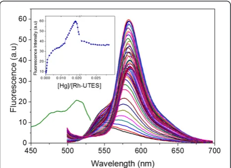
The photoluminescent properties of Rh-UTES deriva-tive in solution were investigated toward the metal ion complexation. Figure 4a shows the excitation and emis-sion spectra of Rh-UTES derivative with peaks centered at 513 and 583 nm, respectively. In the figure we can no-tice that the organic receptor exhibited a slight fluores-cence emission. Upon the addition of increasing amount of Hg2+ ions (0.166 to 27.0 μM) to the solution of Rh-UTES receptor, a remarkable enhancement in the emis-sion intensity was observed. This fluorescent enhance-ment is attributed to the formation of the Rh-UTES-Hg2+ complex. Thus, it is clear that the addition of Hg2+ ions ‘turns-on’ the fluorescence whereby the colorless weak fluorescent derivative changed to a colored highly fluores-cent complex, as was also shown in Figure 3. Additionally, we found that the Rh-UTES-Hg2+ complex presents a maximum emission at 11.9 μM Hg2+ concentration, after which a fluorescent quenching phenomenon was observed. The fluorescent intensity is reduced since some molecules of the complex act as a quencher (be-cause the high concentration of the complex may induce a
self-absorption process) which in turn decreases the num-ber of molecules that can emit. Finally, after addition of 24.2μM Hg2+concentration, the fluorescent emission of complex remains constant, which is attributed to the de-pletion of Rh-UTES derivative.
The fluorophore selectivity was also investigated by measuring the changes in the fluorescent emission pro-duced by the addition of the following metal ions: Ag+, Hg2+, Ca2+, Pb2+, Li2+, Zn2+, Fe2+, Ni2+, K+, Cu2+, Na+, and Mn2+to various solutions of Rh-UTES. The results are dis-played in Figure 5; it is clear that the presence of these ions led to increases in the fluorescence intensity to varying de-grees. It was observed that only Li2+ions promote small fluorescence intensity changes, while the other metal ions did not cause any significant changes under identical con-ditions. The fluorescent emission intensity observed for Hg2+over the other ions is remarkably high pointing out the high selectivity of Rh-UTES toward Hg2+.
Reflectance spectra
[image:4.595.58.289.574.677.2]The reflectance spectra of the PSiMc were recorded after each modification step using the UV-vis spectrophotom-eter. Figure 6 compares reflectance spectra taken before and after PSiMc functionalization and a metal capture. It is observed that Rh-UTES derivative binding produces a red shift (12 nm) in the PSiMc reflectance spectrum; we also found that this process is repeatable showing a stand-ard deviation (SD) of ±2.12 nm. The red shift can be at-tributed to the effective refractive index (ȵ) changes after infiltration of the fluorescent molecule into the PSi pores [18]. After exposition of PSiMc/Rh-UTES sensor to Hg2+ solution, surprisingly and contrary to the expectation, a blue shift was observed in the specular reflectance
Figure 3Colorimetric changes in the Rh-UTES derivative solutions. (a)Before Hg2+addition and after Rh-UTES-Hg2+complex formation at the following molar ratios:(b)1:1,(c)1:6, and(d)1:10, respectively. Rh-UTES concentration remained fixed at 1.16μM in ACN solution.
Figure 4Fluorescence response of Rh-UTES derivative in liquid phase at different metal concentration.Fluorescence response of Rh-UTES derivative in liquid phase (1 mM in ACN) upon addition of different concentrations of Hg2+ions (0.166 to 27.0
spectrum (9 nm, SD ± 3.35 nm). Normally, this drift in signal (blue shifts) can be associated to the degradation (or oxidation) of PSi [21]. However, in this work, the ob-served negative shift is attributed to the derivative-metal binding. This was confirmed by the negative con-trols that were carried out to ensure the specificity of the linking chemistry. These results showed a negligible drift in the PSi sensor reflectance spectrum over the same incubation periods used to collect data in the per-formed experiments. It seems that the metal capture
produces a decrease ofȵ. Nevertheless, to have a better understanding of the metal-ligand-substrate interac-tions and their effect on the optical properties of the PSiMc structure, more studies are being conducted in our research group. Thus, the capture of the metal ions for the PSi/Rh-UTES sensor was confirmed using com-plementary analytical techniques.
Monitoring molecular infiltration
PSi nanostructured devices were analyzed by FTIR be-fore and after derivative functionalization and the metal capture. Riikonen and co-workers reported the typical strong absorptions of oxidized PSi (OxPSi) [22]. Bands characteristic of the stretching mode of silanol groups ν(SiO-H) were observed at around 3400 cm−1
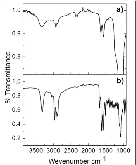
[image:5.595.57.291.88.275.2], the δ(SiO-H) bending mode at 1640 cm−1, and theν(Si-OH) stretch-ing modes at 950 and 887 cm−1. An intense broad peak at around 1085 cm−1was also seen, which may be due to the ν(Si-O) stretching mode for surface silicon-hydroxyl spe-cies. All of these bands are consistent with FTIR spectrum of our thermally (OxPSi) device [19]. The immobilization of Rh-UTES derivative into the PSiMc surface was carried out and confirmed by FTIR spectroscopy (Figure 7a); the hybrid sensor owns the next characteristics bands:ν(N-H) stretching modes at 3344 cm−1,ν(C = O) stretching modes at 2924 cm−1, δ(N-H) bending mode at 1571 cm−1 of secondary amide,ν(C-H) stretching modes of methylene
[image:5.595.306.539.424.706.2]Figure 6Specular reflectance spectra of PSiMc devices.(a) Thermally oxidized sample (black line), (b) after Rh-UTES immobilization (red line), and (c) after metal coordination (blue line). [Hg2+] = 3.48μM.
Figure 5Maximum fluorescence emission of Rh-UTES after metal capture.Maximum fluorescence emission of Rh-UTES (10μM in ACN) derivative upon addition of 100μM of Ag+, Hg2+, Ca2+, Pb2+, Li2+, Zn2+, Fe2+, Ni2+, K+, Cu2+, Na+, and Mn2+, respectively. The emission spectra were recorded under identical experimental conditions at excitation wavelength of 485 nm.
[image:5.595.57.294.504.684.2]groups at 3008 to 2861 cm−1, and mainly the siloxane (Si-O) bands of OxPSi at 1054 cm−1. These bands are similar to those belonging to the pure Rh-UTES deriva-tive reported in the ‘Methods’section (Figure 7b), thus confirming that incorporation of Rh-UTES into the PSiMc was successful. The hybrid sensor was then ex-posed in a Hg2+solution (1.16μM) for 12 h, and the FTIR analysis of the PSiMc/Rh-UTES-Hg2+sample showed no significant changes in the infrared bands (not shown) compared with the reference spectrum of Figure 7b.
[image:6.595.58.289.368.694.2]Morphological analysis
Figure 8 shows cross-sectional SEM images of PSiMc devices before (a) and after (b) functionalization with Rh-UTES derivative. The top view of unmodified PSiMc device (image not shown) shows a high porosity struc-ture composed of well-defined pores with an average size distribution of 19.25 ± 4 nm. In these PSi structures, the pore sizes were big enough to allow the molecular infiltration as demonstrated by specular reflectance spec-trometry. The lateral view of the unmodified sample (Figure 8a) shows the high (white line) and low porosity
(black line) layers together with the defect layer (cen-tered in the middle of the structure). The morphology of the PSiMc structures after chemical modification is shown in Figure 8b, and we observed a homogeneous layer of organic derivative covering the first layers of the PSi struc-ture, which confirms the infiltration of Rh-UTES deriva-tive into the porous device.
Photoluminescence properties
[image:6.595.304.538.504.674.2]In solid phase, photoluminescence (PL) measurements were used to characterize the performance of the fluor-escent sensor under λexc= 490 nm. Figure 9 shows the fluorescent emission of (a) thermally oxidized PSiMc, (b) PSiMc/Rh-UTES functionalized device [1.16 μM of de-rivative (3)], and (c, d) PSiMc/Rh-UTES sensors after ex-posure to solutions contaminated with Hg2+ (3.45 and 6.95μM, respectively). The amount of infiltrated derivative into the PSi pores was obtained by calculating the concen-tration of the residual supernatant (recovered after the ex-posure time of the sample was completed) and making a mass balance. The obtained concentration value was of 1.4058 ± 0.35 nmol of Rh-UTES/cm2of etched area, which corresponds at approximately 20% of the initial solution concentration (1.16 μM) [19]. By comparing the optical features of bare PSiMc with that obtained after device functionalization, it is clear that the emission spectra show important optical changes. The most remarkable is the well-defined emission curve in the 525 to 625-nm range at-tributed to the fluorescent emission of Rh-UTES derivative, which confirms the attachment of the derivative molecule on the PSi surface. Exposure of PSiMc/Rh-UTES sensor at a heavy metal solution produced two new changes: first, an increase in the integrated emission intensity of 0.13-fold and secondly, a 16-nm red shift (552 to 568 nm) of the
Figure 8Cross-sectional SEM micrographs of PSiMc before and after derivative immobilization. (a)Thermally oxidized sample.
(b)PSiMc/Rh-UTES hybrid device.
Figure 9Emission spectra of PSiMc devices (λexc= 490 nm)
main peak position. As we mentioned before, some studies have demonstrated that the spirolactam-rhodamine deriva-tives can be used to develop liquid phase OFF-ON metal ion-fluorescent chemosensors, mainly because their chem-ical structure may change in the presence of metal ions. In agreement with those contributions, we believe that the enhanced emission observed when the PSiMc/Rh-UTES sensor captured the Hg2+ions is produced by the forma-tion of metal-ligand coordinaforma-tion bonds, which in turn in-duces the spirolactam ring opening [23]. Thus, based on this coordination mechanism, the red shift in the fluores-cent emission may be attributed to the electronic interac-tions of PSiMc/Rh-UTES-Hg2+ complex (Figure 9c). A similar optical behavior was found in the liquid phase che-mosensor; however, our solid device presents several ad-vantages that are related with (i) the easy operation of the device, (ii) special solvents that are not needed, (iii) the higher stability of the fluorescent derivative when immobi-lized in the solid support, and (iv) the possibility of port-ability. Then, by comparing spectra (c) and (d) which correspond at the sensing of two different Hg2+ion con-centrations (3.45 and 6.95 μM, respectively), a 6-nm red shift (from 568 to 574 nm) and a fluorescent emission en-hancement of 0.12-fold was observed. In this case, the red shift may be attributed to PSi-derivative-Hg2+ interaction processes produced in the reduced space of PSi pores. Our hypothesis is that after increasing the metal ion concentra-tion, the derivative Rh-UTES receptor changed its chem-ical structure, provoking a molecular reorganization inside the pore. According to Tu and co-workers [24], the chem-ical change can reduce the distances between neighboring molecules limiting their free stretching movement and leading to their self-interaction, which may reduce their ex-cited state energy and produce the red shift in the spectra.
On other hand, the enhancement of the emission intensity observed when the PSiMc/Rh-UTES device coordinates higher amount of Hg2+ions confirms that the fluorescent intensity of the PSiMc hybrid device is metal concentration dependent [25,26]. This is important to notice if the sensing principle of the PSiMc/Rh-UTES sensor is based on the fluorescence spectroscopy measurements. Finally, to inves-tigate the optical contribution of PSi devices in the fluores-cence response, we compared the fluoresfluores-cence emission of Rh-UTES derivative in liquid (ACN) and immobilized on PSi structures. We observed a 277-fold fluorescence in-crease in the case of PSi/Rh-UTES nanostructure, and it is important to keep in mind that the derivative concentration in the solid device is three orders of magnitude lower than in the solution (1.4058 ± 0.35 nmol cm−2 compared with 1.16μM). Therefore, these results highlight the benefits of use PSi optical device as support of the organic receptor.
Figure 10 shows a proposed mechanism of the coordin-ation mode of Hg2+ions. Several proposed binding modes have been reported on which oxygen, sulfur, and nitrogen atoms have provided higher affinity toward Hg2+[11]. In our study and as the FTIR spectra have showed, two carbonyl oxygen atoms as well as the amide oxygen can provide a binding pocket for Hg2+. To confirm the pro-posed mechanism, further studies need to be completed (X-ray diffraction).
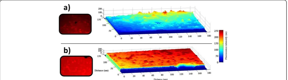
[image:7.595.57.543.515.706.2]An analysis using fluorescence microscopy was also carried out to characterize the emission intensity over the entire surface of the hybrid sensor. The samples were ex-cited using a mercury lamp with 510 to 560-nm filter in a Nikon Optiphot-2 (G2-A) microscope coupled with 3CCD MTI 8-bit camera. The emission intensities are shown in the Figure 11. The image in the Figure 11a is presenting a real view of the PSiMc/Rh-UTES hybrid sensor and its
corresponding tridimensional fluorescence profile over the entire surface, on which we can see the emission intensity produced for the immobilized Rh-UTES derivative. After metal sensor exposure, the hybrid sensor showed a strong brilliant red light (Figure 11b), and the fluorescence en-hancement was 0.22-fold (integrated emission). This value coincided well with the fluorescent enhancement observed on the fluorescent spectroscopy analysis (0.25-fold for the same metal concentration).
Conclusions
In this work we have proposed a novel method for detec-tion of Hg2+ions using rhodamine fluorescent derivative as the recognizing element. We studied the fluorescent performance of the derivative receptor in liquid and solid phases. In solution, after the Hg2+ addition to the Rh-UTES receptor, it was observed that the colorless solution becomes colored (pink) and a remarkable enhancement in the emission intensity. We found that both the color in-tensity and the fluorescent inin-tensity of the solution are linearly dependent on the metal concentration. This dis-tinct color and fluorescent change due to the spirolactam ring opening makes this derivative valuable for sensing ions through fluorescent or naked-eye detection. Add-itionally, a new sensing strategy was evaluated by immo-bilizing the Rh-UTES derivative on porous silicon devices. We found that after immobilization procedure, the Rh-UTES derivate maintained its fluorescent properties. PSi/ Rh-UTES' sensing capabilities for Hg2+ detection were studied. It was observed that metal-hybrid sensor coordin-ation produces a 0.25-fold enhancement in the integrated fluorescent emission at 6.95μM Hg2+ ion concentration. By comparing the fluorescence response of Rh-UTES de-rivative in liquid and solid phases, we found that the immobilization procedure produced a 277-fold integrated fluorescence increasing which highlights the benefits of using PSi optical devices as support of the organic
receptor. This work may open the door to the develop-ment of optical fluorescence-based sensors that can be easily used in field without the need of complicated instru-mentation, allowing the fast diagnosis of the quality of natural water sources or water from the industrial waste.
Abbreviations
ACN:acetonitrile; APTES: 3-aminopropyltriethoxysilane; ATR: attenuated total reflectance; au: arbitrary unities; AuNPs: gold nanoparticles; C: carbon; CDCl3: deuterated chloroform; d: double; DMSO: dimethyl sulfoxide; FTIR: Fourier transform infrared; H: high current density; H: proton; IR: infrared spectroscopy; L: low current density; m: multiplet; NMR: nuclear magnetic resonance; PL: photoluminescence; ppm: parts per million; Psi: porous silicon; PSiMc: porous silicon microcavity; q: quartet; Rh-UTES: rhodamine
organosilane derivative (3); SD: standard deviation; SEM: scanning electron microscopy; s: singlet; t: triplet.
Competing interests
The authors declare no competing interests.
Authors' contributions
GP designed the project, coordinated, reviewed and drafted the manuscript. MDC carried out the main experimental work, and performed the characterizations of interferometry, Infrared, fluorescent spectroscopy, fluorescent microscopy and SEM, and wrote the in liquid phase discussion of fluorescence spectroscopy. AA carried out the organic synthesis, NMR experiments, FTIR and NMR discussion, organized and drafted the manuscript. LHA participated in the PL characterization and results discussion, analysis data, and in drafting the manuscript. ABF performed the fluorescence microscopy analysis and made the tridimensional emission profile through computing data processing. FJMR participated in infrared measurements. All the authors read and approved the manuscript.
Acknowledgements
This work was supported by the National Council for Science and Technology of Mexico (CONACYT), Project No. CB-153161. We thank CONACYT for the following student scholarships: MDG No. 237466, LHA No. 270040, ABF No. 229949, and AA postdoctoral scholarship 2013 (3). We would like to thank the University of Guanajuato for NMR support via the CONACYT-UGTO National Laboratory (Grant 123732). We acknowledge to I.Q. Olga Dávalos Montoya for her technical support during FTIR studies and Dr. Jaime Ruiz Garcia (Physics Institute-UASLP) for the facilities given for use the fluorescence microscope.
Author details
1
[image:8.595.57.541.90.225.2]Biopolymers and Nanostructures Laboratory, Faculty of Chemical Sciences, Universidad Autónoma de San Luis Potosí, Av. Manuel Nava No. 6, San Luis
Potosí, San Luis Potosí 78210, México.2Materials Laboratory, Faculty of
Chemical Sciences, Universidad Autónoma de San Luis Potosí, Av. Manuel Nava No. 6, San Luis Potosí, San Luis Potosí 78210, México.3Colloids and
Interfaces Laboratory, Institute of Physics, Universidad Autónoma de San Luis Potosí, Av. Manuel Nava No. 6, San Luis Potosí, San Luis Potosí 78210, México.
Received: 1 May 2014 Accepted: 12 August 2014 Published: 26 August 2014
References
1. Bryan AJ, de Silva AP, De Silva SA, Rupasinghe RADD, Sandanayake KRAS: Photo-induced electron transfer as a general design logic for fluorescent molecular sensors for cations.Biosensors1989,4:169–179.
2. Woodroofe CC, Lippard SJ:A novel two-fluorophore approach to ratiometric sensing of Zn2+.J Am Chem Soc2003,125:11458–11459. 3. Kim SK, Lee SH, Lee JY, Lee JY, Bartsch RA, Kim JS:An excimer-based,
binuclear, on-off switchable calix[4]crown chemosensor.J Am Chem Soc 2004,126:16499–16506.
4. Lee SJ, Jung JH, Seo J, Yoon I, Park KM, Lindoy LF, Lee SS:A chromogenic macrocycle exhibiting cation-selective and anion-controlled color change: an approach to understanding structure-color relationships. Org Lett2006,8:1641–1643.
5. Metivier R, Leray I, Lebeau B, Valeur B:A mesoporous silica functionalized by a covalently bound calixarene-based fluoroionophore for selective optical sensing of mercury(II) in water.J Mater Chem2005,15:2965–2973. 6. Coronado E, Galan-Mascaros JR, Mart-Gastaldo C, Palomares E, Durrant JR,
Vilar-Ramn, Gratzel M, Nazeeruddin M:Reversible colorimetric probes for mercury sensing.J Am Chem Soc2005,127:12351–12356.
7. Marsella MJ, Newland RJ, Carroll PJ, Swager TM:Ionoresistivity as a highly sensitive sensory probe: investigations of polythiophenes functionalized with calix[4]arene-based ion receptors.J Am Chem Soc1995,
117:9842–9848.
8. Bartsch RA, Chapoteau E, Czech BP, Krzykawski J, Kumar A, Robison TW: Chromogenic diaza-crown ether dicarboxylic acids for determination of calcium ions.J Org Chem1994,59:616–621.
9. De Silva AP, Gunaratne HQN, Gunnlaugsson T, Huxley AJM, McCoy CP, Rademacher JT, Rice TE:Signaling recognition events with fluorescent sensors and switches.Chem Rev1997,97:1515–1566.
10. Amendola V, Fabbrizzi L, Foti F, Licchelli M, Mangano C, Pallavicini P, Poggi A, Sacchi D, Taglietti A:Light-emitting molecular devices based on transition metals.Coordin Chem Rev2006,250:273–299.
11. Chen X, Pradhan T, Wang F, Kim JS, Yoon J:Fluorescent chemosensors based on spiroring-opening of xanthenes and related derivatives. Chem Rev2011,112:1910–1956.
12. Huang CC, Chang HT:Selective gold-nanoparticle-based“Turn-On” fluorescent sensors for detection of mercury(II) in aqueous solution. Anal Chem2006,78:8332–8338.
13. Lee MH, Lee SJ, Jung JH, Lim H, Kim JS:Luminophore-immobilized mesoporous silica for selective Hg2+sensing.Tetrahedron2007,63:12087– 12092.
14. Zhou P, Meng Q, He G, Wu H, Duan C, Quan X:Highly sensitive fluorescence probe based on functional SBA-15 for selective detection of Hg2+in aqueous media.J Environ Monit2009,11:648–653.
15. Childress ES, Roberts C, Sherwood DY, LeGuyader CLM, Harbron EJ: Ratiometric fluorescence detection of mercury ions in water by conjugated polymer nanoparticles.Anal Chem2012,84:1235–1239. 16. Wang M, An X, Gao J:An off-on Hg(II) sensor excited by near-infrared to
visible upconversion nanorods.J Lumin2013,144:91–97. 17. Deng G:Principles of chemical and biological sensors.Mater Manuf
Processes1999,14:623–625.
18. Palestino G, Agarwal V, Aulombard R, Perez E, Gergely C:Biosensing and protein fluorescence enhancement by functionalized porous silicon devices.Langmuir2008,24:13765–13771.
19. Márquez J, Cházaro-Ruiz LF, Zimányi L, Palestino G:Immobilization strategies and electrochemical evaluation of porous silicon based cytochrome c electrode.Electrochim Acta2014, doi:10.1016/ j. electacta.2014.05.065.
20. Shiraishi Y, Miyamoto R, Zhang X, Hirai T:Rhodamine-based fluorescent thermometer exhibiting selective emission enhancement at a specific temperature range.Org Lett2007,9:3921–3924.
21. Karacali T, Cakmak B, Efeoglu H:Aging of porous silicon and the origin of blue shift.Opt Express2003,11:1237–1242.
22. Riikonen J, Salomaki M, van Wonderen J, Kemell M, Xu W, Korhonen O, Ritala M, MacMillan F, Salonen J, Lehto VP:Surface chemistry, reactivity, and pore structure of porous silicon oxidized by various methods. Langmuir2012,28:10573–10583.
23. Zhang X, Xiao Y, Qian X:A ratiometric fluorescent probe based on FRET for imaging Hg2+ions in living cells.Angewandte Chemie International Edition2008,47:8025–8029.
24. Tu J, Li N, Chi Y, Qu S, Wang C, Yuan Q, Li X, Qiu S:The study of photoluminescence properties of Rhodamine B encapsulated in mesoporous silica.Mater Chem Phys2009,118:273–276. 25. Yang H, Zhou Z, Huang K, Yu M, Li F, Yi T, Huang C:Multisignaling
optical-electrochemical sensor for Hg2+based on a rhodamine derivative with a ferrocene unit.Org Lett2007,9:4729–4732.
26. Yang YK, Yook KJ, Tae J:A rhodamine-based fluorescent and colorimetric chemodosimeter for the rapid detection of Hg2+ions in aqueous media. J Am Chem Soc2005,127:16760–16761.
doi:10.1186/1556-276X-9-431
Cite this article as:De la Cruz-Guzmanet al.:A turn-on fluorescent
solid-sensor for Hg(II) detection.Nanoscale Research Letters20149:431.
Submit your manuscript to a
journal and benefi t from:
7Convenient online submission
7Rigorous peer review
7Immediate publication on acceptance
7Open access: articles freely available online
7High visibility within the fi eld
7Retaining the copyright to your article