Comparative genome and phenotypic analysis of
Clostridium difficile
027 strains provides insight into the evolution of a hypervirulent
bacterium
Richard A Stabler
*
, Miao He
†
, Lisa Dawson
*
, Melissa Martin
*
,
Esmeralda Valiente
*
, Craig Corton
†
, Trevor D Lawley
†
,
Mohammed Sebaihia
†
, Michael A Quail
†
, Graham Rose
†
, Dale N Gerding
‡
,
Maryse Gibert
§
, Michel R Popoff
§
, Julian Parkhill
†
, Gordon Dougan
†
and
Brendan W Wren
*
Addresses: *London School of Hygiene and Tropical Medicine, Keppel Street, London, WC1E 7HT, UK. †Wellcome Trust Genome Campus, Hinxton, Cambridge, CB10 1SA, UK. ‡Hines VA Hospital, Hines, IL 60141, USA. §Institut Pasteur, rue du Dr Roux, 75724, Paris, France.
Correspondence: Brendan W Wren. Email: [email protected]
© 2009 Stabler et al.; licensee BioMed Central Ltd.
This is an open access article distributed under the terms of the Creative Commons Attribution License (http://creativecommons.org/licenses/by/2.0), which permits unrestricted use, distribution, and reproduction in any medium, provided the original work is properly cited.
Clostridium difficile virulence evolution
<p>A genome comparison of non-epidemic and epidemic strains of Clostridium difficile reveals gene gains that could explain how a hyper-virulent strain has emerged</p>
Abstract
Background: The continued rise of Clostridium difficile infections worldwide has been accompanied by the rapid emergence of a highly virulent clone designated PCR-ribotype 027. To understand more about the evolution of this virulent clone, we made a three-way genomic and phenotypic comparison of an 'historic' non-epidemic 027 C. difficile (CD196), a recent epidemic and hypervirulent 027 (R20291) and a previously sequenced PCR-ribotype 012 strain (630).
Results: Although the genomes are highly conserved, the 027 genomes have 234 additional genes compared to 630, which may contribute to the distinct phenotypic differences we observe between these strains relating to motility, antibiotic resistance and toxicity. The epidemic 027 strain has five unique genetic regions, absent from both the non-epidemic 027 and strain 630, which include a novel phage island, a two component regulatory system and transcriptional regulators.
Conclusions: A comparison of a series of 027 isolates showed that some of these genes appeared to have been gained by 027 strains over the past two decades. This study provides genetic markers for the identification of 027 strains and offers a unique opportunity to explain the recent emergence of a hypervirulent bacterium.
Background
Clostridium difficile, a spore-forming anaerobic bacillus that often resides in the gut of mammals, is the causative agent of
C. difficile infection (CDI) (reviewed in [1]). The hospital
environment and patients undergoing antibiotic treatment
provide a discrete ecosystem where C. difficile persists and
selected virulent clones thrive. Consequently, C. difficile is the most frequent cause of nosocomial diarrhea worldwide, Published: 25 September 2009
Genome Biology 2009, 10:R102 (doi:10.1186/gb-2009-10-9-r102)
Received: 8 June 2009 Revised: 29 June 2009 Accepted: 25 September 2009 The electronic version of this article is the complete one and can be
where patients exhibit a range of symptoms from mild diarrhea to life threatening pseudomembranous colitis (PMC) [2,3]. In most cases of CDI antibiotic therapies disrupt the protective gut microbiota, whereupon ingested or existent
C. difficile spores germinate, colonize the gastrointestinal tract and produce toxins. Another feature of CDI is the high relapse rate due to re-infection or reactivation of infection [2,3]. The population at risk for CDI includes not only patients on antimicrobial and other therapies that can alter the balance of the gut microbiota (for example, antacid/pro-ton pump inhibitors and non-steroidal anti-inflammatory drugs), but also the immunocompromised and the elderly. Given the continued use of broad-spectrum antibiotics and the rising numbers of immunocompromised and elderly patients, the problems associated with CDI are unlikely to recede.
Alarmingly, in the past 5 years a new group of highly virulent
C. difficile strains has emerged to cause outbreaks of increased disease severity in North America and Europe. Sev-eral studies have shown that patients infected with these PCR-ribotype 027 strains have more severe diarrhea, higher mortality and more recurrences [4-8]. Prior to 2003, only a handful of these strains were isolated in the UK, whereas cur-rently most typed isolates are PCR-ribotype 027. This is also mirrored in Canada, where 027 strains were undetected in 2000, but reached 75.2% of all PCR-ribotyped strains by 2003 [9]. The emergence of 027 strains might partially explain the 72% annual increase in mortality in the UK due to CDI to 6,500 cases in 2006 [7]. The CDI outbreaks at the Stoke Mandeville hospital, Buckinghamshire, marked the arrival of the epidemic 027 isolates to the UK. Between April
2003 and March 2006 a total of 498 patients acquired C.
dif-ficile at the hospital (measured by onset of symptoms 72 hours after admission), of which 127 died [10].
PCR-ribotype 027 strains are genetically highly uniform, which is confirmed by the application of diverse genotyping methods. For example, 027 strains are invariably designated as BI by restriction endonuclease analysis, NAP1 (North American pulsotype 1) by pulse field gel electrophoresis, are exclusively toxinotype III by toxinotyping and are indistin-guishable by multi-locus sequence analysis [11]. The earliest retrospective recorded PCR-ribotype 027 isolate was strain CD196 in 1985, which is a non-epidemic strain isolated from a single patient with CDI in a Paris hospital [12]. The next ret-rospective recorded 027 isolate was a non-epidemic strain designated BI-1, which was from a patient with CDI in a Min-neapolis hospital in 1988 [13]. Since 1988 a further 19 BI des-ignated strains (all PCR-ribotype 027) have been isolated and characterized by Gerding and colleagues representing a use-ful time-line of the evolution of 027 strains (DN Gerding, per-sonal communication).
Comparative phylogenomics (whole genome comparisons of bacteria using DNA microarrays combined with
Bayesian-based algorithms to model the phylogeny) was recently applied to 75 C. difficile strains of diverse origin, including 19 strains confirmed as PCR-ribotype 027 (16 BI strains from the US, CD196, a strain from a recent Canadian outbreak and a representative strain from the Stoke Mandeville outbreak designated R20291). All 027 strains formed a tight clade, which was distinct from the other 56 strains analyzed [14]. Closer inspection of the 027 clade revealed micro-evolution among strains with the historic non-epidemic CD196 and BI-1 strains as progenitors compared to their recently isolated counterparts [14]. These studies confirm the clonal nature of PCR-ribotype 027 strains and that they are continuing to evolve.
C. difficile is known to produce two related glucosylating tox-ins, named toxin A and toxin B, which are encoded on the pathogenicity locus (PaLoc) [15]. For some time, toxin pro-duction has been the main focus of study when addressing
virulence of C. difficile. However, in the hamster model of
infection toxin B plays the most significant role in infection [16]. A recent report has shown the binding domain of toxin B
in 027 strains to be highly divergent compared to other C.
dif-ficile strains [8]. However, the significance of the difference of the 027 toxin B gene sequence has yet to be investigated. The PaLoc also includes toxin regulatory components, including
tcdR, a sigma factor, and tcdC, a negative regulator that
destabilizes the TcdR-holoenzyme to prevent transcription of the PaLoc [17]. It has been reported that some 027 strains can
produce more toxin in vitro [18], which was initially
attrib-uted to deletions in the negative regulator tcdC. Further char-acterization has revealed that the 18-bp in-frame deletion was found to have no effect on toxin production [19]. Two
addi-tional deletions have been identified within tcdC, a 39 and
single base-pair deletion. The single base-pair deletion results in the formation of a stop codon downstream and truncation of the protein, thus leading to increased toxin pro-duction. However, various deletions have been identified in
tcdC in non-epidemic PCR-ribotypes as well [20], suggesting the increased virulence cannot solely be attributed to these deletions. This has stimulated debate on the mode of hyper-virulence in the epidemic 027 strains. Apart from classic vir-ulence determinants such as toxin production, other factors such as antibiotic resistance, increased motility and adher-ence in the gut, increased resistance to bile salts and increased transmissibility manifested through sporulation might explain the emergence of epidemic 027 strains. A recent report comparing three 'historical' 027 strains from Sweden with an epidemic strain concluded that the epidemic strain sporulated more readily than its three non-epidemic counterparts [21].
Given the medical and economic importance of CDI and the difficulties in studying the genetics of C. difficile, we recently
reported the complete genome sequence of a pathogenic C.
PMC at a hospital in Zurich in 1982 [22]. The full sequence revealed a 4.29 Mb chromosome with a mosaic of potential mobile genetic elements, antibiotic resistance genes and vir-ulence determinants [22].
The rapid international emergence of the C. difficile 027
strain lineage provides a unique opportunity to understand the recent emergence of a highly virulent bacterium. In this study we undertake a three-way genome comparison of an
'historic' non-epidemic 027 C. difficile strain (CD196), a
recent epidemic and hypervirulent 027 strain (R20291) and the previously published PCR-ribotype 012 strain (630). Where possible we relate genetic differences to phenotypic differences observed in these strains with respect to motility, survival, antibiotic resistance and toxicity.
Results and discussion
Genome comparison of the PCR-ribotype 027 strains (CD196 and R20291) and strain 630
The two newly sequenced genomes of the PCR-ribotype 027 strains (CD196 historic and R20291 modern; Table 1) were compared with the previously sequenced strain 630 (PCR-ribotype 012). The three strains share 3,247 core genes, including those encoding determinants important for patho-genesis, such as antimicrobial resistance,
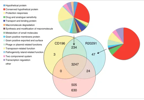
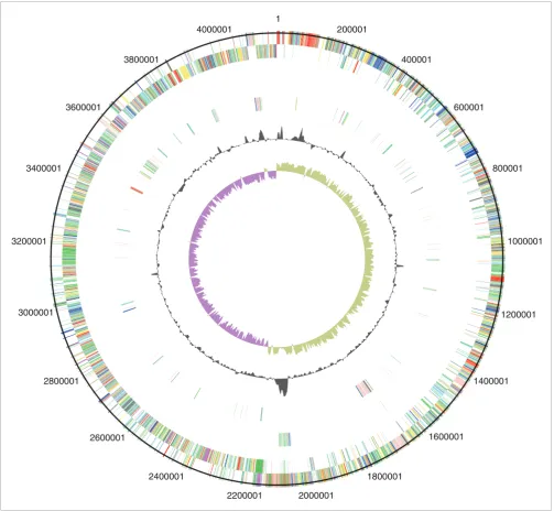
ethanolamine/pro-panediol metabolism, sporulation, a beta-lactamase-inducing penicillin-binding protein, a quaternary ammonium compound-resistance protein, tellurium resistance proteins, a putative nogalamycin resistance protein and L-rhamnose biosynthesis (Figure 1). There are 505 coding sequences (CDSs) unique to 630 compared to the 027 strains, whereas there are 47 CDSs unique to R20291 and three CDSs unique to CD196 (Figure 1). The locations of regions of genetic differ-ence between the three strains are highlighted in the concen-tric circular chromosome representations of the three genomes (Figure 2). There are 234 genes unique to both 027 ribotypes spread among at least 50 regions of genetic differ-ence (Figure 2; Additional data file 1). These include a phage island, transposon genes, two-component response regula-tors, drug resistance genes, transporter genes and type I restriction enzyme/restriction modification genes (Addi-tional data file 1).
There are 14 CDSs that have been disrupted by an insertion in both 027 strains but are intact in 630; conversely, 12 CDSs are intact in both 027 strains but have been disrupted in 630 (Additional data file 2). All three genomes have multiple cop-ies of genes for transposase-like proteins that have been inserted both intragenically and intergenically. In C. difficile
630 there are eight full transposon copies and two remnant copies; all eight functional copies have inserted within CDSs. In both 027 strains there are 17 transposon copies, of which only 6 inserted within CDSs. Only three CDSs are interrupted by transposons in all three strains. Furthermore, three CDSs have been truncated by sequence loss in both 027 strains but are intact in 630 and 10 CDSs are truncated in 630 but not 027 strains (Additional data file 2). Finally, point mutations have resulted in frameshifts exclusively in three 630 CDSs and 10 of the 027 strain CDSs (Additional data file 2).
Toxin-related genes specific to 027
Variation within the PaLoc region (containing toxins A and B
and their associates genes) [15] between C. difficile strains
has been observed frequently and has been used to develop the toxinotyping method to distinguish strains [23-25]. PCR-ribotype 027 isolates are invariably toxinotype III, whereas 630 (PCR-ribotype 012) is toxinotype 0. A comparison of the PaLoc sequences from 630, R20291 and CD196 confirms the
previous data, indicating that the tcdB sequence varies
among strains, particularly at the 3' region, which encodes the toxin-binding domain [26]. However, there is a high level of
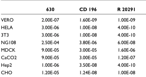
sequence conservation in tcdB between the 027 strains
[image:3.612.54.298.424.734.2]CD196 and R20291 compared to strain 630. Examination of the relative in vitro cytotoxicity of these three strains on sev-eral cell lines confirms differences in both toxicity and cell line specificity (Table 2). Purified toxin B from R20291 has more potent activity than that from 630 in all eight cell lines tested whereas the historic 027 (CD196) is more potent in six of the eight cell lines tested (Table 2). Given the recent dem-onstration in the hamster model of CDI that toxin B, and not toxin A, is essential for virulence, the observation that toxin B Table 1
Strains used in this study
Isolates Date isolated/recorded City, state/province
630 1982 Zurich, Switzerland
CD196 1985 Paris, France
R20291 2006 Aylesbury, UK
BI-1 26/2/1988 Minneapolis, MN
BI-2 14/1/1991 Tucson, AZ
BI-3 14/12/1990 Minneapolis, MN
BI-4 10/2/1993 Minneapolis, MN
BI-5 25/8/1995 Albany, NY
BI-6 20/5/2003 Portland, OR
BI-6p 09/9/2004 Atlanta, GA
BI-6p2 09/9/2004 New Jersey
BI-7 20/5/2003 Portland, OR
BI-8 22/1/2004 Portland, ME
BI-10 10/8/2001 Pittsburgh, PA
BI-11 10/8/2001 Pittsburgh, PA
BI-12 09/9/2004 Camp Hill, PA
BI-13 09/9/2004 New Jersey
BI-14 09/9/2004 New Jersey
BI-15 09/9/2004 New Jersey
BI-16 01/9/2004 Augusta, ME
from strain R20291 has a more potent activity over a broader range of cell types may indicate that this is a contributory fac-tor to the hypervirulence of R20291 [16].
Regulation of toxin expression has also been shown to vary
between strains of C. difficile, which has been attributed to
mutations in the negative regulator tcdC [9]. The most
nota-ble of these mutations is the 1-bp deletion present in 027 strains that results in a frame shift and truncation of TcdC, causing de-repression of the PaLoc [17]. A single base dele-tion at posidele-tion 117, resulting in truncadele-tion of TcdC at the 66th amino acid, was confirmed in both R20291 and CD196 but absent from 630. The presence of the 18-bp deletions in both R20291 and CD196 but their absence from 630 was con-firmed.
The binary ADP-ribosyltransferase toxin, first identified in
1988 in the strain CD196 [12], consists of two genes, cdtA and
cdtB. Surveys have identified the binary toxin in up to 8.6% of
C. difficile strains [27-30]; recently, however, binary toxin positive PCR-ribotype 027 incidence has reached 41.3% in the UK [31]. Additionally, the binary toxin has been linked with
increased severity of disease [32-34]. Sequence analysis con-firms the presence of full-length cdtA and cdtB genes in both CD196 and R20291, which by contrast have accumulated sequence deletions, several frameshift mutations and in-frame stop codons in 630. Recently, the CDS upstream has been identified as the binary toxin response regulator, desig-nated cdtR [35]. C. difficile 630 contains a functional copy of
cdtR despite lacking binary toxin, and CdtR is 96% identical to the homologues found in both 027 isolates.
Differences in antibiotic resistance between 630 and PCR-ribotype 027 strains
In contrast to strain 630, the epidemic 027 strains are highly resistant to fluoroquinolones due to point mutations in the
DNA gyrase genes [36]. Comparison of the gyrA gene
identi-fied seven point mutations in DNA gyrase genes between C.
difficile 630 and both 027 strains. Four mutations are silent and two substitutions - Leu406Ile and Asp468Asn - served. Interestingly, the previously described Thr82Ile con-version was only present in the epidemic 027 [36]. Two silent point mutations (A1458G and C1890T) were identified in the
gyrB gene of the 027 strains. Three fluoroquinolones
[image:4.612.56.554.86.431.2](gati-Distribution of orthologues CDSs in C. difficile strains 630, CD196 and R20291 Figure 1
Distribution of orthologues CDSs in C. difficile strains 630, CD196 and R20291. The Venn diagram shows the number of genes unique, shared or core between the three strains. The associated pie charts show the breakdown of the functional categories assigned to these CDS.
CD196
630
R20291
3247
234
24
505
3
47
Hypothetical protein
Conserved hypothetical protein Protection responses
Drug and analogue sensitivity Transport and binding protein Macromolecule degradation
Synthesis and modification of macromolecule Metabolism of small molecules
Gram positive membrane protein Gram positive exported and surface Phage or plasmid related functions Transposon-related function Pathogenicity island-related function Two componenet system
Transcription regulation other
floxacin, moxifloxacin and lexofloxacin) were used and results showed that R20291 was highly resistant to the
fluor-oquinolones (≤ 32 mg/l for all three fluoroquinolones), but
CD196 was fluoroquinolone sensitive (gatifloxacin minimum inhibitory concentration (MIC) 1.5 mg/l, moxifloxacin MIC 2 mg/l and lexofloxacin MIC 3 mg/l) and 630 was sensitive or had intermediate resistance to fluoroquinolones (gatifloxacin
MIC 2 mg/l, moxifloxacin MIC 1.5 mg/l and lexofloxacin 6 mg/l).
Sequence data revealed that both 027s have acquired two
unique conjugative transposons absent in C. difficile 630.
One of these transposons (CTn-027) encodes a novel
chlo-ramphenicol resistance gene (CDR20291_3461). R20291 and CD196 demonstrated intermediate resistance (MIC 16 mg/l),
[image:5.612.54.557.86.550.2]Circular representations of C. difficile chromosomes Figure 2
Circular representations of C. difficile chromosomes. From the outside (scale in bp): circles 1 and 2 show the position of R20291 CDS transcribed in a clockwise and anti-clockwise direction colored according to predicted function; circle 3 shows CDS unique to R20291; circle 4 shows CDS unique to both R20291 and CD196; circle 5 shows GC content; circle 6 shows GC deviation (> 0%, olive; < 0%, purple). Color coding for CDS functions: dark blue, pathogenicity/adaptation; black, energy metabolism; red, information transfer; dark green, surface-associated; cyan, degradation of large molecules; magenta, degradation of small molecules; yellow, central/intermediary metabolism; pale green, unknown; pale blue, regulators; orange, conserved hypothetical; brown, pseudogenes; pink, phage and IS (Insertion Sequence) elements; grey, miscellaneous.
1
200001
400001
600001
800001
1000001
1200001
1400001
1600001
1800001
2000001 2200001
2400001 2600001
2800001 3000001 3200001
3400001
3600001
3800001
but 630 was sensitive to chloramphenicol (MIC 12 mg/l) (P < 0.05).
C. difficile strain 630 has two copies of the erythromycin
resistance gene (ermB1/CD2007 and ermB2/CD2010) on a
mobile transposon Tn5398 (CD2001-2010b), which was
absent in CD196 and R20291. However, experimental data
showed that C. difficile 630 and R20291 were erythromycin
resistant (MIC ≥ 256 mg/l) whereas CD196 had intermediate
resistance (MIC 2 mg/l). Additionally, strain 630 has a
tetra-cycline resistance gene (tetM; CD0508) on CTn3/Tn5397
(CD0496-511), which is absent in CD196 and R20291. C.
diff-icile 630 demonstrated tetracycline resistance (MIC 64 mg/l) in contrast to CD196 and R20291, which were tetracycline
sensitive (MIC 0.17 mg/l and 0.22 mg/l, respectively) (P <
0.001).
The difference between drug resistance patterns may reflect changes in antibiotic policy. For example, both CD196 and
630 predate 1992 when Golledge et al. [37] demonstrated
clindamycin not to be a risk factor; subsequently, clindamy-cin use has been strongly associated with PCR-ribotype 027 outbreaks [38-40]. This demonstrates that antibiotic usage may be driving the evolution of drug resistance and the pre-dominance of certain isolates.
027-specific genes involved in flagella biosynthesis, glycosylation and motility
Flagella have been found to be important for motility in sev-eral enteric pathogens as a prerequisite to traverse the mucous layer of the gut to interact with gut epithelial cells [41-43]. Additionally, chemotaxis mediated through motility is important in survival, to enable movement towards nutri-ent-rich sources and movement away from noxious environ-ments. Flagella have been observed in some C. difficile strains [44,45]. Post-translational modification of flagellin proteins by glycosylation has been shown to be prevalent in several bacterial pathogens and the loci encoding these modifications are frequently located adjacent to the structural flagellin genes [46]. Such modifications are important in subverting
host immune defenses [47], autoagglutination [48] and adhe-sion and colonization [49].
In 630, flagella-associated genes are found in two loci, F1 (CD0226-CD0240) and F3 (CD0245-CD0271), which are separated by an inter-flagella locus F2 (CD0241-CD0244). Loci F2 encodes a phosphoserine phosphatase, two conserved hypothetical proteins and a putative CDP-Glycerol:Poly (glyc-erophosphate) glycerophosphotransferase [14] (Figure 3). Microarray analysis of this region previously showed a loss of, or high divergence in, F1 and F2 in all 027 isolates tested [14]. The sequence data from both R20291 and CD196 show that the F1 locus has been retained, but with only 84 to 90% sequence identity, whereas the four genes present in the inter-flagella F2 locus of 630 have been replaced by six differ-ent genes encoding a glycosyl transferase (family 2), two putative uncharacterized proteins, a putative carbamoyl-phosphate-synthetase and a putative ornithine cyclodeami-nase (Figure 3).
The variation in the F1 region between 630 and the 027 ribotypes may be important in motility, as there are clear phe-notypic differences in the motility of 630 and the 027
ribotypes CD196, R20291 and BI-16 (Figure 4). C. difficile
630 is less motile than the 027 ribotypes, whereas M120 is non-motile (Figure 4). Microarray data have shown the absence/divergence of the complete F3 region in M120 [14]. Recent sequence data for M120 have confirmed the deletion of the entire F3 region in this strain [50], explaining the lack of motility for strain M120. The subtle differences in motility between the 630 and the 027 ribotypes may be due to the lev-els of sequence conservation over the F1 region.
The different genes present in the F2 region of 630 and the 027 ribotypes may be important in the glycosylation of the flagella, as the six genes present in R20291 and CD196 con-tain glycosyl transferases. Studies in other enteric bacteria
such as Campylobacter jejuni have shown that both Flagellin,
encoded by FlaA, as well as post-translational modifications
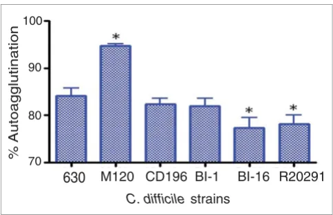
of it are required for autoagglutination, which is linked to vir-ulence [48]. Significant differences in autoagglutination between 630 and the more recent 027 isolates R20291 and
BI-16 (P < 0.05) were observed, whereas the more historic
027 isolates BI-1 and CD196 show no significant difference in autoagglutination compared to 630 (Figure 5). The differ-ences in autoagglutination observed between 630 and the recent 027 isolates are likely to be multifactorial as, in addi-tion to flagella and glycosylaaddi-tion, other surface phenomena can contribute to autoagglutination.
[image:6.612.53.296.116.244.2]Four 027 unique genes upstream of the flagella F1 region (CDR20291_0223-0226 and CD196_0236-0239) that are absent in 630 may be involved in virulence. The four CDSs encode DTDP-4-dehydrorhamnose reductase, glucose-1-phosphate thymidylyltransferase, DTDP-4-dehydrorham-nose 3,5-epimerase and DTDP-glucose 4,6-dehydratase. Table 2
Toxin B cytotoxicity assay
630 CD 196 R 20291
VERO 2.00E-07 1.60E-09 1.00E-09
HELA 3.00E-06 1.00E-08 4.00E-10
3T3 3.00E-06 1.00E-08 4.00E-10
NG108 2.50E-04 3.80E-06 6.00E-08
MDCK 9.00E-05 3.00E-05 1.60E-06
CaCO2 9.00E-05 3.00E-05 1.20E-07
Hep2 1.00E-06 3.50E-08 4.00E-10
CHO 1.20E-05 1.24E-08 1.00E-08
These four enzymes (RlmA, B, C and D) are involved in the synthesis of L-rhamnose. Carbohydrates such as L-rhamnose can act as structural elements as well as energy sources [51] and can be important virulence factors in both Gram-positive
and Gram-negative bacteria. In Vibrio cholerae, Escheichia
coli and Salmonella enterica, L-rhamnose is an important
residue in the O-antigen of lipopolysaccharides. In
Strepto-coccus mutans, L-rhamnose is part of an antigen involved in colonization of tooth surfaces [52] and mutations in this path-way have been shown to prevent initiation and maintenance
of infection [53]. In Mycobacterium tuberculosis,
L-rham-nose links peptidoglycan and arabinogalactan to form the unique cell wall. Given their co-location in the F regions, it is possible that these genes may play a role in flagellin glycosyla-tion in the 027 strains.
027 specific regulatory genes that may be important in survival
Regulatory genes form a large proportion of the 027-specific genes, with 8 two-component regulators and 15 other tran-scriptional regulators. One of the most striking regions of
genetic difference was an additional complete copy of the agr
[image:7.612.55.555.85.545.2]ACT comparison of flagellin and flagellin glycosylation-associated loci Figure 3
ACT comparison of flagellin and flagellin glycosylation-associated loci. F1 genes are CD0226-240 (630), CDR20291_0227- 241 (R20291), and CD196_0240-254 (CD196). F2 genes are CD0241-244 (630), CDR20291_0242-247 (R20291), and CD196_0255-260 (CD196). F3 genes are CD0245-271 (630), CDR20291_0248-275 (R20291), and CD196_0261-288 (CD196). Red bars indicate > 84% DNA sequence identity.
F1
F2
F3
630
R 20291
regulatory locus (termed agr2), consisting of agrA
(CDR20291_3189/CD196_3143), agrB (CDR20291_3187/
CD196_3141), agrC (CDR20291_3188/CD196_3142) and
agrD (CDR20291_3187a/CD196_3141a). The agr1 locus from C. difficile 630 contains only single copies of agrB and
agrD, with the response regulator (agrA) and histidine
pro-tein kinase (agrC) genes absent. The agr1 locus was present
in both 027 strains. The complete agr locus (agrA to agrD)
has been identified as a key regulatory system involved in
multiple aspects of virulence and quorum sensing in
Staphy-lococcus aureus [54]. Downstream of the agr2 locus are three 027-specific CDSs that encode two putative membrane pro-teins and an ABC transporter ATP-binding protein.
One of the additional transcriptional regulators in the 027 ribotypes is a PadR-like transcriptional regulator (CDR20291_2964/CD196_2917). The PadR family regulates phenolic acid metabolism, which may be important in sur-vival of bacteria in the gut, where energy sources are limited. The CDS is found within a region of six 027specific genes transcribed on the opposite strand to the other five CDSs -that encode a predicted enoate reductase, a nitrate/nitrite transporter and a conjugative transposon site-specific recom-binase. The PadR regulator may also be important in
toler-ance or production of p-cresol, a phenolyic agent produced by
C. difficile from the degradation of tyrosine. The p-cresol operon CD0153-155 was conserved within both 027s and in 630. However, there are clear phenotypic differences between
the tolerance to p-cresol between the recent 027 isolates and
630 [55], which may be due to PadR or another transcrip-tional regulator.
Genetic differences between the historic CD196 strain and the R20291 hypervirulent strain
Sequence data show that there are at least five genetic regions unique to the epidemic 027 (R20291) compared to the non-epidemic 027 strain (CD196) (Table 3). We hypothesize that these newly identified R20291 genetic elements contribute to the virulent phenotype of this clone. These genetic regions include a unique approximately 20-kb phage island of high G+C DNA content termed SMPI1 inserted into a 027 unique
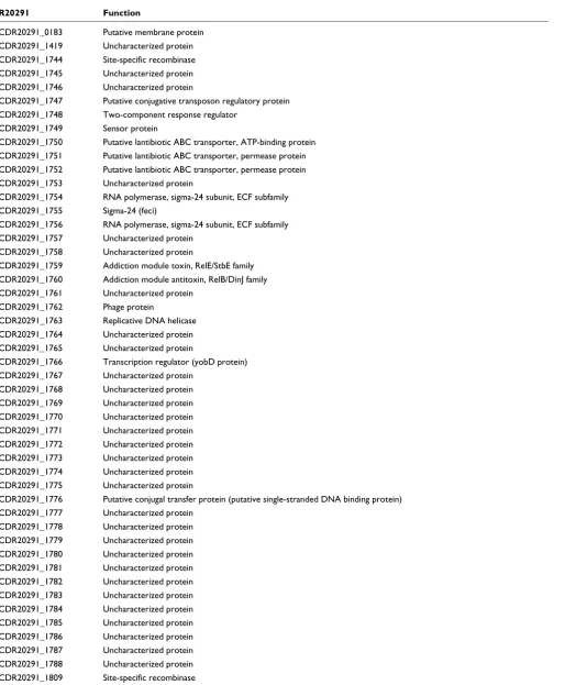
conjugative transposon (named CTn027; Figure 6). This
phage island insertion sequence disrupts the R20291 CDS CDR20291_1744 and carries a number of cargo genes present only in R20291, including a two-component response regula-tor (CDR20291_1748), a putative lantibiotic ABC transporter (CDR20291_1752), a putative cell surface protein along with a number of hypothetical and conserved hypothetical pro-teins. CDR20291_1755 is a unique R20291 gene encoding a transcriptional regulator (σ24). The phage island also encodes
a toxin-antitoxin system (RelE/StbE family) that is important in maintaining the stability of mobile elements [56]. RelE encodes a stable toxin that inhibits translation by cleaving mRNAs on translating ribosomes [57]. The toxin is inhibited by an unstable anti-toxin (RelB). This toxin-antitoxin system has been linked to translation moderation under amino-acid starvation stress [58].
Both 027 strains share a similar prophage (prophage phi-027), which has integrated between the orthologues of 630 CDSs CD1566-7. These prophages (CDR20291_1415-1465, CD196_1438-89) are identical apart from one small region. CD196 contains three strain-specific adjacent CDSs, the only CD196-specific CDS in the whole genome, which encode a
Comparative motility assays for C. difficile strains Figure 4
Comparative motility assays for C. difficile strains. The motility of strain 630 was compared to that of both recent and historic 027 ribotypes, R20291, BI-16 and CD196; M120 was the non-motile control. Strains were inoculated into 0.05% BHI agar and incubated for 24 hours in an anaerobe chamber. The motility is visualized as stalactite projections.
630 CD196 R20291 BI -16 M120
[image:8.612.54.296.455.611.2]Autoagglutination of C. difficile strains Figure 5
Autoagglutination of C. difficile strains. C. difficile strains were grown on BHI plates for 1 to 2 days, then inoculated into pre-equilibrated phosphate-buffered saline to an OD600 nm of 1.0 (± 0.1). These were incubated for 24 hours in pre-equilibrated glass tubes, then the OD600 nm was measured. The percentage of autoagglutination was normalized to the starting OD ((Starting OD - Final OD)/Final OD × 100). The bars indicate the percentage of cells autoagglutinating. Significant differences in autoagglutination are marked with an asterisk; P < 0.05, Students t-test. M120 is a non-motile strain thar autoagglutinates to a significantly higher level than 630 (P < 0.05).
630
M120 CD196 BI-1 BI-16 R20291C. difficile strains
% Autoagglutination 70
R20291 Function
CDR20291_0183 Putative membrane protein CDR20291_1419 Uncharacterized protein
CDR20291_1744 Site-specific recombinase
CDR20291_1745 Uncharacterized protein CDR20291_1746 Uncharacterized protein
CDR20291_1747 Putative conjugative transposon regulatory protein
CDR20291_1748 Two-component response regulator
CDR20291_1749 Sensor protein
CDR20291_1750 Putative lantibiotic ABC transporter, ATP-binding protein
CDR20291_1751 Putative lantibiotic ABC transporter, permease protein CDR20291_1752 Putative lantibiotic ABC transporter, permease protein
CDR20291_1753 Uncharacterized protein
CDR20291_1754 RNA polymerase, sigma-24 subunit, ECF subfamily CDR20291_1755 Sigma-24 (feci)
CDR20291_1756 RNA polymerase, sigma-24 subunit, ECF subfamily
CDR20291_1757 Uncharacterized protein CDR20291_1758 Uncharacterized protein
CDR20291_1759 Addiction module toxin, RelE/StbE family
CDR20291_1760 Addiction module antitoxin, RelB/DinJ family CDR20291_1761 Uncharacterized protein
CDR20291_1762 Phage protein
CDR20291_1763 Replicative DNA helicase CDR20291_1764 Uncharacterized protein
CDR20291_1765 Uncharacterized protein
CDR20291_1766 Transcription regulator (yobD protein) CDR20291_1767 Uncharacterized protein
CDR20291_1768 Uncharacterized protein
CDR20291_1769 Uncharacterized protein
CDR20291_1770 Uncharacterized protein CDR20291_1771 Uncharacterized protein
CDR20291_1772 Uncharacterized protein CDR20291_1773 Uncharacterized protein
CDR20291_1774 Uncharacterized protein
CDR20291_1775 Uncharacterized protein
CDR20291_1776 Putative conjugal transfer protein (putative single-stranded DNA binding protein) CDR20291_1777 Uncharacterized protein
CDR20291_1778 Uncharacterized protein
CDR20291_1779 Uncharacterized protein CDR20291_1780 Uncharacterized protein
CDR20291_1781 Uncharacterized protein
CDR20291_1782 Uncharacterized protein CDR20291_1783 Uncharacterized protein
CDR20291_1784 Uncharacterized protein
CDR20291_1785 Uncharacterized protein CDR20291_1786 Uncharacterized protein
CDR20291_1787 Uncharacterized protein
[image:9.612.56.570.98.721.2]putative phage anti-repressor and two putative uncharacter-ized proteins. R20291 appears to have lost these three CDSs and replaced them with a single putative uncharacterized protein that has 88% identity at the 5' end to one of the lost uncharacterized proteins and may represent a pseudogene. In addition, there is a unique R20291 region encoding six genes,
including matE (CDR20291_1779), a member of the
Multi-antimicrobial extrusion family drug/sodium antiporters. This region also shows a high G+C content, indicating recent acquisition.
Acquisition of R20291-specific genes in other PCR-ribotype 027 strains over time
In order to validate the presence of the R20291-specific genes and to monitor their acquisition over time, PCR analysis was undertaken on 19 PCR-ribotype 027 strains that have been isolated over a 16-year period across the US (Table 1). These isolates were typed by restriction endonuclease analysis as BI, which is equivalent to PCR-ribotype 027; however, each iso-late represents a unique small variation found in the BI restriction endonuclease analysis patterns. Strains BI-1 to -5 are considered 'historic' and were isolated between 1988 and 1995. BI-6 to -17 are considered 'modern' and were isolated from 2001 to 2004. Strains 630 and CD196 (ribotypes 012 and 027, respectively) were used as negative controls (Table 4).
Eleven R20291-specific genes were chosen for PCR analysis (Table 4). Four genes (CDR20291_1744, CDR20291_1751 to _1753) are found on the R20291-specific phage island; gene CDR20291_1744 is a site specific recombinase, CDR20291_1751 and CDR20291_1752 are putative lantibi-otic ABC transporters and CDR20291_1753 is unknown. In addition, the R20291-specific transcriptional regulator (YobD protein) is also present in the 'modern' BI strains (6p, 8, 12, 16 and 17) but absent from the earlier BI strains. Only one R20291-specific gene (CDR20291_1419; BRO protein family) was amplified in the early BI strains (BI-1, -2 and -5), showing the acquisition of R20291 genes was more prevalent in the epidemic 027 BI strains (Table 4). Furthermore, recent data demonstrate that the epidemic 027 strain, named BI-6, is more virulent in the hamster infection model than early strains such as BI-1 [13].
Conclusions
C. difficile is the most frequent cause of nosocomial diarrhea worldwide, in part due to the rapid and dramatic worldwide emergence of the PCR-ribotype 027 strains. We show that 027 strains have considerable genetic differences compared to strain 630 that may relate to observed phenotypic differ-ences in motility, survival, antibiotic resistance and toxicity. Additionally, five genetic regions appear to have accumulated in the modern day epidemic 027 strain R20291 compared to the historic CD196 counterpart. This includes a unique approximately 20-kb phage island of high G+C content DNA
(SMPI1) inserted into a 027 unique conjugative transposon. However, the role of individual determinants through
muta-genesis and the testing of mutants in appropriate in vivo
models is required to provide conclusive evidence. Some of these elements appear to have accumulated in 027 strains over the past 16 years and may therefore be useful genetic markers for epidemic 027 strains. The observed gene differ-ences between these strains might individually or collectively explain why modern 027 strains are more likely to be epi-demic and could explain the higher case-fatality ratio and persistence associated with infection by these strains. These studies facilitate pinpointing the genetic and phenotypic attributes that may explain the emergence of the hyperviru-lent 027 strain and contribute in general to our understand-ing of the evolution of bacterial virulence.
Materials and methods
Bacterial strains and growth conditions
C. difficile 027 isolates designated BI-1 to -17 were provided
by Dale Gerding (Hines VA Hospital, Hines, IL, USA). C.
dif-ficile 630 [59] was isolated from a patient with PMC in Zurich, 1982 and has been fully sequenced by the Wellcome Trust Sanger Institute [22]. Strain 630 was provided by Peter Mullany, Eastman Dental Institute, London, UK. The 027 strain CD196 is a non-epidemic strain isolated from a patient with PMC in Paris, 1985 and was provided by Michel Popoff, Institut Pasteur, Paris, France. The hypervirulent 027 R20291 was isolated from a recent outbreak in Stoke Man-deville, UK and was provided by Jon Brazier, Anaerobe Ref-erence Laboratory, Cardiff, UK.
C. difficile was routinely cultured on Braziers agar (Biocon-nections, Leeds, South Yorkshire, UK) containing 4% egg
yolk, C. difficile supplement (Bioconnections) and 2%
defibri-nated horse blood or in brain heart infusion (BHI) broth
con-taining C. difficile supplement (Oxoid, Basingstoke,
Hampshire, UK) and 0.04% cysteine. All cultures were grown
in an anaerobic atmosphere (10% CO2, 10% H2, 80% N2) at
37°C.
DNA isolation and PCR amplification
Genomic C. difficile DNA was isolated by cell lysis, phenol
NanoDrop1000 spectrophotometer and by running the sam-ples on 1.0% agarose gel, 100 mV for 45 minutes.
PCR amplifications were performed using primers described in Additional data file 3. Reactions were performed using 35 cycles at 94°C for 15 seconds, 50°C for 1 minute, 72°C for 1 minute, followed by a final extension of 72°C for 7 minutes. PCR products were analyzed on 1% agarose gels run at 100 mV for 1 hour and stained with ethidium bromide.
DNA sequencing and assembly
Genomic sequences were generated by combining data from 454/Roche technology (using GS20 for R20291 and FLX for CD196) with shotgun capillary reads from ABI 3730xl
analyz-ers (Table 5). Reads from the 454 platform were assembled de
novo (without guidance from a reference sequence) into con-tigs using newbler (Roche, Welwyn Garden City,
Hertford-shire, UK), then shredded into artificial reads of comparable lengths to capillary reads. An assembly was created with data from both platforms using Phrap. For each combined assem-bly the order of contigs was estimated by comparing them to strain 630 genomic sequence using ABACAS [60]. To further correct homopolymer tract errors inherent in early 454 sequencing data, Solexa (Illumina, Saffron Walden, Essex) sequence data were generated for isolate R20291. The
Illu-mina sequences were assembled de novo using Velvet [61]
and the resulting contigs were incorporated with the com-bined 454 and capillary assembly. Closing gaps between con-tigs for both CD196 and R20291 was either by primer walking on subclones from the capillary shotgun or by sequencing PCR products covering gaps between adjacent contigs. The final contiguous sequence for CD196 was mostly from com-bined data but small regions were covered with only 454 data (a total of less than 2.6% of the sequence) or with only
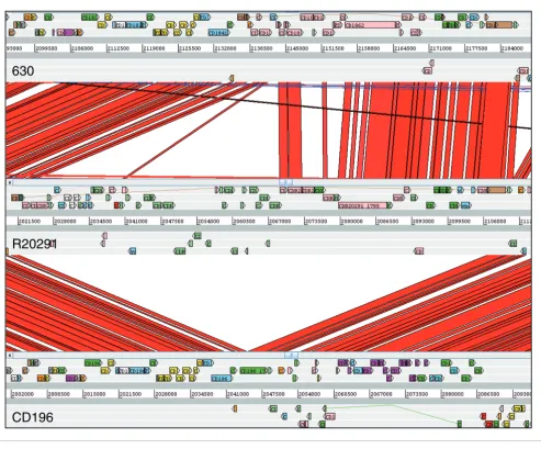
[image:11.612.59.552.89.498.2]capil-Comparison of phage island SMPI (Stoke Mandeville phage island) between C. difficile strains 630, CD196 and R20291 Figure 6
Comparison of phage island SMPI (Stoke Mandeville phage island) between C. difficile strains 630, CD196 and R20291.
630
R20291
lary reads, giving a consensus confidence of < 41 (< 0.3% of the sequence), and rRNA repeats were represented as consen-sus sequences (Table 5). All regions of the final finished R20291 assembly are covered by high quality capillary reads or by combinations of data from at least two sequencing tech-nologies, although three gaps remain where ribosomal rRNA operons have not been bridged by read-pairs. All regions of the final finished R20291 assembly are covered by high qual-ity capillary reads or by combinations of data from at least two sequencing technologies.
Genome annotation, comparison and orthologue identification
Genome annotation of C. difficile strains CD196 and R20291
was based on previously published annotations of C. difficile
strain 630 [22]. The genomic sequences of strains CD196 and R20291 were compared against the database of strain 630 proteins by blastx, and a CDS feature in the query genome was created when a hit of over 90% identity was found. Glimmer3 [62] was used to predict CDSs in genomic regions where no significant hits were found. Any unique genomic regions left were examined and annotated manually in Artemis [63]. The genome comparisons were visualized in Artemis and ACT (Artemis Comparison Tool) [64]. The sequences of CD196 and R20291 can be accessed using the accession numbers [EMBL: FN538970] and [EMBL: FN545816], respectively.
The reciprocal-best-hit fasta search algorithm was used to identify orthologues among strains 630, CD196 and R20291. All CDSs in the query genome were searched in the database of subject CDSs by FASTA [65]. When a hit of over 30% iden-tity and over 80% length was found, the hit CDS in the subject genome was searched again in the database of query CDSs in a similar fashion. If the hit of the second search is the same as the original query CDS, the two CDSs are considered as ortho-logues by this method. These identified orthoortho-logues were manually curated to account for inaccuracies caused by inserted elements, frameshifts and pseudogenes.
Toxin B toxicity assay
Toxins were produced by the dialyzing cultivation method [66] with BHI broth (Oxoid) as outer medium and 10% NaCl as inner medium. Cultures were performed at 37°C for 4 days. Toxin B was purified as previously described [67] using ion exchange chromatography (DEAE-Sephacel, GE Healthcare Life Sciences, Little Chalfont, Buckinghamshire UK) and gel filtration (Superdex G200, GE Healthcare Life Sciences). Toxin B preparations were analyzed by SDS-PAGE and the band corresponding to toxin B for each strain was quantified by gel densitometry (Additional data file 4).
[image:12.612.53.557.119.281.2]Cytotoxicity assays were performed as previously described [68]. Subconfluent cell monolayers were obtained in 96 well plates and were inoculated with serial dilutions of toxin B Table 4
Presence of R20291 specific genes in a time course of 19 PCR ribotype 027 strains designated BI-1 to -17
SM unique 630 SM CD196 1 2 3 4 5 6 6p 6p2 7 8 10 11 12 13 14 15 16 17
1419 X X X X X X X X X X X X X X X X X
1744 X
1751* X X X
1752* X X X X X X X
1753* X X
1766 X X X X X X
1784 X
1788 X
1772 X X X X
1775 X X X X X X X X
1779 X
*Phage island. X = PCR positive, blank = PCR negative. 630 = C. difficile 630 sequence strain, SM = R20291 (epidemic 027), CD196 = original, non-epidemic 027, BI-1 to -5 = 'historic' ribotype 027 strains, BI-6 to -17 = 'modern' ribotype 027 strains.
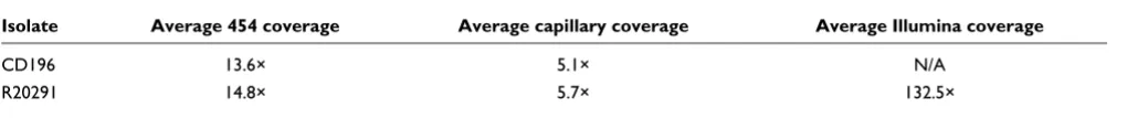
Table 5
Average sequence coverage from aligned reads
Isolate Average 454 coverage Average capillary coverage Average Illumina coverage
CD196 13.6× 5.1× N/A
[image:12.612.54.564.681.734.2]samples. The cells were monitored for 24 hours after inocula-tion for morphological alterainocula-tion. The cytotoxicity titer corre-sponds to the reciprocal of the greater dilution giving rounding up in 50% of the cells and is expressed as toxin molarity (corresponding to toxin specific activity).
Motility assay
Cultures were grown anaerobically for 1 to 2 days on Braziers media from glycerol stocks. BHI agar (0.05%) was poured into 30 ml glass vials that were then pre-equilibrated for 4 hours in the anaerobe chamber. Three single colonies were picked with a loop and inoculated into the top 2 to 5 mm of BHI agar in the glass vial. These were then left overnight in the anaerobe chamber; the vials were then removed from the anaerobe chamber and photographed to record the motility.
Minimum inhibitory concentration determination using Etest
Chloramphenicol, erythromycin, tetracycline and fluoroqui-nolones (gatifloxacin, mocifloxacin and lexofloxacin) MICs were determined using Etest strips (Biomérieux, Marcy
l'Etoile, France). C. difficile was cultured overnight on
Bra-zier's CCEY agar (Bioconnections) with 1% defibrinated horse
blood (Oxoid) and C. difficile supplement (Bioconnections).
All cultures were undertaken at 37°C in an anaerobe chamber
(10% CO2, 10% H2, 80% N2). A bacterial suspension in BHI
(no. 3 McFarland standard) was inoculated onto the surface of Brazier's agar and was then dried for 15 to 30 minutes. Etest strips were placed onto agar surface. Agar plates were incubated anaerobically (37°C) for 24 hours and 48 hours, and MICs were determined following the manufacturer's instructions.
Statistical analysis
Chloramphenicol, erythromycin, tetracycline and
fluoroqui-nolone MICs for C. difficile 630, CD196 and R20291 were
analyzed by Tukey test using GraphPad Prism 4 software (La Jolla, CA, USA). P-value < 0.05 was considered statistically significant.
Abbreviations
BHI: brain heart infusion; CDI: C. difficile infection; CDS:
coding sequence; IS: Insertion Sequence; MIC: minimum inhibitory concentration; PaLoc: pathogenicity locus; PMC: pseudomembranous colitis; SMPI: Stoke Mandeville phage island.
Authors' contributions
RAS, JP, GD and BWW conceived of the study. MAQ and GR performed the sequencing experiments. MH, CC, MS, TDL and JP performed the data analysis. MG and MRP performed toxin B toxicity assays, LD performed motility and autoagglu-tination assays, EV performed MIC assays, MM performed PCR analysis, and DNG provided strains and critical analysis.
RAS, LD, JP, MH and BWW drafted the manuscript. All authors contributed to and approved the final manuscript.
Additional data files
The following additional data are available with the online version of this paper: CDSs specific to PCR-ribotype 027 iso-lates (Additional data file 1); CDSs that have been disrupted by an insertion in both 027 strains but are intact in 630, and CDSs that are intact in both 027 strains but have been dis-rupted in 630 (Additional data file 2); R20291-specific gene primers used in this study (Additional data file 3); SDS-PAGE of toxin B preparations (Additional data file 4).
Additional data file 1
CDS specific to PCR-ribotype 027 isolates CDS specific to PCR-ribotype 027 isolates. Click here for file
Additional data file 2
CDSs that have been disrupted by an insertion in both 027 strains but are intact in 630, and CDSs that are intact in both 027 strains but have been disrupted in 630
Fourteen CDSs have been disrupted by an insertion in both 027 strains but are intact in 630, and, conversely, 12 CDSs are intact in both 027 strains but have been disrupted in 630.
Click here for file Additional data file 3
R20291-specific gene primers used in this study R20291-specific gene primers used in this study. Click here for file
Additional data file 4
SDS-PAGE of toxin B preparations
Toxin B was quantified by gel densitometry. 1 = VPI10463, 2 = CD196, 3 = 630.
Click here for file
Acknowledgements
We thank Jon Brazier for provision of strain R20291 and Frances Smith for assistance with library making. We acknowledge the Wellcome Trust for funding this research.
References
1. Bartlett JG: Historical perspectives on studies of Clostridium difficile and C. difficile infection. Clin Infect Dis 2008, 46(Suppl 1):S4-11.
2. Bartlett JG: Clostridium difficile: history of its role as an enteric pathogen and the current state of knowledge about the organism. Clin Infect Dis 1994, 18(Suppl 4):S265-272.
3. Kuijper EJ, van Dissel JT, Wilcox MH: Clostridium difficile: chang-ing epidemiology and new treatment options. Curr Opin Infect Dis 2007, 20:376-383.
4. Goorhuis A, Kooi T Van der, Vaessen N, Dekker FW, Berg R Van den, Harmanus C, Hof S van den, Notermans DW, Kuijper EJ: Spread and epidemiology of Clostridium difficile polymerase chain reaction ribotype 027/toxinotype III in The Nether-lands. Clin Infect Dis 2007, 45:695-703.
5. Hubert B, Loo VG, Bourgault AM, Poirier L, Dascal A, Fortin E, Dionne M, Lorange M: A portrait of the geographic dissemina-tion of the Clostridium difficile North American pulsed-field type 1 strain and the epidemiology of C. difficile-associated disease in Quebec. Clin Infect Dis 2007, 44:238-244.
6. Loo VG, Poirier L, Miller MA, Oughton M, Libman MD, Michaud S, Bourgault AM, Nguyen T, Frenette C, Kelly M, Vibien A, Brassard P, Fenn S, Dewar K, Hudson TJ, Horn R, Rene P, Monczak Y, Dascal A: A predominantly clonal multi-institutional outbreak of Clostridium difficile-associated diarrhea with high morbidity and mortality. N Engl J Med 2005, 353:2442-2449.
7. Mooney H: Annual incidence of MRSA falls in England, but C. difficile continues to rise. BMJ 2007, 335:958.
8. Redelings MD, Sorvillo F, Mascola L: Increase in Clostridium diffi-cile-related mortality rates, United States, 1999-2004. Emerg Infect Dis 2007, 13:1417-1419.
9. MacCannell DR, Louie TJ, Gregson DB, Laverdiere M, Labbe AC, Laing F, Henwick S: Molecular analysis of Clostridium difficile PCR ribotype 027 isolates from Eastern and Western Can-ada. J Clin Microbiol 2006, 44:2147-2152.
10. Healthcare Commission: Investigation into outbreaks of Clostridium difficile at Stoke Mandeville Hospital, Bucking-hamshire Hospitals NHS Trust. [http://www.cqc.org.uk/_db/ _documents/Stoke_Mandeville.pdf].
12. Popoff MR, Rubin EJ, Gill DM, Boquet P: Actin-specific ADP-ribo-syltransferase produced by a Clostridium difficile strain. Infect Immun 1988, 56:2299-2306.
13. Razaq N, Sambol S, Nagaro K, Zukowski W, Cheknis A, Johnson S, Gerding DN: Infection of hamsters with historical and epi-demic BI types of Clostridium difficile. J Infect Dis 2007, 196:1813-1819.
14. Stabler RA, Gerding DN, Songer JG, Drudy D, Brazier JS, Trinh HT, Witney AA, Hinds J, Wren BW: Comparative phylogenomics of Clostridium difficile reveals clade specificity and microevolu-tion of hypervirulent strains. J Bacteriol 2006, 188:7297-7305. 15. Hammond GA, Johnson JL: The toxigenic element of Clostridium
difficile strain VPI 10463. Microb Pathog 1995, 19:203-213. 16. Lyras D, O'Connor JR, Howarth PM, Sambol SP, Carter GP,
Phu-moonna T, Poon R, Adams V, Vedantam G, Johnson S, Gerding DN, Rood JI: Toxin B is essential for virulence of Clostridium diffi-cile. Nature 2009, 458:1176-1179.
17. Matamouros S, England P, Dupuy B: Clostridium difficile toxin expression is inhibited by the novel regulator TcdC. Mol Microbiol 2007, 64:1274-1288.
18. Warny M, Pepin J, Fang A, Killgore G, Thompson A, Brazier J, Frost E, McDonald LC: Toxin production by an emerging strain of Clostridium difficile associated with outbreaks of severe dis-ease in North America and Europe. Lancet 2005, 366:1079-1084.
19. Dupuy B, Govind R, Antunes A, Matamouros S: Clostridium difficile toxin synthesis is negatively regulated by TcdC. J Med Micro-biol 2008, 57:685-689.
20. Spigaglia P, Mastrantonio P: Molecular analysis of the patho-genicity locus and polymorphism in the putative negative regulator of toxin production (TcdC) among Clostridium dif-ficile clinical isolates. J Clin Microbiol 2002, 40:3470-3475. 21. Akerlund T, Persson I, Unemo M, Noren T, Svenungsson B, Wullt M,
Burman LG: Increased sporulation rate of epidemic Clostrid-ium difficile Type 027/NAP1. J Clin Microbiol 2008, 46:1530-1533. 22. Sebaihia M, Wren BW, Mullany P, Fairweather NF, Minton N, Stabler R, Thomson NR, Roberts AP, Cerdeno-Tarraga AM, Wang H, Holden MT, Wright A, Churcher C, Quail MA, Baker S, Bason N, Brooks K, Chillingworth T, Cronin A, Davis P, Dowd L, Fraser A, Feltwell T, Hance Z, Holroyd S, Jagels K, Moule S, Mungall K, Price C, Rabbinow-itsch E, et al.: The multidrug-resistant human pathogen Clostridium difficile has a highly mobile, mosaic genome. Nat Genet 2006, 38:779-786.
23. Rupnik M, Avesani V, Janc M, von Eichel-Streiber C, Delmee M: A novel toxinotyping scheme and correlation of toxinotypes with serogroups of Clostridium difficile isolates. J Clin Microbiol 1998, 36:2240-2247.
24. Rupnik M, Braun V, Soehn F, Janc M, Hofstetter M, Laufenberg-Feld-mann R, von Eichel-Streiber C: Characterization of polymor-phisms in the toxin A and B genes of Clostridium difficile. FEMS Microbiol Lett 1997, 148:197-202.
25. Rupnik M, Kato N, Grabnar M, Kato H: New types of toxin A-neg-ative, toxin B-positive strains among Clostridium difficile iso-lates from Asia. J Clin Microbiol 2003, 41:1118-1125.
26. Stabler RA, Dawson LF, Phua LT, Wren BW: Comparative analy-sis of BI/NAP1/027 hypervirulent strains reveals novel toxin B-encoding gene (tcdB) sequences. J Med Microbiol 2008, 57:771-775.
27. Alonso R, Martin A, Pelaez T, Marin M, Rodriguez-Creixems M, Bouza E: Toxigenic status of Clostridium difficile in a large Spanish teaching hospital. J Med Microbiol 2005, 54:159-162.
28. Goncalves C, Decre D, Barbut F, Burghoffer B, Petit JC: Prevalence and characterization of a binary toxin (actin-specific ADP-ribosyltransferase) from Clostridium difficile. J Clin Microbiol 2004, 42:1933-1939.
29. Pituch H, Rupnik M, Obuch-Woszczatynski P, Grubesic A, Meisel-Mikolajczyk F, Luczak M: Detection of binary-toxin genes (cdtA and cdtB) among Clostridium difficile strains isolated from patients with C. difficile-associated diarrhoea (CDAD) in Poland. J Med Microbiol 2005, 54:143-147.
30. Stubbs S, Rupnik M, Gibert M, Brazier J, Duerden B, Popoff M: Pro-duction of actin-specific ADP-ribosyltransferase (binary toxin) by strains of Clostridium difficile. FEMS Microbiol Lett 2000, 186:307-312.
31. Brazier JS, Patel B, Pearson A: Distribution of Clostridium difficile PCR ribotype 027 in British hospitals. Euro Surveill 2007, 12:E070426. 070422
32. Barbut F, Decre D, Lalande V, Burghoffer B, Noussair L, Gigandon A,
Espinasse F, Raskine L, Robert J, Mangeol A, Branger C, Petit JC: Clin-ical features of Clostridium difficile-associated diarrhoea due to binary toxin (actin-specific ADP-ribosyltransferase)-pro-ducing strains. J Med Microbiol 2005, 54:181-185.
33. Barbut F, Gariazzo B, Bonne L, Lalande V, Burghoffer B, Luiuz R, Petit JC: Clinical features of Clostridium difficile-associated infec-tions and molecular characterization of strains: results of a retrospective study, 2000-2004. Infect Control Hosp Epidemiol 2007, 28:131-139.
34. McEllistrem MC, Carman RJ, Gerding DN, Genheimer CW, Zheng L: A hospital outbreak of Clostridium difficile disease associated with isolates carrying binary toxin genes. Clin Infect Dis 2005, 40:265-272.
35. Carter GP, Lyras D, Allen DL, Mackin KE, Howarth PM, O'Connor JR, Rood JI: Binary toxin production in Clostridium difficile is regulated by CdtR, a LytTR family response regulator. J Bac-teriol 2007, 189:7290-7301.
36. Drudy D, Kyne L, O'Mahony R, Fanning S: gyrA mutations in fluo-roquinolone-resistant Clostridium difficile PCR-027. Emerg Infect Dis 2007, 13:504-505.
37. Golledge CL, Carson CF, O'Neill GL, Bowman RA, Riley TV: Cipro-floxacin and Clostridium difficile-associated diarrhoea. J Anti-microb Chemother 1992, 30:141-147.
38. McCusker ME, Harris AD, Perencevich E, Roghmann MC: Fluoro-quinolone use and Clostridium difficile-associated diarrhea. Emerg Infect Dis 2003, 9:730-733.
39. Muto CA, Pokrywka M, Shutt K, Mendelsohn AB, Nouri K, Posey K, Roberts T, Croyle K, Krystofiak S, Patel-Brown S, Pasculle AW, Paterson DL, Saul M, Harrison LH: A large outbreak of Clostrid-ium difficile-associated disease with an unexpected propor-tion of deaths and colectomies at a teaching hospital following increased fluoroquinolone use. Infect Control Hosp Epi-demiol 2005, 26:273-280.
40. Patel NS: Fluoroquinolone use is the predominant risk factor for the development of a new strain of Clostridium difficile -associated disease. BJU Int 2007, 99:1333-1334.
41. Rosey EL, Kennedy MJ, Yancey RJ Jr: Dual flaA1 flaB1 mutant of Serpulina hyodysenteriae expressing periplasmic flagella is severely attenuated in a murine model of swine dysentery. Infect Immun 1996, 64:4154-4162.
42. Allen-Vercoe E, Woodward MJ: The role of flagella, but not fim-briae, in the adherence of Salmonella enterica serotype Enteritidis to chick gut explant. J Med Microbiol 1999, 48:771-780.
43. Postnova T, Gomez-Duarte OG, Richardson K: Motility mutants of Vibrio cholerae O1 have reduced adherence in vitro to human small intestinal epithelial cells as demonstrated by ELISA. Microbiology 1996, 142:2767-2776.
44. Delmee M, Avesani V, Delferriere N, Burtonboy G: Characteriza-tion of flagella of Clostridium difficile and their role in sero-grouping reactions. J Clin Microbiol 1990, 28:2210-2214. 45. Tasteyre A, Karjalainen T, Avesani V, Delmee M, Collignon A,
Bour-lioux P, Barc MC: Phenotypic and genotypic diversity of the flagellin gene (fliC) among Clostridium difficile isolates from different serogroups. J Clin Microbiol 2000, 38:3179-3186. 46. Logan SM: Flagellar glycosylation - a new component of the
motility repertoire? Microbiology 2006, 152:1249-1262.
47. Taguchi F, Ogawa Y, Takeuchi K, Suzuki T, Toyoda K, Shiraishi T, Ich-inose Y: A homologue of the 3-oxoacyl-(acyl carrier protein) synthase III gene located in the glycosylation island of Pseu-domonas syringae pv. tabaci regulates virulence factors via N-acyl homoserine lactone and fatty acid synthesis. J Bacteriol 2006, 188:8376-8384.
48. Guerry P, Ewing CP, Schirm M, Lorenzo M, Kelly J, Pattarini D, Majam G, Thibault P, Logan S: Changes in flagellin glycosylation affect Campylobacter autoagglutination and virulence. Mol Microbiol 2006, 60:299-311.
49. Howard SL, Jagannathan A, Soo EC, Hui JP, Aubry AJ, Ahmed I, Kar-lyshev A, Kelly JF, Jones MA, Stevens MP, Logan SM, Wren BW: A Campylobacter jejuni glycosylation island important in cell charge, legionaminic acid biosynthesis and colonisation of chickens. Infect Immun 2009, 77:2544-2556.
50. Clostridium difficile [http://www.sanger.ac.uk/Projects/C_difficile] 51. Dong C, Major LL, Srikannathasan V, Errey JC, Giraud MF, Lam JS,
52. Tsukioka Y, Yamashita Y, Oho T, Nakano Y, Koga T: Biological function of the dTDP-rhamnose synthesis pathway in Strep-tococcus mutans. J Bacteriol 1997, 179:1126-1134.
53. Yamashita Y, Tomihisa K, Nakano Y, Shimazaki Y, Oho T, Koga T: Recombination between gtfB and gtfC is required for sur-vival of a dTDP-rhamnose synthesis-deficient mutant of Streptococcus mutans in the presence of sucrose. Infect Immun 1999, 67:3693-3697.
54. Novick RP: Autoinduction and signal transduction in the reg-ulation of staphylococcal virulence. Mol Microbiol 2003, 48:1429-1449.
55. Dawson LF, Stabler RA, Wren BW: Assessing the role of p-cresol tolerance in Clostridium difficile. J Med Microbiol 2008, 57:745-749.
56. Hayes F: A family of stability determinants in pathogenic bac-teria. J Bacteriol 1998, 180:6415-6418.
57. Christensen SK, Gerdes K: RelE toxins from bacteria and Archaea cleave mRNAs on translating ribosomes, which are rescued by tmRNA. Mol Microbiol 2003, 48:1389-1400.
58. Christensen SK, Mikkelsen M, Pedersen K, Gerdes K: RelE, a global inhibitor of translation, is activated during nutritional stress. Proc Natl Acad Sci USA 2001, 98:14328-14333.
59. Wust J, Hardegger U: Transferable resistance to clindamycin, erythromycin, and tetracycline in Clostridium difficile. Antimi-crob Agents Chemother 1983, 23:784-786.
60. Assefa S, Keane TM, Otto TD, Newbold C, Berriman M: ABACAS: algorithm-based automatic contiguation of assembled sequences. Bioinformatics 2009, 25:1968-1969.
61. Zerbino DR, Birney E: Velvet: algorithms for de novo short read assembly using de Bruijn graphs. Genome Res 2008, 18:821-829.
62. Delcher AL, Harmon D, Kasif S, White O, Salzberg SL: Improved microbial gene identification with GLIMMER. Nucleic Acids Res 1999, 27:4636-4641.
63. Rutherford K, Parkhill J, Crook J, Horsnell T, Rice P, Rajandream MA, Barrell B: Artemis: sequence visualization and annotation. Bio-informatics 2000, 16:944-945.
64. Carver TJ, Rutherford KM, Berriman M, Rajandream MA, Barrell BG, Parkhill J: ACT: the Artemis Comparison Tool. Bioinformatics 2005, 21:3422-3423.
65. Kuijper EJ, Coignard B, Brazier JS, Suetens C, Drudy D, Wiuff C, Pituch H, Reichert P, Schneider F, Widmer AF, Olsen KE, Allerberger F, Notermans DW, Barbut F, Delmee M, Wilcox M, Pearson A, Patel BC, Brown DJ, Frei R, Akerlund T, Poxton IR, Tull P: Update of Clostridium difficile-associated disease due to PCR ribotype 027 in Europe. Euro Surveill 2007, 12:E1-2.
66. Syuto B, Kubo S: Isolation and molecular size of Clostridium botulinum type C toxin. Appl Environ Microbiol 1977, 33:400-405. 67. von Eichel-Streiber C, Harperath U, Bosse D, Hadding U:
Purifica-tion of two high molecular weight toxins of Clostridium diffi-cile which are antigenically related. Microb Pathog 1987, 2:307-318.