Open Access
Vol 13 No 3Research
Anemia and red blood cell transfusion in neurocritical care
Andreas H Kramer
1and David A Zygun
21Departments of Critical Care Medicine & Clinical Neurosciences, University of Calgary, Foothills Medical Center, 1403 29thSt. N.W., Calgary, AB,
Canada, T2N 2T9
2Departments of Critical Care Medicine, Clinical Neurosciences, & Community Health Sciences, University of Calgary, Foothills Medical Center, 1403
29thSt. N.W., Calgary, AB, Canada, T2N 2T9
Corresponding author: Andreas H Kramer, andreas.kramer@albertahealthservices.ca
Received: 26 Jan 2009 Revisions requested: 3 Mar 2009 Revisions received: 9 Apr 2009 Accepted: 11 Jun 2009 Published: 11 Jun 2009
Critical Care 2009, 13:R89 (doi:10.1186/cc7916)
This article is online at: http://ccforum.com/content/13/3/R89 © 2009 Kramer and Zygun; licensee BioMed Central Ltd.
This is an open access article distributed under the terms of the Creative Commons Attribution License (http://creativecommons.org/licenses/by/2.0), which permits unrestricted use, distribution, and reproduction in any medium, provided the original work is properly cited.
Abstract
Introduction
Anemia is one of the most common medical
complications to be encountered in critically ill patients. Based
on the results of clinical trials, transfusion practices across the
world have generally become more restrictive. However,
because reduced oxygen delivery contributes to 'secondary'
cerebral injury, anemia may not be as well tolerated among
neurocritical care patients.
Methods
The first portion of this paper is a narrative review of
the physiologic implications of anemia, hemodilution, and
transfusion in the setting of brain-injury and stroke. The second
portion is a systematic review to identify studies assessing the
association between anemia or the use of red blood cell
transfusions and relevant clinical outcomes in various
neurocritical care populations.
Results
There have been no randomized controlled trials that
have adequately assessed optimal transfusion thresholds
specifically among brain-injured patients. The importance of
ischemia and the implications of anemia are not necessarily the
same for all neurocritical care conditions. Nevertheless, there
exists an extensive body of experimental work, as well as human
observational and physiologic studies, which have advanced
knowledge in this area and provide some guidance to clinicians.
Lower hemoglobin concentrations are consistently associated
with worse physiologic parameters and clinical outcomes;
however, this relationship may not be altered by more
aggressive use of red blood cell transfusions.
Conclusions
Although hemoglobin concentrations as low as 7
g/dl are well tolerated in most critical care patients, such a
severe degree of anemia could be harmful in brain-injured
patients. Randomized controlled trials of different transfusion
thresholds, specifically in neurocritical care settings, are
required. The impact of the duration of blood storage on the
neurologic implications of transfusion also requires further
investigation.
Introduction
A key paradigm in the management of neurocritical care
patients is the avoidance of 'secondary' cerebral insults [1-3].
The acutely injured brain is vulnerable to systemic
derange-ments, such as hypotension, hypoxemia, or fever, which may
further exacerbate neuronal damage [4-7]. Thus, critical care
practitioners attempt to maintain a physiologic milieu that
min-imizes secondary injury, thereby maximizing the chance of a
favorable functional and neurocognitive recovery.
Anemia is defined by the World Health Organization as a
hemoglobin (Hb) concentration less than 12 g/dl in women
and 13 g/dl in men [8]. It is one of the most common medical
complications encountered in critically ill patients, including
those with neurologic disorders. About two-thirds of patients
have Hb concentrations less than 12 g/dl at the time of
inten-sive care unit (ICU) admission, with a subsequent decrement
of about 0.5 g/dl per day [9-12]. The etiology of ICU-acquired
anemia is multifactorial. Systemic inflammation reduces red
CBF: cerebral blood flow; CaO2: arterial oxygen content; CMRO2: cerebral metabolic rate; CO: cardiac output; CO2: carbon dioxide; CPP: cerebral perfusion pressure; DO2: oxygen delivery; Hb: hemoglobin; HBBS: hemoglobin-based blood substitutes; ICH: intracerebral hemorrhage; ICU:
inten-sive care unit; LPR: lactate to pyruvate ratio; MRI: magnetic resonance imaging; NO: nitric oxide; O2: oxygen; OEF: oxygen extraction fraction; PbtO2:
blood cell (RBC) development by blunting the production of
erythropoietin and interfering with the ability of erythroblasts to
incorporate iron [13-17]. RBC loss is accelerated by frequent
phlebotomy, reduced RBC survival, and occasional
hemor-rhage. Large volumes of fluid used during resuscitation, with
resultant hemodilution, may also contribute to early reductions
in Hb levels [18-22].
Anemia can easily be corrected with the use of allogeneic
RBC transfusions. The proportion of patients receiving blood
during their ICU stay varies from 20 to 44%, and those who
are transfused receive an average of as many as five units
[10,11,23,24]. However, two multi-center, randomized
con-trolled trials (RCTs) and two large observational studies have
shown the liberal use of blood transfusions, with the goal of
maintaining relatively arbitrary Hb concentrations (e.g. 10 g/
dl), to not only be ineffective at improving outcomes, but also
potentially harmful [10,11,25,26]. Still, because impaired
oxy-gen (O
2) delivery is thought to be an important factor in
sec-ondary brain injury, it remains uncertain whether these findings
can be broadly applied to neurocritical care patients.
Accord-ingly, it remains common practice for clinicians to set target
Hb levels at a minimum of 9 to 10 g/dl in this setting [27-29].
Materials and methods
To describe the physiologic and clinical implications of anemia
and transfusion in neurocritical care patients, we used the
OVID interface to search MEDLINE from its inception until
March 9, 2009. We combined the following MESH headings:
(anemia OR blood transfusion OR hemodilution OR
hemat-ocrit OR hemoglobins) AND (stroke OR craniocerebral trauma
OR subarachnoid hemorrhage OR cerebral hemorrhage OR
cerebrovascular circulation OR cardiac surgical procedures
OR coronary artery bypass). This search yielded 2137 English
language publications dealing primarily with adults (>18 years
old). Each abstract was reviewed, and both human and animal
studies assessing the impact of anemia, hemodilution, or the
use of RBC transfusions on a physiologic or clinical outcome
were chosen for more detailed review. Relevant review articles
and case reports were also included, and the references of
selected papers were screened for additional publications.
Clinical studies involving specific groups of neurocritical care
patients were selected for inclusion in evidentiary tables.
Results and discussion
Physiologic implications of anemia
Cerebral blood flow and oxygen delivery
The amount of oxygen reaching specific organs is the product
of local blood flow and the arterial oxygen content (C
aO
2). The
latter is dependent on the Hb concentration and the degree to
which it is saturated with O
2(S
aO
2), with a small amount of O
2also dissolved in blood. Thus, global systemic O
2delivery can
be expressed by the following equation:
O
2delivery to the brain can be conceptualized using the same
equation, but by substituting cerebral blood flow (CBF) for
cardiac output (CO). Flow through the cerebral vasculature is
determined by the cerebral perfusion pressure (CPP), the
length and caliber of the vessels, and the viscosity of blood, as
described by the Hagen-Poiseuille equation:
Regulation of CBF and cerebral O
2delivery in response to
physiologic stressors is achieved largely by homeostatic
varia-tions in the caliber of cerebral vessels (the 'r' in the above
equation; Figure 1).
CPP is the difference between mean arterial pressure and
cer-ebral venous pressure; intracranial pressure is widely used as
a surrogate for the latter. The response of the cerebral
vascu-lature to changes in CPP is referred to as CBF autoregulation
('pressure-reactivity'). Cerebral arterioles vasoconstrict in
response to raised CPP and vasodilate when there are
reduc-tions, thereby maintaining constant CBF (Figure 1a).
Autoreg-ulation is sometimes impaired in neurocritical care patients,
such that CBF becomes directly dependent on CPP, making
the brain more vulnerable to both hypo- and hyperperfusion
[30-32].
There are numerous other stimuli that may modify cerebral
vas-cular resistance and CBF. Both global and regional CBF are
tightly coupled to metabolism. Thus, physiologic changes that
lead to a reduction in cerebral metabolic rate (CMRO
2) (e.g.
hypothermia or sedation) will also proportionally reduce CBF
(Figure 1b). In addition, CBF is influenced by variations in the
partial pressures of carbon dioxide (PCO
2; 'CO
2-reactivity'),
and to a lesser degree, O
2(PO
2) (Figures 1c, d). CBF
increases in response to a decrease in PO
2, although this
effect is probably minimal until the level approaches 60 mmHg
[30].
In response to worsening anemia, neuronal O
2delivery is
ini-tially preserved both by the systemic cardiovascular response
and mechanisms that are more specifically neuroprotective.
Cardiovascular response to anemia
A falling Hb concentration is sensed by aortic and carotid
chemoreceptors, resulting in stimulation of the sympathetic
nervous system, which in turn raises heart rate and
contractil-ity, thereby augmenting CO [33-35]. The reduction in blood
viscosity results in a corresponding reduction in afterload, as
well as enhanced flow through post-capillary venules, greater
venous return, and increased preload [36-38]. Thus, stroke
volume, CO, and blood pressure (as well as CPP) increase in
response to isovolemic anemia. Tissues are further protected
from falling O
2delivery because of their capacity to increase
O
2extraction and maintain constant O
2consumption. In the
brain, irreversible ischemia may not occur until the O
2extrac-DO ml O2( 2/min)=cardiac output L min( / ) (×Hb g L( / ) (×S Oa 2(%)×1 3.99(ml O2/g Hb)) ( .+0 003×PO2))
tion fraction (OEF) exceeds 75% [39-43]. Systemic anaerobic
metabolism does not develop until the Hb concentration falls
well below 5 g/dl in otherwise healthy individuals [44]. On the
other hand, many neurocritical care patients have concomitant
cardiac disease and left ventricular dysfunction which may
prevent an appropriate increase in CO in response to
sympa-thetic stimulation. This is commonly the case even in the
absence of pre-existing heart disease; for example, among
patients with acute 'high-grade' aneurysmal subarachnoid
hemorrhage (SAH) (Hunt-Hess grades 3 to 5), more than
one-third have regional left ventricular wall motion abnormalities
detectable by echocardiography [45].
Cerebrovascular response to anemia
Apart from the increased flow produced by higher CPP and
lower blood viscosity, anemia also induces cerebral
vasodila-tation [46-48]. When Hb (and therefore C
aO
2) falls, there
appears to be a disproportionate increase in CBF in relation to
other organs (Figure 1d) [49]. The mechanisms underlying this
increase in vessel caliber are still being clarified, but include
some of the same factors involved in CBF
pressure-autoregu-lation; these have recently been reviewed in detail [46].
Impor-tantly, anemia results in upregulation of nitric oxide (NO)
production by perivascular neurons and vascular smooth
mus-cle surrounding cerebral blood vessels. The importance of
these pathways is supported by the observation that inhibition
of NO synthase blunts hypoxia- and anemia-induced cerebral
vasodilatation [50-52]. However, additional factors are
undoubtedly involved [53-55]. Sympathetic
β
2 receptor
stim-ulation is an example of one such mechanism that contributes
to vasodilatation and maintenance of CBF [56]. Other
bio-chemical mediators that are upregulated in the brain in
response to anemia include vascular endothelial growth factor,
hypoxia inducible factor 1
α
, and erythropoietin [46,57].
Although it seems likely that these mediators are
neuroprotec-tive, it remains possible that they could also have harmful
pathophysiologic effects [46].
Compensatory mechanisms eventually fail
[image:3.612.54.538.90.348.2]As anemia worsens, the resultant increases in CBF and OEF
eventually become insufficient to overcome the reduced C
aO
2produced by a low Hb concentration (Figure 2). The point at
which this threshold is reached is not clear and probably varies
somewhat between patients. A sophisticated mathematical
model based on animal data suggested that CMRO
2is well
preserved in normal brain, even with severe reductions in Hb
concentration. In contrast, penumbral brain appears to be
much more vulnerable, with O
2delivery and CMRO
2progres-sively declining as Hb falls below 10 to 12 g/dl [58-62]. As
with cerebral ischemia, impairment of the usual protective
mechanisms induced by anemia has also been demonstrated
as a result of brain trauma [63].
Figure 1
Physiologic parameters influencing cerebral blood flow
Physiologic parameters influencing cerebral blood flow (a) The effects of mean arterial blood pressure (MAP) (solid line = normal autoregulation; dashed line = deranged autoregulation), (b) cerebral metabolic rate (CMRO2), (c) partial pressure of carbon dioxide (PCO2), (d) partial pressure of
A study of euvolemic hemodilution in healthy human volunteers
confirmed that even profound anemia (Hb about 5 g/dl) was
relatively well tolerated; however, subtle abnormalities in
neu-rocognitive testing began to emerge when Hb concentrations
fell below 7 g/dl [64,65]. The co-existence of other physiologic
stressors may also make anemia less tolerable; for example,
experimental studies have found that cerebral O
2delivery is
preserved in the presence of both severe anemia and
hypoten-sion individually, but not when they are both present [66,67].
Additionally, anemia-induced cerebral vasodilatation appears
to interfere with the usual response to variations in PCO
2[47,68-70]. These observations raise concerns that relatively
inadequate O
2delivery could occur at Hb levels well above 7
g/dl in critical care patients with cerebrovascular disease,
pre-existing central nervous system pathology (e.g. an ischemic or
'traumatic' penumbra) or deranged regulation of CBF. Thus,
there is strong physiologic rationale for believing that a
restric-tive transfusion threshold of 7 g/dl, although clearly safe in
many critical care patients [25,26], may not be without risk in
neurocritical care patients.
Risks of red blood cell transfusion
Even if anemia is harmful, this does not necessarily prove that
liberal use of allogeneic RBCs to normalize Hb concentrations
is justified. Emerging data indicates that stored blood has
important differences from patients' own blood. A number of
changes occur over time as RBCs are being stored; some of
these alterations could have important implications after
trans-fusion, and they are collectively referred to as the 'storage
lesion'. Biochemical changes include reductions in ATP, loss
of membrane phospholipids, and oxidative damage to
pro-teins. The consequence is a gradual change in RBC
appear-ance from the usual biconcave discs to irreversibly deformed
and stellate-shaped spheroechinocytes [71,72]. Loss of RBC
membrane function, as well as an increased tendency to
adhere to endothelium, may interfere with microcirculatory
flow [72,73]. RBC 2-3-diphosphoglycerate levels become
depleted to the point of being essentially undetectable after
one week of storage. Although levels are usually restored
within 24 to 72 hours after transfusion, the transiently
increased binding affinity of Hb interferes with the release of
O
2for use by tissues [74].
Thus, although blood transfusions are generally given with the
intention of raising O
2delivery, the storage-induced changes
may prevent RBCs from achieving their intended purpose. For
example, studies using gastric tonometry parameters as a
sur-rogate for mesenteric perfusion have not shown improvements
following transfusion [75,76]. Similarly, RBCs also appear to
have little effect on skeletal muscle O
2tension in postoperative
patients or on global O
2consumption in the critically ill
[77,78].
Transfusion-related acute lung injury is now the most common
cause of transfusion-related mortality reported to the Food and
Drugs Administration [79]. Transfusion may have
immunosup-pressive effects, which are thought to be due to concomitant
white blood cell transmission. Several studies have suggested
a link between the use of allogeneic RBCs and both
nosoco-mial infections and acute respiratory distress syndrome
[80-83]. Alternatively, RCTs, where well-matched groups were
transfused with differing intensities, have not yet convincingly
confirmed these associations [25,26]. Furthermore, the risk of
Effects of falling hemoglobin concentration on cerebral oxygen delivery
[image:4.612.66.469.92.300.2]complications may be less since the implementation of
univer-sal leukoreduction in many jurisdictions [84].
It has been suggested that the use of fresher blood might
fur-ther minimize the risks of transfusion, while also maximizing
their physiologic effect. Results have been conflicting, and
there is little data specifically in neurocritical care patients
[71,75,76]. A recent animal study found fresh blood to be
more effective at raising brain tissue oxygen tension (P
btO
2)
and preserving CBF in comparison to stored blood [85].
Alter-natively, Weiskopf and colleagues performed isovolemic
hemodilution to Hb concentrations of 55 to 74 g/L in healthy
volunteers and then transfused them with autologous blood
stored for either less than five hours or more than 14 days;
neurocognitive test performance did not differ between the
two groups [86].
Anemia and RBC transfusion in specific neurocritical
care settings
The importance of ischemia in causing secondary brain injury
appears to vary for different neurocritical care conditions. For
example, cerebral vasospasm and delayed infarction are major
causes of neurologic deterioration in the two weeks following
a ruptured cerebral aneurysm [87,88]. In contrast, the
fre-quency and relevance of cerebral ischemia in the
pathophysi-ology of traumatic brain injury (TBI) or intracerebral
hemorrhage (ICH) continue to be debated [40,89-91].
Accordingly, the significance of anemia and optimal
transfu-sion thresholds may not be consistent from one condition to
the next.
Lessons from cardiac surgery
A great deal of what is known about the neurologic effects of
anemia has been reported in the cardiac surgical literature. A
substantial proportion of patients undergoing cardiac surgery
receive blood transfusions, even though large volume
hemor-rhage is comparatively less common [92]. Perioperative stroke
occurs in 1 to 6% of patients and is strongly associated with
greater morbidity and mortality [93,94]. An even larger
propor-tion (
≥
50%) develops at least transient neurocognitive
dys-function that is likely to be, at least in part, due to cerebral
ischemia [95,96]. Thus, the prevention and treatment of
cere-bral ischemia is of major concern in the perioperative period.
We identified 12 studies assessing the association between
perioperative Hb concentrations and subsequent neurologic
complications (Table 1). When defined as an Hb
concentra-tion less than 12.5 g/dl, about one-quarter of patients are
ane-mic preoperatively [97]. Blood loss and hemodilution during
cardiopulmonary bypass usually lead to nadir intraoperative
Hb concentrations of 7.0 to 8.5 g/dl; levels at ICU admission
are typically 8.5 to 9.5 g/dl [98]. Several, but not all, studies
have suggested that the degree of Hb reduction is an
inde-pendent predictor of stroke, delirium, neurocognitive
dysfunc-tion, and other adverse outcomes [97-108] (Table 1).
Although it has not been proven with certainty that these
rela-tions are causative, it seems prudent to avoid major reducrela-tions
in Hb as best as possible with relevant blood-conservation
strategies [109-113].
A recent RCT involving 121 elderly patients undergoing
coro-nary artery bypass compared two intraoperative hematocrit
targets (15 to 18% vs.
≥
27%) [102]. The study was
termi-nated early because of high complication rates in both groups;
however, a greater degree of postoperative neurocognitive
dysfunction was observed among patients managed with
more extreme hemodilution. In addition, although not
neces-sarily directly applicable to adults, further evidence that
exces-sive hemodilution may have harmful neurologic effects comes
from the neonatal literature. Combined data from two RCTs
suggested that hematocrit levels below 23.5% during
cardi-opulmonary bypass were associated with impaired
psychomo-tor development at one year of age [114-116].
Whether using RBC transfusions to maintain higher
perioper-ative Hb levels helps avoid neurologic complications remains
uncertain. For example, although Karkouti and colleagues
found nadir hematocrit levels during cardiopulmonary bypass
to be a predictor of stroke in a multivariable analysis, the same
was also true for the perioperative use of transfusions [105].
An association between transfusion and focal or global
neuro-logic deficits has been confirmed in numerous other studies
(Table 2) [117-125].
One study compared clinical outcomes, including the risk of
perioperative stroke, between 49 Jehovah's Witnesses who
underwent cardiac surgery without blood products and a
matched control group of 196 patients, in whom RBC
transfu-sions were used. No significant differences were observed;
however, only nine patients in total experienced a stroke, such
that this study lacked statistical power to detect a difference.
The severity of anemia in Jehovah's Witness patients was not
reported [123].
In a large, single-center, retrospective study, Koch and
col-leagues explored whether the association between RBCs and
worse outcomes could be related to the duration of blood
stor-age. Outcomes were compared among cardiac surgical
patients depending on whether they were transfused with
exclusively 'newer' (
≤
14 days old; median 11 days) or 'older'
(>14 days old; median 20 days) blood during the
periopera-tive period [126]. In-hospital mortality and postoperaperiopera-tive
com-plications, including sepsis, renal failure, and need for
mechanical ventilation, were greater among patients receiving
older blood. However, there was no significant difference in
the incidence of stroke and coma.
Adult studies assessing the association between anemia and the development of perioperative stroke or cognitive dysfunction among patients undergoing cardiac surgery
Study Patients Design and
setting
Multivariable analysis
Exposure Outcome Main result
Karkouti and colleagues [97]
10,179 Retrospective(pro spective database) Single-center
Logistic regression
Maximum decrease intraoperative Hb compared with baseline
Composite of in-hospital death, stroke (new persistent postoperative neurologic deficit), or dialysis-dependent renal failure
>50% decrement in Hb
independently associated with composite outcome
Bell and colleagues [98]
36,658 (CABG) Retrospective (prospective database) Multi-center
Logistic regression
Preoperative Hb Postoperative stroke (not further defined)
No significant association between Hb and stroke
Karkouti and colleagues [99]
3286 (CABG)
Retrospective Multi-center
Logistic regression and propensity scores
Preoperative anemia (Hb <12.5 g/dl)
Postoperative stroke
(new neurologic deficit)
- Risk of stroke 1.1% in non-anemic pts vs. 2.8% in anemic patients - Trend towards more stroke among anemic patients in propensity-matched analysis
Chang and colleagues [100]
288 Retrospective
Single-center
Logistic regression
Postoperative Hct <30%
Delirium (DSM-IV criteria)
Postoperative hct <30% associated with development of delirium (OR = 2.2, P = 0.02)
Kulier and colleagues [101]
4804 Retrospective
(prospective database) Multi-center
Logistic regression
Preoperative Hb 'Cerebral outcomes' = stroke or encephalopathy (not further defined)
- Each 10 g/L Hb reduction associated with 15% increase in risk of non-cardiac (renal or CNS) complications - Association stronger for renal complications
Matthew and colleagues [102]
121
(CABG; age >65)
Prospective RCT Single-center
Logistic regression
Comparison of hemodilution to hct of ≥27% vs. 15 to 18%
Six-week postoperative neurocognitive function (battery of 5 tests)
- Trial stopped early because of unusually high rate of complications in both groups - Significant interaction between age and hct; more neurocognitive deficits among older patients with low hct
Cladellas and colleagues [103]
201 (VR) Retrospective (prospective database) Single-center
None Preoperative
anemia (Hb <12 g/dl)
New permanent stroke or transient ischemic attack (not further defined)
- Risk of TIA or stroke 9.5% in anemic patients vs. 4.4% in non-anemic
Giltay and colleagues [104]
8139 (CABG) Retrospective Single-center
Logistic regression
Lowest hematocrit first 24 hours ICU
Psychotic symptoms (hallucinations and/or delusions)
[image:6.612.48.557.115.750.2]transfusion threshold of 7 g/dl, although this may not be the
optimum Hb level for the avoidance of neurologic
complica-tions. By necessity, the recommendations of published
con-sensus guidelines are relatively non-specific, and state that it
is "not unreasonable to transfuse red cells in certain patients
with critical noncardiac end-organ ischemia whose Hb levels
are as high as 10 g/dl" [111]. Funding was recently secured
in the UK for a multi-center RCT comparing transfusion
trig-gers of 7.5 vs. 9 g/dl [92].
Traumatic brain injury
The majority of patients dying from severe TBI have histologic
evidence of ischemic damage [127]. Early global CBF
reduc-tions occur in many patients, often to levels that are
consid-ered to be in the ischemic range [128,129]. Reductions in
both jugular venous O
2saturation (S
jvO
2) and P
btO
2are not
only common, but their frequency and depth are predictive of
worse outcomes [130-133]. However, the fall in CBF may be
appropriate for a corresponding drop in metabolic rate
[134,135]. Recent studies using positron emission
tomogra-phy (PET) have suggested that although ischemia does occur,
it is less common than previously thought. Furthermore, much
of the 'metabolic distress' detected by multimodal monitoring
(S
jvO
2, P
btO
2, and microdialysis parameters) is not necessarily
attributable to classical ischemia [39,134,135].
On the other hand, there appears to be a great deal of regional
heterogeneity in CBF and CMRO
2[136]. Even if the overall
ischemic brain volume is relatively small, certain vulnerable
regions may still benefit from enhanced O
2delivery [137]. As
with cardiac surgical patients, relatively extreme reductions in
Hb are likely to be deleterious. A recent animal model found
that although isovolemic hemodilution to Hb concentrations of
5 to 7 g/dl resulted in an overall increase in CBF, it produced
larger contusion volumes, more apoptosis, and lower P
btO
2[138].
Potentially beneficial physiologic effects of transfusion have
been shown in four studies of patients with severe TBI
[139-142], each of which demonstrated that P
btO
2increases
fol-lowing the administration of RBCs (Table 3) [139]. However,
this increment was inconsistent, relatively small and often of
questionable clinical importance. Of concern, in some cases
there was even a reduction in P
btO
2. It is possible that some of
the variation in the cerebral effects of transfusion could be, in
part, attributable to the variable age of transfused blood.
Leal-Karkouti andcolleagues [105]
10,949 Retrospective (prospective database) Single-center
Logistic regression
Nadir
intraoperative hct
Postoperative stroke (new persistent postoperative neurologic deficit) that was present on emergence from anesthesia
Each 1% hct reduction associated with OR = 1.1 for stroke (P = 0.002)
Habib and colleagues [106]
5000 Retrospective
(prospective database) Single-center
None Nadir
intraoperative hct
Transient or permanent postoperative stroke (not further defined)
Risk of TIA or stroke 5.4% in quintile with lowest hct vs. 1.3% in quintile with highest hct (P < 0.001)
DeFoe and colleagues [107]
6980 (CABG) Retrospective (prospective database) Multi-center
Logistic regression
Nadir
intraoperative hct
Intra- or postoperative stroke (new focal neurologic deficit which appears and is still at least partially evident more than 24 hours after onset; occurs during or following CABG)
No statistically significant association between hct and stroke
Van
Wermeskerken and colleagues [108]
2804 (CABG) Retrospective Single-center
Logistic regression
Nadir
intraoperative hct
Adverse neurologic outcomes: stroke, coma, or TIA; verified
retrospectively by neurologist
No significant association between hct and outcome
[image:7.612.59.556.116.425.2]CABG = coronary artery bypass grafting; CI = confidence interval; CNS = central nervous system; Hb = hemoglobin; hct = hematocrit; ICU = intensive care unit; OR = odds ratio; RCT = randomized controlled trial; TIA = transient ischemic attack; VR = valve replacement
Table 1 (Continued)
Adult studies assessing the association between transfusion and the development of perioperative stroke or cognitive dysfunction among patients undergoing cardiac surgery
Study Patients Design and
setting
Multivariable analysis
Exposure Outcome Main result
Brevig and colleagues [117]
2531 Retrospective
(prospective database) Single-center
None Any blood product transfusion
Postoperative CVA (not further defined)
Despite reduction in proportion of patients transfused over time (43% in 2003 vs. 18% in 2007), no change in proportion of patients with CVA (0.8 to 1.5%)
Ngaage and colleagues [118]
383 (≥80 years old)
Retrospective (prospective database) Single-center
Logistic regression
Any blood product transfusion
Neurologic complications (confusion/ agitation, seizures, TIA, RIND, stroke, or coma)
Transfusion associated with neurologic complications (OR = 3.6 vs. no transfusion, P = 0.003)
Murphy and colleagues [119]
8518 Retrospective
Single-center
Logistic regression and propensity scores
Any perioperative RBC transfusion
Composite of MI, stroke (permanent or transient), or renal failure
RBC transfusion was associated with composite outcome (OR = 3.35 for transfusion vs. no transfusion; P < 0.0001)
Whitson and colleagues [120]
2691 Retrospective
(prospective database) Single-center
Logistic regression
Any RBC transfusion
CVA (not further defined)
RBC transfusion was associated with CVA (OR = 1.7, P = 0.01)
Norkiene and colleagues [121]
1367 Retrospective
Single-center
Logistic regression
Any RBC transfusion
Delirium (DSM-IV criteria)
Postoperative RBC transfusion was associated with delirium (OR = 4.6, P < 0.001)
Koch and colleagues [122]
11,963 (CABG) Retrospective (prospective database) Single-center
Logistic regression
Total number of units of RBCs transfused
Focal or global neurologic deficits or death without awakening
RBC transfusion was associated with stroke (OR = 1.73 for each unit RBCs; P < 0.0001)
Stamou and colleagues [123]
49 JW patients Retrospective Single-center
196 controls Logistic regression and propensity scores
Any RBC transfusion Nadir Hb not reported
Perioperative stroke
No statistically significant difference in risk of stroke between JWs refusing RBCs and transfused control patients
Karkouti and colleagues [105]
10,949 Retrospective (prospective database) Single-center
Logistic regression
Total number of units of blood product
New perioperative persistent postoperative neurological deficit
Transfusion was associated with stroke (OR = 1.02 for each unit RBCs; P = 0.01)
Bucerius and
colleagues [124] 16,184 Retrospective (prospective database) Single-center
Logistic
regression Any perioperative RBC transfusion Temporary or permanent focal or global neurologic deficit
[image:8.612.57.556.123.720.2]Noval and colleagues recently found that only those patients
having received RBCs less than 14 days old had a statistically
significant improvement in P
btO
2one hour after transfusion
[141]. Although these results are intriguing, they are too
pre-mature to influence clinical practice and require confirmation
in larger studies. Just because P
btO
2rises, does not
necessar-ily mean that CMRO
2has increased. On the contrary, Zygun
and colleagues found no improvement in cerebral lactate to
pyruvate ratio (LPR – a marker of ischemia and 'metabolic
dis-tress') in response to transfusion, despite an increment in
P
btO
2[142].
In a retrospective study of 169 patients with TBI, Carlson and
colleagues found nadir hematocrit levels to be associated with
a worse Glasgow Outcome Scale at hospital discharge.
How-ever, the association between RBC transfusion and poor
out-come was even stronger [143]. Other observational studies
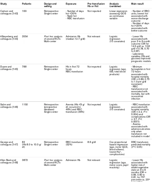
have reached similar conclusions (Table 4) [144-151].
Unfor-tunately, there are no large RCTs to guide practice at this time.
The TRICC trial enrolled only 67 patients with severe TBI
[150]. Although no statistically significant benefit from a liberal
transfusion strategy was observed, this subgroup was too
small to reach meaningful conclusions. Thus, the optimal use
of RBCs in patients with severe TBI remains unclear. A recent
survey found that practice across the USA is variable, and that
the majority of clinicians believe a threshold of 7 g/dl to be too
restrictive, especially in the presence of intracranial
hyperten-sion [27].
Subarachnoid hemorrhage
Narrowing of the cerebral vasculature (angiographic
vasos-pasm) complicates about two-thirds of cases of SAH.
Vasos-pasm most often emerges between days 3 and 14 after SAH
and is the most important cause of secondary brain injury [87].
Evidence of cerebral infarction that was not present initially is
observed in as many as 50 to 70% of survivors using magnetic
resonance imaging (MRI) [152,153]. Unlike other forms of
stroke, the predictable risk of vasospasm and cerebral
ischemia provides a unique opportunity for the provision of
neuroprotection prior to the insult.
Three studies have assessed the association between daily
Hb concentrations and eventual neurologic outcome
[154-156]. Each of these demonstrated that patients with an
unfa-vorable outcome consistently have lower Hb levels throughout
much of the first two weeks in hospital (Table 5). The degree
of decrement in Hb levels over time was also highly predictive
of outcome [154]. Despite the use of multivariable analyses,
there were numerous potentially confounding variables that
could not be adjusted for. For example, patients who are
'sicker' tend to have more blood drawn for laboratory tests,
have more invasive procedures performed, and tend to receive
more intravenous fluids, all of which could contribute to lower
Hb concentrations. Thus, the association between lower Hb
and poor outcome has not conclusively been proven to be
causative.
As in other settings, several studies have also shown a strong
association between transfusion and unfavorable outcomes
following SAH (Table 5) [28,157-160]. One unconfirmed
report suggested that the use of RBCs could contribute to the
development of cerebral vasospasm, perhaps by promoting
inflammation or depleting endogenous NO supplies [160]. A
recent observational study found no difference in
complica-tions based on the transfusion of older (>21 days) compared
with newer (
≤
21 days) units of blood, although this
assess-ment was based on only 85 transfused patients [28].
Hemodilution, together with hypervolemia and hypertension,
has been used as part of 'triple H therapy', a therapeutic
strat-egy to improve CBF in patients with vasospasm [161]. One
study used
133Xenon injections to assess global CBF in eight
patients with SAH. As expected, isovolemic hemodilution from
a mean Hb of 11.9 to 9.2 g/dl produced an increase in global
CBF and a reduction in cerebral vascular resistance. However,
the increase in CBF was not sufficient to overcome the
reduc-tion in C
aO
2, such that global O
2delivery fell and ischemic
brain volume actually increased [162]. Complimentary findings
were subsequently reported by Muench and colleagues, who
used aggressive volume expansion on days 1, 3, and 7, which
produced a concomitant reduction in Hb concentration
rang-ing from of 1.3 to 2.0 g/dl. Although this intervention
consist-ently produced a small increment in CBF, it actually caused a
proportionally larger decline in P
btO
2(Table 3) [163].
More recently, Dhar and colleagues assessed the effects of
transfusion in patients with SAH using PET [164]. PET scans
were performed before and after the administration of one unit
of RBCs to patients with pre-transfusion Hb concentrations
less than 10 g/dl. Although no change in CMRO
2was
D'Ancona andcolleagues [125]
9916 (CABG) Retrospective (prospective database) Single-center
Logistic regression
Any blood product transfusion
New temporary or permanent, focal or global neurologic deficit
Transfusion was associated with stroke (OR = 1.59 vs. no transfusion; P = 0.002)
[image:9.612.53.555.120.165.2]CABG = coronary artery bypass grafting; CVA = cerebrovascular accident; Hb = hemoglobin; JW = Jehovah's Witness; MI = myocardial infarction; OR = odds ratio; RBC = red blood cell; RIND = reversible ischemic neurologic deficit; TIA = transient ischemic attack. Table 2 (Continued)
Clinical studies assessing the impact of anemia or RBC transfusions on PbtO2 and other physiologic parameters in brain-injured
patients
Study Patients Design Baseline Intervention Main findings
Smith and colleagues [139]
23 TBI 12 SAH
Retrospective (prospective database)
Hb = 8.7 g/dl PbtO2 = 24.4 mmHg
Any RBC transfusion (number of units not specified a priori; 80% received ≥1 unit; mean Hb increased to 10.2 g/dl)
General transfusion threshold Hb <10 g/ dl or hct <30% (no protocol)
- Mean increment in PbtO2 3.2 mmHg (15%)
- Increment not related to baseline PbtO2
- PbtO2 decreased in 9/35 patients (26%)
Leal-Noval and colleagues [140]
51 TBI Prospective
observational
Hb = 9.0 g/dl PbtO2 = 24.4 mmHg
1 or 2 units RBCs (number of units not specified a priori; 52% received 2 units; mean Hb increased to 10.6 g/dl)
General transfusion threshold Hb <10 g/ dl (no protocol)
- Mean increment in PbtO2 3.8 mmHg
(16%)
- Increment larger at lower baseline PbtO2
- PbtO2 decreased in
13/51 patients (25%)
Leal-Noval and colleagues [141]
66 TBI (males) Prospective observational
Hb = 8.9 g/dl PbtO2 = 21.3 to 26.2 mmHg
1 or 2 units RBCs number of units not specified a priori; 59% received 2 units; mean Hb increased to 10.2 g/dl)
General transfusion threshold Hb <9.5 g/ dl (no protocol)
- Newer units of blood (≤14 days) resulted in greater mean increment in PbtO2 (3.3 mmHg (16%) vs. 2.1 mmHg (8%)) - PbtO2 decreased only in patients receiving older blood (>19 days)
Zygun and colleagues [142]
30 TBI Prospective RCT Hb = 8.2 g/dl PbtO2 = 18.8 mmHg
Randomized to transfusion thresholds of 8, 9, or 10 g/dl; 2 units RBCs administered over 2 hours (mean Hb increased to 10.1 g/ dl)
- Mean increment in PbtO2 2.2 mmHg
(12%)
- Increment in PbtO2
most prominent when LPR >25
- PbtO2 decreased in
13/30 patients (43%) - No effect on SjvO2 or
microdialysis parameters
Ekelund and colleagues [162]
8 SAH
(TCD-vaso-spasm)
Prospective interventional
Hb = 11.9 g/dl Isovolemic hemodilution (venesection with infusion of dextran 70 and 4% albumin) to mean Hb of 9.2 g/dl
- Outcomes (using
133Xenon and
SPECT): - Increased global CBF
(52.3 to 58.6 ml/100 g/min)
- Reduced cerebral vascular resistance - Reduced oxygen delivery
- Increased ischemic brain volume
Muench and colleagues [163]
10 SAH Prospective
interventional
Hb = 10.6 g/dl PbtO2 = 24.8 mmHg
Volume expansion with HES ± crystalloid to achieve ITBVI >1000 ml/m2;
this produced a decline in Hb of 1.3 to 2.0 g/dl
(on various days)
- Although hypervolemia/ hemodilution produced a slight increment in CBF, PbtO2 decreased by an average of 0 to 5 mmHg
[image:10.612.51.557.126.734.2]observed, OEF dropped from 49 to 41%. Thus, it is possible
that in vulnerable regions of the brain with relatively high OEF,
RBC transfusions could help avoid irreversible infarction.
Another recent study of 20 SAH patients found Hb
concentra-tions less than 9 g/dl to be associated with lower P
btO
2and
higher LPR [165].
In summary, there is now extensive data to suggest that even
moderate degrees of anemia are associated with worse
phys-iologic parameters and clinical outcomes in patients with SAH.
However, it is not clear that the use of RBC transfusions can
modify these associations. An adequately powered, RCT
com-paring different transfusion thresholds is urgently required,
especially in light of the vulnerability of these patients to
delayed cerebral ischemia and the frequency with which they
develop anemia.
Ischemic stroke
Because of the known inverse relation between hematocrit
and CBF, there has long been interest in the clinical use of
hemodilution in the management of acute ischemic stroke
[166]. Some studies have suggested that relatively high Hb
concentrations may predispose to the development of strokes
[167-173], as well as contribute to worse outcomes when
cer-* Dhar and colleagues[164]
8 SAH Prospective
interventional
Hb = 8.7 g/dl One unit RBCs (mean Hb increased to 9.9 g/dl)
- Outcomes assessed using PET:
- No significant change in CBF - Reduced O2
extraction ratio (49 to 41%; P = 0.06) - No significant change in CMRO2 - Reduction in oxygen extraction ratio observed also in territories with vasospasm and low oxygen delivery
Oddo and colleagues [165]
20 SAH Retrospective
(prospective database)
Not applicable None - Hb <9 g/dl associated with higher risk of PbtO2
<20 mmHg (OR 7.2, P < 0.01) and LPR >40 (OR 4.2, P = 0.02)
Chang and colleagues [237]
27 TBI Retrospective Not applicable None - 13.7% of PbtO2
readings <20 mmHg - No significant association between PbtO2 and Hb
Naidech and colleagues [238]
6 SAH Prospective
observational
Not reported 14 RBC transfusions (no protocol)
- Hb correlated with cerebral oximetry (rO2)
- rO2 increased
following 11/14 transfusions, but not statistically significant
Sahuquillo and colleagues [239]
28 TBI Prospective Not applicable None - Critical LOI
(suggestive of ischemia/infarction) associated with lower Hb
(11.7 g/dl vs. 13.1 g/ dl)
Cruz and colleagues [240]
62 TBI Retrospective
(prospective data)
Not applicable None - Cerebral extraction of oxygen was highest when Hb <10 g/dl
* published only as abstract.
CBF = cerebral blood flow; CMRO2 = cerebral metabolic rate; Hb = hemoglobin; HES = hydroxyethyl starch; ITBVI = intrathoracic blood volume
[image:11.612.60.555.117.542.2]index; LOI = jugular venous lactate:oxygen index; LPR = lactate:pyruvate ratio; PbtO2 = brain tissue oxygen tension; PET = positron emission tomography; RBC = red blood cell; RCT = randomized controlled trial; rO2 = cerebral oximetry; SAH = subarachnoid hemorrhage; SjvO2 = jugular venous oxygen saturation; SPECT = single photon emission computed tomography; TBI = traumatic brain injury; TCD = transcranial Doppler. Table 3 (Continued)
Clinical studies assessing the impact of anemia or RBC transfusions on PbtO2 and other physiologic parameters in brain-injured
Clinical studies assessing the association between hemoglobin concentrations, anemia, or transfusion and subsequent outcomes among patients with traumatic brain injury
Study Patients Design and
setting
Exposure Pre-transfusion Hb or Hct
Analysis (variables)
Main result
Carlson and colleagues [143]
169 Retrospective
Single-center
- Number of days hct <30% - Nadir hct - RBC transfusion
Not reported Linear regression assessing GOS as continuous variable
- Number of RBC units, lowest hct associated with worse discharge outcome - Number of days hct <30% associated with better outcome
‡Steyerberg and colleagues [144]
3554 Post hoc analysis of several RCTs Multi-center
Admission Hb (median 12.7 g/dl)
Not relevant Logistic regression (10 covariates)
- Lower Hb associated with poor 3 to 6 month outcome (OR for 14.3 g/dl vs. 10.8 g/dl = 0.78, 0.70 to 0.87) - Laboratory variables (Hb and glucose) improved prognostic models
Duane and colleagues [145]
788 Retrospective
Single-center
Hb in first 72 hours
RBC transfusion
Not reported Logistic regression (age, ISS, total blood products)
- Minimum hemoglobin in first 72 hours associated with hospital mortality (OR = 0.86, 0.73 to 1.0 per g/dl increment) - RBC transfusions not associated with mortality, but with nosocomial infection
Salim and colleagues [146]
1150 Retrospective
(prospective database) Single-center
Anemia (Hb <9 g/ dl; occurred in 46%) and RBC transfusion (46%)
Not reported Logistic regression (10 covariates)
- RBC transfusion associated with hospital mortality (OR = 2.2, P = 0.004) and complications (OR = 3.7, P < 0.0001) - Anemia associated with adverse outcomes only when transfusion not included in model
George and colleagues [147]
82
(Hb 8.0 to 10.0 g/ dl)
Retrospective Single-center
RBC transfusion (52%)
8.6 g/dl Cox proportional hazard regression (age, motor GCS, blood ethanol, lowest Na+,
complications)
RBC transfusion predicted mortality (P < 0.05)
‡Van Beek and colleagues [148]
3872 Post hoc analysis of several RCTs Multi-center
Admission Hb Not relevant Logistic regression (age, motor score, pupil reactivity)
- Lower Hb associated with higher risk of death/vegetative state at 3 to 6 months (OR = 0.69, 0.60 to 0.81, for 75th
percentile vs. 25th
[image:12.612.59.556.123.719.2]ebral ischemia occurs [174-177]. It is conceivable that
increased viscosity could have a particularly deleterious effect
on microvascular flow through the ischemic penumbra.
Con-sistent with this notion, Allport and colleagues performed
serial MRI scans in 64 stroke patients and found that a higher
baseline hematocrit was independently associated with infarct
growth and less chance of successful reperfusion [178].
The deleterious association with a higher hematocrit has,
how-ever, been inconsistent and largely observed at levels in
excess of 45% (Table 6). Indeed, several studies have shown
a U-shaped relation where low hematocrit levels are also
asso-ciated with larger infarct size and worse outcomes
[175,177,179-184]. The lowest risk of stroke and the best
outcomes have generally been observed with mid-range
hematocrit levels of about 42 to 45% [172,175]. This range
was also supported by a study using
133Xe to assess CBF in
stroke patients, with the finding that cerebral O
2delivery was
optimized at a hematocrit level of 40 to 45% [185].
Con-versely, several animal studies have suggested that cerebral
O
2delivery and neuroprotection are optimized at slightly lower
hematocrit or Hb values, in the range of 30 to 36% and 10 to
12 g/dl, respectively [58,186,187]. Greater degrees of
hemodilution consistently appear to be deleterious [188].
Some case reports have even described patients with
rela-tively stenotic cerebral vessels who may have developed
ischemic strokes directly attributable to anemia [189-191].
Several RCTs and a meta-analysis have not shown any clear
benefit to using hemodilution as a therapeutic strategy in acute
ischemic stroke [192]. However, there was a great deal of
het-erogeneity in the methodology of these studies (timing of
treat-ment, specific type and dose of plasma expander, target
hematocrit). Although each study deliberately produced
reductions in hematocrit with the use of colloids and/or
phle-botomy, the reductions were relatively modest, generally not
beyond 37 to 38% [192-196].
More recently, several animal studies and phase II human trials
have suggested that hemodilution with relatively high doses of
albumin may reduce infarct size and enhance the efficacy of
thrombolytic therapy [197-200]. It is likely that this effect was
observed, in part, because of the unique properties of albumin,
rather than only hemodilution. In a phase II dose-finding study,
the reduction in hematocrit induced by the highest doses of
albumin averaged 6 to 10% [198,199].
In summary, there is currently no routine role for hemodilution
in the management of acute ischemic stroke. Whether
trans-fusing anemic stroke patients with Hb concentrations lower
than 9 to 11 g/dl is beneficial has not been well evaluated.
Intracerebral hemorrhage
There has been controversy regarding the importance of
cere-bral ischemia in causing secondary brain injury after ICH. Early
studies had suggested that an expanding intracerebral
hematoma could cause mechanical compression and
vaso-constriction of the surrounding vasculature, thereby producing
a 'perihematomal penumbra' [201-203]. Imaging with PET, CT
perfusion scans, and MRI have confirmed that the majority of
patients with ICH have a surrounding rim of hypoperfusion
[91,204-206]. The biochemistry of this region appears to be
similar to that of traumatic cerebral contusions [207].
How-ever, OEF is not increased in the perihematomal tissues,
sug-gesting that this hypoperfusion is due to reduced cerebral
metabolism, rather than true ischemia [91]. Thus, mild
reduc-tions in Hb concentration are unlikely to have a major impact
in contributing to neuronal death. Nevertheless, it remains
Schirmer-Makalsen and colleagues [149]
133 Retrospective
Single-center
Hb ever <8 g/dl (22%)
Not reported Logistic regression (10 covariates)
A single Hb <8 g/ dl did not predict adverse outcome
McIntyre and colleagues [150]
67 Post hoc analysis of RCT
Multi-center
Comparison of transfusion thresholds of 7.0 g/dl vs. 10.0 g/dl
Not reported Logistic regression (age, APACHE II, PAC use)
- 30-day mortality 17% in restrictive group vs. 13% in liberal group (P = 0.64)
- Development of MOD and ICU LOS similar in both groups
Robertson and colleagues [151]
102 Prospective
Single-center
Hb at time of CBF determination
Not reported Logistic regression (age, CBF, GCS, CPP, CMRO2)
- Lower Hb associated with unfavorable GOS after 3 months
‡ Based, in part, on same datasets
APACHE = Acute Physiology and Chronic Health Evaluation; CBF = cerebral blood flow; CMRO2 = cerebral metabolic rate; CPP = cerebral
[image:13.612.55.555.118.281.2]perfusion pressure; GCS = Glasgow Coma Scale; GOS = Glasgow Outcome Scale; Hb = hemoglobin; hct = hematocrit; ICU = intensive care unit; ISS = injury severity score; LOS = length of stay; MOD = multiple organ dysfunction; OR = odds ratio; PAC = pulmonary artery catheter; RBC = red blood cell; RCT = randomized controlled trial.
Table 4 (Continued)
Clinical studies assessing the association between hemoglobin concentrations, anemia, or transfusion and subsequent outcomes among patients with aneurysmal subarachnoid hemorrhage
Study Patients Design and setting
Exposure Mean
pre-transfusion Hb/Hct
Analysis (variables) Main result
‡Kramer and colleagues [28]
245 Retrospective Single-center
- Anemia
(nadir Hb <10 g/dl) - RBC transfusion (35%)
9.5 g/dl No transfusion protocol
Logistic regression (WFNS score, age, vasospasm, modified Fisher score)
- Anemia and transfusion associated with poor 6 week outcome
(association stronger for transfusion)
- RBCs associated with nosocomial infection - Age of blood not associated with complications
‡Kramer and colleagues [154]
245 Retrospective Single-center
Daily nadir Hb over 2 weeks
9.5 g/dl No transfusion protocol
GEE to account for correlated data (WFNS score, age, vasospasm, modified Fisher score)
- Hb and decline in Hb over time predict poor outcome
- Association between Hb and outcome stronger among high grade patients
†Naidech and colleagues [155]
611 Retrospective (prospective database) Single-center
- Mean and nadir Hb over 2 weeks - 35% transfused
Not reported No transfusion protocol
Multinomial regression (Hunt-Hess, age, cerebral infarction)
Higher nadir (but not mean) Hb associated with better outcome after 3 months (OR = 0.83 per 10 g/dl increase; P = 0.04)
Naidech and colleagues [156]
103 Retrospective (prospective database) Single-center
- Mean Hb over 2 weeks
- 47% transfused
9.2 g/dl No transfusion protocol
Logistic regression (Hunt-Hess, age, angiographic vasospasm)
Higher 2 week mean Hb associated with better outcome at discharge (OR = 0.57 per 10 g/dl increase; P = 0.04)
Tseng and colleagues [157]
160 Post hoc analysis 2 RCTs) Single-center
RBC transfusion (19%)
Not reported Logistic regression (age, WFNS, IVH,
postoperative deficits, sepsis, DIDs)
- Transfusion associated with poor outcome at discharge (OR = 4.5, P = 0.04) but not 6 months
- More colloid use predicted lower hct and need for transfusion
†Wartenberg and colleagues [158]
576 Retrospective (prospective database) Single-center
Anemia (Hb <9 g/ dl treated with transfusion; 36% of cohort)
Not reported No transfusion protocol
Logistic regression (Hunt-Hess, age, cerebral infarction, re-bleeding, aneurysm size >10 mm)
Anemia associated with worse 3 month outcome (OR = 1.8; P = 0.02)
* DeGeorgia and colleagues [159]
166 Retrospective Single-center
RBC Transfusion (49%)
Not reported No transfusion protocol
Logistic regression (Hunt-Hess, APACHE II)
Transfusion associated with worse outcome at discharge among patients with vasospasm, not without
(OR = 2.9, CI = 1.1 to 7.8)
Smith and colleagues [160]
441 Retrospective (prospective database) Single-center
RBC transfusion (61%)
Intra-operative: 39.6% Post-operative: 32.0% No transfusion protocol
Logistic regression (Hunt-Hess, Fisher, smoking, intra-operative rupture, delay to surgery)
- Intraoperative transfusion associated with poor 6 month outcome
(OR = 2.4, CI = 1.3 to 4.5) - Postoperative transfusion associated with angiographic vasospasm (OR = 1.7, CI = 1.0 to 2.8))
‡ & †: studies used same datasets; *: published only as abstract
[image:14.612.57.564.126.658.2]Table 6
Studies assessing the association between hemoglobin concentrations or anemia and subsequent clinical outcomes among patients with acute ischemic stroke
Study Patients Design and
setting
Exposure Outcome Main result Comment
Sacco and colleagues [174] 3481 ischemic stroke Retrospective (prospective data-base) Multi-center Baseline hct (patients divided into quartiles)
Death at 28 days Hct >46% associated with death, but only among women
Hct ≤40% represented lowest quartile; effects of more extreme anemia not reported Diamond and colleagues [175] 1012 ischemic stroke Retrospective Single-center Baseline hct Median 41%; inter-quartile range 38 to 44%
Discharge home (rather than nursing facility)
High and low hct associated with worse outcome (U shaped curve) Optimal hct 45%
Only 2% of patients had hct <30% at time of their stroke Lowe and colleagues [177] 270 ischemic stroke Retrospective Single-center
Baseline hct Death in hospital Patients with high hct (≥50%) had higher mortality (66 to 71%)
Elderly (≥75) with hct <40% also had higher mortality (65%) Allport and colleagues [178] 64 hemispheric ischemic stroke Prospective Single-center Baseline hct Median 42%; range 33 to 48%
Reperfusion, infarct growth on serial MRI
Higher hct associated with less reperfusion (OR = 0.53, P < 0.0001) and more infarct growth (OR = 1.26, P < 0.05)
This was a study of the effects of high hct; few patients were anemic †Huang and colleagues [179] 774 ischemic stroke Prospective Single-center
Anemia (Hb <13 g/dl for men, <12 g/dl for women) (21%)
Death and mRS
≥3 at 3 years
Anemic patients more likely to die (OR = 2.2, P = 0.02) and to have a poor
neurological outcome (67% vs. 60%, P = 0.07)
Numerous potential confounders not adjusted for; severity of anemia not well characterized †Huang and colleagues [180] 66 ischemic stroke (complicating ICA occlusion) Prospective Single center
Anemia (Hb <13 for men, <12 for women)
Death or recurrent stroke at 2 years
Anemia associated with death or recurrent stroke at 2 years (OR = 5.1, P = 0.012)
Numerous potential confounders not adjusted for; severity of anemia not well characterized Nybo and colleagues [181] 250 ischemic stroke Retrospective Single-center
Anemia (Hb <13 g/dl for men, <12 g/dl for women) (15%)
Death at 6 months Anemia associated with greater risk of death
(OR = 3.6, CI = 1.4 to 9.3)
Numerous potential confounders not adjusted for; severity of anemia not well
characterized
Bhatia and colleagues [182]
116 ischemic or hemorrhagic stroke
Retrospective Single-center
Baseline Hb Death at 30 days Hb not associated with risk of death
Degree of anemia relatively mild
Wade and
colleagues [183] 1377 symptomatic cerebrovascular disease
Retrospective (post hoc review of prospective RCT) Multi-center
Hb >15 g/dl vs.
≥15 g/dl at study entry
Stroke Patients with Hb
≥15 had similar outcomes to patients with Hb <15 g/dl
This was a study of the effects of high Hb; few patients were anemic
LaRue and colleagues [184]
2077 ischemic or hemorrhagic stroke Retrospective (prospective database) Multi-center Baseline hct (patients divided into quartiles)
Death in hospital Hct not predictive of death (neither when high nor low)
Neurologic outcomes (other than death) not reported
uncertain whether perihematomal tissues tolerate anemia as
well as healthy brain.
Use of hemoglobin-based blood substitutes
Hb-based blood substitutes (HBBS) have theoretical
advan-tages over other fluids in the resuscitation of neurocritical care
patients, because they have the potential to achieve the
CBF-enhancing effects of hemodilution, while concomitantly
main-taining, or even raising, C
aO
2. Several animal studies
per-formed in the setting of experimental ischemic stroke, TBI, and
SAH-induced vasospasm have supported this concept
[208-221]. Alternatively, free Hb may also have numerous
deleteri-ous effects, probably mediated, in large part, by scavenging of
NO [222]. Although not all products are identical, a recent
meta-analysis of RCTs suggested that their use is associated
with an increased risk of death and myocardial infarction
[223]. One phase II RCT involving 85 patients with ischemic
stroke reported worse neurological outcomes with the use of
diaspirin cross-linked Hb [224]. Of the five RCTs involving
trauma patients, none specifically assessed the subgroup of
patients with TBI, although the largest study reported no
sta-tistically significant interaction between HBBS and admission
Glasgow coma scale on mortality [225-229]. Two of the three
RCTs in the setting of cardiac surgery reported the
occur-rence of perioperative stroke; there were no diffeoccur-rences
between HBBS-treated and control patients [230,231]. Thus,
although the use of HBSS in neurocritical care should be
fur-ther investigated, fur-there is currently no role for the routine use
of these products.
Conclusions
Anemia is common in neurocritical care patients, is associated
with worse outcomes, and should be avoided as much as
pos-sible with blood conservation strategies. Although Hb
concen-trations as low as 7 g/dl are well tolerated by most critically ill
patients [25], there is ample data from animal studies, as well
as human physiologic and observational studies to suggest
that such a severe degree of anemia could be harmful in the
brain-injured patient. Thus, in our practice, we frequently
trans-fuse selected patients with Hb concentrations less than 8 to 9
g/dl. However, because allogeneic RBCs have multiple
poten-tially deleterious effects, it cannot be assumed that the use of
transfusions to 'correct' Hb levels alters the association
between anemia and adverse outcomes. The impact of the
duration of blood storage on the neurologic implications of
transfusion requires further investigation. Unfortunately,
exist-ing guidelines provide little guidance to clinicians in decidexist-ing
when to transfuse anemic stroke and neurocritical care
patients [232-236]; clearly, RCTs are needed.
Competing interests
The authors declare that they have no competing interests.
Authors' contributions
AHK was responsible for the conception and design of the
study, the analysis and interpretation of the data, and the
draft-ing and revision of the manuscript. DAZ was responsible for
the analysis and interpretation of data, and the revision of the
manuscript. Both authors approved the final version of the
manuscript.
References
1. Al Thanayan E, Bolton C, Hichici D, Savard M, Teitelbaum J, Young B, Zygun D: Neurocritical care in Canada: evolving streams in a new discipline. Can J Neurol Sci 2008, 35:405-408. 2. Elf K, Nilsson P, Enblad P: Outcome after traumatic brain injury
improved by an organized secondary insult program and standardized neurointensive care. Crit Care Med 2002, 30:2129-2134.
3. Patel HC, Menon DK, Tebbs S, Hawker R, Hutchinson PJ, Kirk-patrick P: Specialist neurocritical care and outcome from head injury. Intensive Care Med 2002, 28:547-553.
4. Miller JD, Sweet RC, Narayan R, Becker DP: Early insults to the injured brain. JAMA 1978, 240:439-442.
5. Chesnut RM, Marshall LF, Klauber MR, Blunt BA, Baldwin N, Eisenberg HM, Jane JA, Marmarou A, Foulkes MA: The role of secondary brain injury in determining outcome from severe head injury. J Trauma 1993, 34:216-222.
6. McHugh GS, Engel DC, Butcher I, Steyerberg EW, Lu J, Mushku-diani N, Hernandez AV, Marmarous A, Maas AI, Murray GD: Prog-nostic value of secondary insults in traumatic brain injury: results from the IMPACT study. J Neurotrauma 2007, 24:287-293.
7. Henzler D, Cooper DJ, Tremayne AB, Rossaint R, Higgins A: Early modifiable factors associated with fatal outcome in patients with severe traumatic brain injury: a case control study. Crit Care Med 2007, 35:1027-1031.
8. Iron Deficiency Anemia Assessment, Prevention, and Control: A guide for programme managers [http://whqlibdoc.who.int/ hq/2001/WHO_NHD_01.3.pdf]
9. Rodriguez RM, Corwin HL, Gettinger A, Corwin MJ, Gubler D, Pearl RG: Nutritional deficiencies and blunted erythropoietin response as causes of anemia of critical illness. J Crit Care 2001, 16:36-41.
10. Vincent JL, Baron JF, Reinhart K, Gattinoni L, Thijs L, Webb A, Meier-Hellmann A, Nollet G, Peres-Bota D: Anemia and blood