IJPSR (2018), Volume 9, Issue 12 (Research Article)
Received on 21 April, 2018; received in revised form, 18 June, 2018; accepted, 29 June, 2018; published 01 December, 2018
PREPARATION AND EVALUATION OF MICROSPHERES OF FLURBIPROFEN
Prashant Patil * 1, Santosh Singh 1 and Janardhan Sarvanan 2
Department of Pharmaceutics 1, School of Pharmacy Suresh Gyan Vihar University Jaipur - 302017, Rajasthan, India.
Department of Pharmaceutical Chemistry 2, PES College of Pharmacy Bengaluru - 560050, Karnataka, India.
ABSTRACT: Microsphere drug delivery systems have used to improve patient
compliance, decreases toxicity, and increase efficacy. Also, the use of microspheres to deliver drugs has many other merits, like control-release of the drug, increase bioavailability and target delivery of the drug to the desired site. In this research shows the utility of encapsulating hydroxypropyl methyl cellulose (Methocel K4M) in biodegradable microsphere delivery system, to be delivered orally via a capsule, to gives desired therapeutic action. Microsphere formulations have advantage over conventional tablet or capsule formulations, since it increases the surface area exposed to the absorption site and thus increasing the absorption of the drug and decreasing the dosing frequency of the drug. Flurbiprofen is a non-steroidal anti-inflammatory agent which is used widely in various colon diseases ulcerative colitis, Crohn’s disease, carcinomas and infections. Flurbiprofen shows maximum absorption in the lower gastrointestinal tract regions, and shows half life 4 - 5 h, it shows low bioavailability orally. The microsphere formulations were evaluated for its production yields, actual drug content, encapsulation efficiency, % Swelling Index release study is done by in vitro release analysis.
INTRODUCTION:
Controlled Release Dosage Forms: Controlled release drug delivery (CRDD) has become the norm in dosage form design and intensive research has been undertaken in achieving better drug product effectiveness, reliability and safety. Controlled drug delivery system is the one, which delivers the drug at a predetermined rate, locally or systemically, for a specified period of time. It releases the bioactive agent (drug) over an extended period of time, the dosage form can control the rate of release of the incorporated bioactive agent & maintained constant drug concentration at biophase: target tissue or cell 1, 2, 3.
QUICK RESPONSE CODE
DOI:
10.13040/IJPSR.0975-8232.9(12).5388-93
Article can be accessed online on: www.ijpsr.com
DOI link: http://dx.doi.org/10.13040/IJPSR.0975-8232.9(12).5388-93
Controlled drug delivery occurs when a polymer, whether natural or synthetic, is judiciously combined with a drug or other active agent in such a way that the active agent is released from the material in a predesigned manner. The purpose behind controlling the drug delivery is to achieve more effective therapies while eliminating the potential for both under and overdosing. Other advantages of using controlled-delivery systems can include the maintenance of drug levels within a desired range, the need for fewer administrations, optimal use of the drug in question, and increased patient compliance. While these advantages can be significant, the potential disadvantages cannot be ignored the possible toxicity or non bio-compatibility of the materials used, undesirable by-products of degradation, any surgery required to implant or remove the system, the chance of patient discomfort due to the delivery device, and the higher cost of controlled-release systems compared with traditional pharmaceutical formulations 4, 5.
Keywords:
Microspheres, Ionic gelation, Flurbiprofen
Correspondence to Author: Mr. Prashant B. Patil
Ph.D Student,
Department of Pharmaceutics, School of Pharmacy Suresh Gyan Vihar University Jaipur - 302017, Rajasthan, India.
MATERIALS AND METHODS: Flurbiprofen gift sample available from Teva Pharma (Pvt.) Ltd., while hydroxypropyl methyl cellulose (Methocel K4M) (Colorcon Ltd., UK). Central composite design was successfully applied from Design Expert software, version 7.0.0, State-Ease, Inc., Minneapolis. Microsoft Excel, DD solver and SPSS 17.0 (SPSS Inc) were used for the assessment of drug release data. Micropsheres formulations were evaluated for release study, percentage yield, actual drug content and encapsulation efficiency.
Preparation Method of Microspheres:
Preparation of Chitosan Alginate Microspheres: The sodium alginate microspheres were prepared by ionotropic gelation method. Sodium alginate microspheres formed as alginate undergoes ionotropic gelation by sodium ions developed from the reaction of alginate and acetic acid.
a) Preparation of Dope Solution: Sodium Alginate was dissolved in distilled water at specific concentration; the solution was stirred thoroughly after drug added.
b) Preparation of Coagulation Solution: Homogeneous aqueous solution of chitosan and calcium chloride in various ratios were prepared. The gelation medium was prepared by dissolving chitosan in 1% glacial acetic acid. Solutions mixed for 2 hr’s before use and pH of Coagulation fluid adjusted 4.5 ± 0.1. The homogenous alginate solution was extruded using a 22G syringe needle into the coagulation / gelation medium. The distance between the edge of the needle and the surface of the gelation medium was about 10 cm. The gel microspheres formed were left in the solution with gentle stirring for specific time at room temperature to be cured. After microspheres were collected, washed with distilled water twice and oven-dried subsequently (40 °C) 6.
Procedure for Preparation of Drug loaded Sodium Alginate Microspheres: The weighed amount of sodium alginate was dissolved in water. The amount of sodium alginate was added in various percentages (2.0% - 4.0%), stirred for 1 h using mechanical stirrer (Remi equipments, Mumbai). The weighed amount of flurbiprofen was dispersed in to polymeric solution and stirred for 30 min. and then allowed to stand in sonicator till the
removal of entrapped air bubbles. After sonication, this solution was added dropwise from the distance 5 cm using syringe fitted with needle (22G) in to coagulation fluid 200 ml consisting of calcium chloride solution (3%) and chitosan (0.5 - 1.5) dissolved in 1% acetic acid in 250 ml beaker kept on magnetic stirrer at room temperature. Microspheres were left for curing for specified time and after curing, microspheres were collected by filtration and washed twice with distilled water and allowed to dry at 40 ºC for 24 h. The dried microspheres were weighed and stored for further evaluation 7, 8, 9, 10, 11, 12, 13.
Selection of Optimize Batch: The criterion for selection of optimum was primarily based on the values of % drug entrapment efficiency, % drug release study and microspheres size. Linear regression plots were obtained using MS-Excel. Prepared flurbiprofen loaded chitosan microspheres were evaluated for microspheres size, sphericity, % entrapment efficiency, % drug content, % swelling index and % drug release.
Selected parameters were as follow,
Conc. of Sodium Alginate (% w/w) : 1-4
Conc. of Drug (% w/w) : 1.5
Conc. of CaCl2 (% w/w) : 3 Chitosan (% w/w) : 0.5-1.5
Curing Time (min) : 10
Needle (G) : 22
Evaluation of Microspheres:
Particle Size and Shape: 14 About 100 microspheres were randomly picked up thrice and their sizes of dried microspheres were measured by using Stage micrometer. Shape of microspheres was observed by visual observation.
A) Percentage Drug Content: The % drug content was calculated by formula:
% Drug content = DW/TW × 100
Where, DW – amount of drug found in total dried microspheres, TW - Total weight of dried microspheres.
B) % Entrapment Efficiency: The encapsulation efficiency was calculated according to the following relationship.
% Entrapment efficiency = AQ/TQ × 100
Where, AQ- actual amount of drug found in the microspheres, TQ- Theoretical amount of drug found in the microspheres.
Equilibrium Swelling studies: 20, 21, 22, 23 The accurately weighed dried microspheres were placed in USP dissolution apparatus II containing 900ml, phosphate buffer (pH 6.8) maintained at 37 ± 2 ºC and allowed to swell upto constant weight. The microspheres were removed, blotted with filter paper, and changes in weight were measured. The experiments were carried out in triplicate. The degree of swelling (Swelling index) was then calculated from the formula,
Swelling index = (Wg –Wo) × 100 / Wo
Where, W o is the initial weight of microspheres
and Wg is the weight of microspheres at
equilibrium swelling in the medium.
In-vitro Dissolution Studies: 7, 8, 9 In-vitro dissolution studies were performed for all the formulation using USP XXIII Dissolution test apparatus II (paddle type). An accurately weighed sample of microspheres containing 150 mg of flurbiprofen drug was dropped into 900 mL of 0.1 N HCl pH 1.2 maintained at a temperature of 37 ºC ± 0.5 ºC and stirred at a speed of 50 rpm.
At different time intervals, a 5 mL aliquot of the sample was withdrawn and the volume was replaced with an equivalent amount of plain dissolution medium kept at 37 ºC. After 2 h same procedure was done into phosphate buffer 6.8 pH. The collected samples were filtered using Whattman’s filter paper (No.041) and analyzed at λmax 247 nm using a UV-visible spectrophotometer
against 0.1N HCl and phosphate buffer 6.8 pH as blank. The results expressed were the mean of three experiments.
TABLE 1: DISSOLUTION TEST DETAILS FOR DISSOLUTION OF MICROSPHERES
S. no. Specification Standard values
1 Apparatus USPXIII dissolution apparatus II
2 Speed 50 rpm
3 Volume of media 900 ml 4 Dissolution Media
used.
0.1 N HCL solution, phosphate buffer 6.8 pH 5 Stirrer Paddle type. 6 Aliquot taken at each
time interval of 1 h
5 ml
7 Temperature 37± 0.5 ºC.
8 λ max 270 nm
Surface Morphology of the Microspheres: 20, 23 The external surface morphology and their internal cross section image of optimized batch of microspheres were obtained by scanning electron microscope (SEM, Philips XL20, Holland) under vacuum. The samples for SEM were prepared by mounting dried microspheres on a double adhesive tape stuck to an aluminium stub.
The stubs were then coated with platinum to a thickness of about 10 Å under an argon atmosphere using a gold sputter module in a high-vacuum evaporator. Afterwards, the stubs containing the coated samples were placed in the scanning electron microscope (JSM-6360A, JEOL and Tokyo, Japan) chamber. The samples were then randomly scanned and photomicrographs were taken at the acceleration voltage of 10 kV.
RESULTS:
[image:3.612.316.566.577.714.2]TABLE 2: PERCENTAGE DRUG RELEASE FROM CHITOSAN ALGINATE MICROSPHERES – A TO I ZERO ORDER BATCHES
Batches/Time (h) A B C D E F G H I
0.5 3.96 2.9 2.58 2.35 2.11 1.98 1.83 1.79 1.72 1 7.93 5.8 6.8 4.7 4.22 3.92 3.66 3.88 3.45 2 19.0 12.9 10.32 9.4 10.34 8.42 9.22 7.16 5.98 4 26.0 23.21 20.64 18.8 16.88 16.01 14.64 14.32 12.2 6 39.0 36.65 32.98 28.2 23.57 23.82 24.54 21.48 22.4 8 52.0 49.0 41.28 32.6 34.38 33.48 29.28 28.64 27.6 10 62.76 58.01 49.58 47.0 45.86 40.05 38.57 35.8 35.73 12 79.0 71.0 61.92 56.45 53.46 47.84 45.87 46.96 42.18 14 91.0 80.0 69.14 62.31 56.91 56.93 51.24 50.12 49.9 16 90.34 80.45 74.23 65.39 64.03 61.79 57.28 55.2
18 90 82.38 74.1 71.87 65.29 64.44 62.8
20 86 84.36 77.1 73.98 71.6 71.0
22 91 85.34 79.12 78.76 75.9
24 92.74 88 86 83
TABLE 3: RESULT OF FORMULATION F OF FLURBIPROFEN CHITOSAN ALGINATE MICROSPHERES
S. no. Batch
Code
% Drug content
% Entrapment % Swelling
Index
Mean Size (μm) Sphericity
1 A 26.85 ± 0.28 67.10 ± 0.97 812.5 ± 2.07 812.5 ± 2.93 Irregular spherical 2 B 27.71 ± 0.19 69.25 ± 0.76 850.0 ± 1.62 850.0 ± 1.23 Irregular spherical 3 C 28.00 ± 0.15 70.00 ± 0.65 812.5 ± 1.51 812.5 ± 2.93 Spherical 4 D 20.50 ± 0.99 75.13 ± 0.08 837.5 ± 0.62 837.5 ± 0.15 Spherical 5 E 29.71 ± 0.02 75.75 ± 0.01 800.0 ± 0.40 800.0 ± 0.37 Spherical 6 F 37.14 ± 0.85 83.25 ± 0.81 837.5 ± 1.37 837.5 ± 0.15 Spherical 7 G 31.71 ± 0.25 77.65 ± 0.19 675.0 ± 2.37 875.5 ± 0.06 Irregular spherical 8 H 31.14 ± 0.18 80.96 ± 0.56 750.0 ± 0.70 850.0 ± 1.23 Irregular spherical 9 I 32.28 ± 0.31 83.90 ± 0.89 775.0 ± 1.81 875.0 ± 1.01 Irregular spherical One formulation, chosen as optimal from A to I
batches, were also evaluated for % drug content, equilibrium % swelling index, sphericity. The formulation that fulfilled maximum requisites because of better drug entrapment efficiency, microspheres size and controlled release of the drug was F.
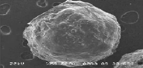
Morphology: Morphology of microspheres was examined by using scanning electron microscopy. The Fig. 2 shows the top view of chitosan microspheres. The top view of the microspheres showed a spherical structure.
FIG. 2: MORPHOLOGY OF MICROSPHERES BY SCANNING ELECTRON MICROSCOPY
DISCUSSION: Optimal formulation of chitosan alginate microspheres were selected by results like % entrapment efficiency, size of microspheres, shape, and % drug release from nine experiments. The concentration of sodium alginate had significant impact on drug entrapment efficiency and particle size.
However, the drug release was greatly retarded at higher concentration of sodium alginate as the concentration increases from 2.0% w/v to 3.0% w/v. Above 4% w/v sodium alginate polymeric solution was too viscous to pass through needle therefore microspheres could not formed. As the concentration increases from 0.5%w/v to 1.5% w/v, (sodium alginate 3 - 4% v) drug release decreases from 88.00% to 83.00% and % entrapment efficiency increases from 77.65% to 80.90%.
[image:4.612.53.565.77.252.2] [image:4.612.48.297.600.719.2]ratio increases from 1.5:3to 1.50:4 the % drug release decreases from 92.74 % to 83.00% due to leaching of drug in to counterion solution. Chitosan, a natural polymer which get degraded in acidic environment, non toxic. Flurbiprofen loaded chitosan microspheres were prepared by using ionotropic gelation technique.
Prepared flurbiprofen loaded chitosan alginate microspheres evaluated for size, shape, % entrapment efficiency, % drug content and % drug release. The concentration of chitosan had significant impact on drug entrapment efficiency and particle size. However the drug release was greatly retarded at higher concentration of chitosan as the concentration increases from 0.5% w/v to 1.0% w/v. 1.5% w/v chitosan polymeric solution was too viscous to pass through needle therefore
microspheres could not formed. As the
concentration increases from 1.0% w/v to 1.5% w/v, drug release decreases and % entrapment efficiency increases. Drug: polymer ratio also showed significant effect on drug entrapment efficiency, drug release and particle size of microspheres, as drug: polymer ratio increases from 1.5:4 to 1.5:3 the % entrapment efficiency increases from 75.13% to 83.90% and % release of drug also decreased from 94.24% to 83.43%. As drug: polymer ratio increases from 0.33:1 to 0.50:1 the % entrapment efficiency decreases from 91.34 % to 59.81% due to leaching of drug in to counter ion solution.
The demonstrated higher values of R2 (0.996 to 0.998), indicating excellent fitting of model. Evaluation of formulations, chosen as optimal from A to I, indicated that the formulation F fulfilled maximum requisites because of better drug entrapment efficiency and % controlled release of the drug. After comparing the evaluation parameter of chitosan alginate formulations optimized microspheres formulation (F) was selected because of better drug entrapment efficiency, and controlled release of the drug. Chitosan alginate optimized formulation (F) was evaluated for SEM, in-vitro drug release.
CONCLUSION: The sodium alginate and chitosan microspheres of flurbiprofen prepared by ionotropic gelation method. Optimization helped to predict the best possible formulation. In-vitro
release study of F indicated that flurbiprofen was released in controlled manner up to 24 h.
ACKNOWLEDGEMENT: Author sincerely thankful to Suresh Gyan Vihar University, Jaipur (Rajasthan) for providing feasibility of all required instruments other facilities as departmental library, reputed journals, Internet access.
CONFLICT OF INTEREST: Nil
REFERENCES:
1. Tiwari S, Krishnamurthy T, Pai M, Mehta P and Choudhary P: Controlled release formulation of tramadol hydrochloride using hydrophilic and hydrophobic matrix system: AAPS Pharma SciTech 2003; 4(3): Article 31. 2. Lachman L, Liberman H and Kanig J: The Theory and
Practice of Industrial Pharmacy. 3rd ed. Varghese Publishing House 1987; 430.
3. Grass G and Robinson J: Sustain and Controlled Drug Release Drug Delivery System. In: Banker G., Rhodes C, Modern Pharmaceutics, Marcel Dekker, New York and Basel, INC; 1990; 40: 635, 647-652.
4. Ghosh T and Jasti B: Oral controlled release solid dosage forms. Theory and Practice of Contemporary pharma-ceutics. CRS press, New York; 2004; 338-355.
5. Anil K and Willem F: Stevens Chitosan–alginate
multilayer beads for controlled release of ampicillin 2005; 290: 45–54.
6. Anal, A, Bhopatkar D, Tokura S, Tamura H and Stevens: Chitosan–alginate multilayer beads for gastric passage and controlled intestinal release of protein. Drug Dev. Ind. Pharm. 2003; 29: 713-724.
7. Kulkarni AR, Soppimath KS and Aminabhavi TM:
Ontrolled release of diclofenac sodium from sodium alginate beads crosslinked with glutaraldehyde Elsevier Science 1999; 74: 29-36.
8. Srinatha A and Pandit J: Ionic Cross-linked Chitosan Beads for Extended Release of Ciprofloxacin: In vitro Characterization. Eur J Pharm Biopharma 2004; 54: 252-263.
9. Khaled A and Taha M: Synthesis of iron-crosslinked chitosan succinate and iron-crosslinkedhydroxamated chitosan succinate and their in vitro evaluation as potential matrix materials for oral theophylline sustained-release beads. Euro. J. Pharma. Sci. 2001; 13: 159-168.
10. Shiraishi S, Imai T and Otagiri M: Controlled release of indomethacin by chitosan-polyelectrolyte complex: optimization and in vivo/in vitro evaluation. J Cont Rel 1993; 25: 217-225.
11. Patel V, Patel H and Kotadiya R: formulation and characterization of chitosan methotrexate beads by Ionotropic gelation. Ind J Pharm Edu Res 2009; 43(1): 71-76.
12. Sinha V, Singla A, Wadhawan S, Kaushik R, Kumria R, Bansal K and Dhawan S: Chitosan microspheres as a potential carrier for drugs 2004; 274: 1-33.
13. George P and Nikolaos B: Swelling studies and in vitro release of verapamil from calcium alginate and calcium chitosan beads. Sci 2006; 323: 34-42.
for delivery of water-soluble macromolecules to colon. Sci Dir 2005; 61: 39-51.
15. Dhole A, Gaikwad P, Bankar VH and Pawar S: A Review
on Floating Multiculate Drug Delivery System–A novel approach to gastric retention. Int J Pharm Sci Rev Res 2011; 6(2): 205-211.
16. Madgulkar A, Bhalekar M and Swami M: In vitro and in-vivo studies on chitosan beads of losartan duolite ap143 complex, optimized by using statistical experimental design. AAPS Pharm Sci Tech 2009; 10(3): 743-751. 17. Chawla G, Gupta P, Koradia V and Bansal AK: Gastro
retention a means to address regional variability in intestinal drug absorption. Pharm Tech 2003; 50-68. 18. Korsmeyer R, Gurny R, Doelker E, Buri P and Peppas N:
Mechanisms of potassium chloride release from
compressed, hydrophilic, polymeric matrices: Effect of entrapped air. J Pharm Sci 1983; 72: 1189-1191.
19. Gander B, Gurny R and Doelker E: Matrices for controlled liberation of drugs from polymers. I. Mechanisms of the penetration of solvents into polymers. Pharm Acta Helv 1986; 61: 130-134.
20. Rania A, Gehanne A, Nahed D and Samia A: Nour, Preparation, in-vitro and in-vivo evaluation of stomach-specific metronidazole-loaded alginate beads as local anti-Helicobacter pylori therapy. Sci Dir 207-214.
21. British pharmacopeia 2009, the department of health, social services and public safety, Published by The Stationery Office on behalf of the Medicines and Healthcare products Regulatory Agency (MHRA), first published 2008, ISBN 978 0 11 322799 0,vol.2.1192
22. Raymond C, Paul J and Paul JW: Handbook of
Pharmaceutical Excipients. 6th Edn. Published by Pharm. press. New York. 2006; 159, 622, 89.
23. Swarbrick J and Boylon J: Encyclopedia of
Pharmaceutical Technology.14nded. New York: Marcel Dekker Inc 2002; 1:.05, 2118, 2722, 402, 2461.
24. Rania A, Gehanne A, Nahed D and Samia A: Nour, Preparation, in-vitro and in-vivo evaluation of stomach-specific metronidazole-loaded alginate beads as local anti-Helicobacter pylori therapy. Sci Dir 207-214.
All © 2013 are reserved by International Journal of Pharmaceutical Sciences and Research. This Journal licensed under a Creative Commons Attribution-NonCommercial-ShareAlike 3.0 Unported License.
This article can be downloaded to ANDROID OS based mobile. Scan QR Code using Code/Bar Scanner from your mobile. (Scanners are available on Google Playstore)
How to cite this article: