INTRODUCTION
Three cell lineages with distinct developmental potentials are formed during mammalian pre-implantation development: the trophectoderm (TE), the epiblast (EPI) and the primitive endoderm (PE) (Cockburn and Rossant, 2010; Rossant and Tam, 2009; Wang and Dey, 2006; Zernicka-Goetz et al., 2009). The first cell fate decision segregates the TE from the inner cell mass (ICM). Prior to implantation, the ICM gives rise to the PE, which is a monolayer separating the blastocoel from the cluster of pluripotent EPI cells. The EPI forms the future fetus, the TE develops into the fetal placenta, and the PE becomes the visceral and parietal endoderm of the yolk sacs. The ICM/EPI is the source of pluripotent embryonic stem cells (ESCs) (Evans and Kaufman, 1981; Martin, 1981).
During the fourth and fifth cleavage divisions of the mouse embryo, polar outer cells retain an apical surface and develop into the TE, while inner apolar cells acquire an ICM fate (Cockburn and Rossant, 2010; Yamanaka et al., 2006). Mutual antagonism between the lineage-specific transcription factors Cdx2 and Oct4 (Pou5f1 – Mouse Genome Informatics) maintains segregation between the TE and ICM lineages (Niwa et al., 2000; Niwa et al., 2005; Strumpf et al., 2005). Cdx2 is required for the specification and differentiation of TE and for suppressing the ICM fate (Ralston and Rossant, 2008; Strumpf et al., 2005). Although segregated from the ICM fate, continued proliferation and maintenance of TE cells depend on signals from the ICM/EPI (Gardner et al., 1973; Rossant and Cross, 2001). Specifically, Fgf4 from the ICM signals
through Fgfr2 in the TE to regulate trophoblast development (Chai et al., 1998; Nichols et al., 1998), possibly via the TE target genes Cdx2and Eomes. Oct4 is essential for the formation of pluripotent ICM cells and for suppressing a TE fate (Nichols et al., 1998). Although Cdx2 and Oct4 are key factors specifying the TE and ICM lineages, respectively, their upstream regulators during early lineage allocation remain largely unknown.
Prior to implantation, the ICM gives rise to the EPI and PE (Cockburn and Rossant, 2010; Rossant, 2004; Rossant and Tam, 2009). Prospective EPI and PE cells express Nanog and Gata6, respectively, and appear randomly within the ICM in mid-blastocysts before the PE morphologically segregates to the outer surface of the ICM (Chazaud et al., 2006; Gerbe et al., 2008; Kurimoto et al., 2006; Plusa et al., 2008). The separation of these two lineages involves cell migration, position induction and apoptosis (Meilhac et al., 2009; Morris et al., 2010; Plusa et al., 2008). Compared with separation of the TE and ICM lineages, relatively little is known about PE and EPI lineage segregation. Gata and Sox transcription factors regulate PE differentiation (Fujikura et al., 2002; Lim et al., 2008; Molkentin et al., 1997; Morrisey et al., 1998; Soudais et al., 1995). Sox7 and Sox17 are both expressed in developing PE and regulate Gata4/6 expression during PE differentiation of ESCs (Kanai-Azuma et al., 2002; Niakan et al., 2010; Seguin et al., 2008; Shimoda et al., 2007). Cells in the pre-implantation embryo with reduced levels of Gata6 or Sox17 are impaired in PE contribution (Kanai-Azuma et al., 2002; Morris et al., 2010). In Xenopus, Sox17 has both Gata-dependent and Gata-inGata-dependent roles during endoderm formation (Sinner et al., 2006). Thus, Sox7 and Sox17 could function redundantly during PE differentiation and could act upstream of, or in parallel to, Gata factors.
Somatic cells can be reprogrammed into a pluripotent state by ectopic expression of Oct4, Sox2, c-Myc and Klf4 (Takahashi and Yamanaka, 2006). Interestingly, among this quartet, Klf4 is Development 137, 3953-3963 (2010) doi:10.1242/dev.054775
© 2010. Published by The Company of Biologists Ltd
1Division of Developmental Biology, 2Division of Gastroenterology, Hepatology and Nutrition, 3Division of Pulmonary Biology, Cincinnati Children’s Hospital Medical Center, 3333 Burnet Avenue, Cincinnati, OH 45229-3039, USA.
*Author for correspondence (james.wells@cchmc.org)
Accepted 22 September 2010
SUMMARY
Kruppel-like transcription factors (Klfs) are essential for the induction and maintenance of pluripotency of embryonic stem cells (ESCs), yet little is known about their roles in establishing the three lineages of the pre-implantation embryo. Here, we show that Klf5 is required for the formation of the trophectoderm (TE) and the inner cell mass (ICM), and for repressing primitive endoderm (PE) development. Although cell polarity appeared normal, Klf5mutant embryos arrested at the blastocyst stage and failed to hatch due to defective TE development. Klf5 acted cell-autonomously in the TE, downstream of Fgf4 and upstream of Cdx2, Eomes and Krt8. In the ICM, loss of Klf5 resulted in reduced expression of pluripotency markers Oct4 and Nanog, but led to increased Sox17 expression in the PE, suggesting that Klf5 suppresses the PE lineage. Consistent with this, overexpression of Klf5 in transgenic embryos was sufficient to suppress the Sox17+PE lineage in the ICM. Klf5 overexpression led to a dose-dependent decrease in Sox17promoter activity in reporter assays in cultured cells. Moreover, in chimeric embryos, Klf5–/–cells preferentially contributed to the Sox17+PE lineage and Cdx2 expression was not rescued in Klf5–/–outer cells. Finally, outgrowths from Klf5–/– embryos failed to form an ICM/pluripotent colony, had very few Oct4+or Cdx2+cells, but showed an increase in the percentage of Sox17+PE cells. These findings demonstrate that Klf5 is a dynamic regulator of all three lineages in the pre-implantation embryo by promoting the TE and epiblast lineages while suppressing the PE lineage.
KEY WORDS: Klf5, Trophectoderm, Epiblast, Embryonic stem cells, Pluripotency, Primitive endoderm, Lineage specification, Mouse
Klf5 regulates lineage formation in the pre-implantation
mouse embryo
Suh-Chin J. Lin1, Maqsood A. Wani2, Jeffrey A. Whitsett3and James M. Wells1,*
D
E
V
E
LO
P
M
E
N
dispensable for the development of pluripotent embryonic lineages despite its crucial role in ESC pluripotency (Guo et al., 2009; Hall et al., 2009; Li et al., 2005). Klf4 is a member of the Kruppel-like transcription factors (Klfs), a family of highly related zinc-finger proteins important for development and physiological homeostasis (Dang et al., 2000; McConnell et al., 2007). In addition to Klf4, Klf1/2/5 also induce pluripotency (Hall et al., 2009; Nakagawa et al., 2008). Although concurrent depletion of Klf2, Klf4 and Klf5 leads to ESC differentiation, knockdown of single Klfs does not have a significant effect (Jiang et al., 2008), suggesting redundancy in their role in ESC self-renewal. Genetic loss-of-function analyses in mice demonstrated that Klf1/2/4 are entirely dispensable for ICM/ESC development (Nuez et al., 1995; Perkins et al., 1995; Segre et al., 1999; Wani et al., 1998). By contrast, Klf5 deficiency leads to arrest at the blastocyst stage, and Klf5is required for ESC derivation from the ICM and for ESC self-renewal (Ema et al., 2008; Parisi et al., 2008). However, the underlying cause of the retarded blastocyst development associated with Klf5deficiency remains largely unknown.
In this study, we show that Klf5 is expressed throughout pre-implantation development and can act both positively and negatively to regulate the development of the three cell lineages in the pre-implantation embryo. Klf5 is expressed in all cells from the 2-cell stage but becomes more highly expressed in the outer cells that form the TE. Consistent with this, Klf5–/–embryos arrested at
the blastocyst stage and failed to hatch, with associated defects in the TE. Expression of TE markers was lost and was not rescued by exogenous FGF4 in culture or by neighboring wild-type cells in chimeric embryos, suggesting that high levels of Klf5 act cell-autonomously to positively regulate TE development. Klf5 was expressed at lower levels in the inner cells that become the EPI and PE lineages. Klf5 has opposite roles in these two lineages. It is required for the formation and expansion of the EPI and promotes pluripotency gene expression. In the PE lineage, loss of Klf5 promotes PE formation and transgenic overexpression of Klf5 represses the Sox17+PE lineage. Taken together, our data show that Klf5 is a multifaceted lineage regulator that acts to both promote and suppress specific cell fates during pre-implantation development.
MATERIALS AND METHODS
Generation of Klf5mutant mice
ESCs were electroporated with a linearized targeting vector (see Fig. S1 in the supplementary material) and selected for hygromycin resistance. Homologous recombinants were verified by PCR and Southern blot
analysis. Selected recombinants were transiently transfected with a Cre expression plasmid and cells carrying a deletion of exons 1/2 (see Fig. S1 in the supplementary material) were used to generate chimeric KDmice. Germline transmission of the KDallele was monitored by PCR. KBmice were generated by our Transgenic Core Facility using a gene-trap ESC line (BayGenomics) with bgeo inserted into the first intron. Mice were housed in the Laboratory Animal Housing Facility of the Cincinnati Children’s Research Foundation and maintained under institutional guidelines.
Embryo genotyping
Genotyping was performed by nested PCR on individual embryos following culture and/or antibody staining. For PCR primers and product size, see Table S1 in the supplementary material. Mice used were wild-type CD1, Klf5+/– (carrying a Klf5 mutant allele, KD or KB), B5/EGFP
(ubiquitously expressing an enhanced green fluorescent protein) (Hadjantonakis et al., 1998), TRE-Klf5(expressing human KLF5from a tetracycline-responsive promoter) (Sur et al., 2006), and R26-M2rtTA [which widely expresses an optimized form of the reverse tetracycline-controlled transactivator (rtTA-M2)] (Hochedlinger et al., 2005).
Immunofluorescent staining
Immunofluorescent staining was performed as described (Ralston and Rossant, 2008). Primary antibodies are listed in Table 1. Secondary antibodies included Cy3-, Cy5- and Alexa 488-conjugated goat anti-rat/mouse/rabbit and donkey anti-goat/mouse/rabbit antibodies (1:200-1:400, Molecular Probes). For BrdU labeling, blastocysts were cultured for 1-2 hours in 20 mM BrdU in CZB (Chemicon). For TUNEL staining, embryos were incubated in the TUNEL reaction mixture (Roche) following anti-Klf5 immunostaining. Nuclei were visualized with DRAQ5 (1:400, Alexis Biochemicals). Images were collected using a Zeiss LSM 510 Meta laser-scanning confocal microscope.
Embryo culture, chimeric embryo aggregation, and doxycycline (Dox)-inducible expression of Klf5
E2.5 and E3.5 embryos from timed intercrosses were flushed from oviducts and uteri, respectively, in M2 (Sigma) and cultured in microdrops of CZB under mineral oil (Sigma) for 48-72 hours at 37°C, 5% CO2 (Nagy et al.,
2003). For outgrowth formation, zona-free blastocysts were cultured in ES medium [knockout-DMEM (Invitrogen) supplemented with 15% fetal bovine serum (FBS), 100 U/100 mg/ml penicillin/streptomycin, 0.1 mM non-essential amino acids, 4.5 mM L-glutamine, 0.2 mM sodium pyruvate, 1000
[image:2.612.50.560.571.742.2]U/ml ESGRO (mLIF, Chemicon) and 0.1 mM b-mercaptoethanol] in gelatin-coated dishes for 48-72 hours. For the FGF4 experiment, zona-free morulae were cultured for 24 hours with and without 100 ng/ml recombinant human FGF4 (R&D). For generating chimeric embryos, 2-cell embryos were collected from a CD1and B5/EGFPcross and a Klf5heterozygote intercross. After removing zonae, CD1/B5/EGFPembryos were aggregated with Klf5 embryos in depression wells. Chimeric embryos were cultured to blastocysts and processed for immunostaining. In some experiments, Klf5heterozygotes
Table 1. Primary antibodies
Antibody (dilution) Source
Rat anti-Klf5 (1:5000) Dr Ryozo Nagai (Shindo et al., 2002) Guinea pig anti-Klf5 (1:2000) Dr Jeffrey Whitsett (Wan et al., 2008) Mouse anti-Oct4 (1:400) C-10, Santa Cruz Biotechnology
Mouse anti-Cdx2 (1:200) Cdx2-88, BioGenex
Rabbit anti-Nanog (1:200) Cosmo Bio
Rabbit anti-Sox2 (1:2000) Chemicon
Rabbit anti-Tbr2 (Eomes) (1:2000) Chemicon
Goat anti-Sox17 (1:500) R&D
Rat anti-Krt8 (1:20) Troma-I, DSHB, University of Iowa Rabbit anti-PKCz (1:400) C-20, Santa Cruz Biotechnology Rabbit anti-b-catenin (1:100) H-102, Santa Cruz Biotechnology Mouse anti-E-cadherin (1:500) BD Transduction Laboratories Rabbit anti-phospho-p44/42 MAPK (1:100) Cell Signaling Technology
Rabbit anti-ZO-1 (1:500) Zymed
Mouse anti-BrdU (1:50) G3G4, DSHB, University of Iowa
Anti-cleaved caspase 3 (Asp175) (1:200) Cell Signaling Technology
D
E
V
E
LO
P
M
E
N
were bred into a B5/EGFPbackground and chimeras were made between CD1and Klf5/B5/EGFPembryos. To induce Klf5 overexpression, 2-cell embryos collected from an intercross between TRE-Klf5and R26-M2rtTA mice were cultured for 72 hours in 20 mg/ml Dox in CZB.
RNA extraction, reverse transcription and quantitative real-time (q) PCR analysis
RNA isolated from individual blastocysts (RNeasy Micro Kit, Qiagen) was converted into cDNA using the Superscript III First-Strand Synthesis System (Invitrogen) in a 20 ml reaction mixture. qPCR was performed on 0.5 ml aliquots in a total reaction volume of 10 ml with SYBR Green QuantiTect Master Mix (Qiagen) and analyzed on an MJ Opticon Monitor (Bio-Rad). The amount of target RNA was determined by the Pfaffl method (Pfaffl, 2001), normalized to b-actin. qPCR primers are listed in Table S1 in the supplementary material.
Statistical analyses
Statistical analyses were performed using Student’s t-test. Data were expressed as mean±s.e.m. The difference between control and mutant samples was considered significant at P<0.05.
Sox17reporter construct, Klf5 expression vectors and luciferase reporter assay
The luciferase reporter plasmid pSox17-5.6kb was constructed by amplifying a 5.6 kb fragment (using the Expand Long PCR Kit, Roche) upstream of the translation start site from FVB/N mouse genomic DNA and cloning into the pGL3-Basic vector (Promega). The mouse Klf5 expression plasmid pBKCMV-Klf5/HA was described previously (Conkright et al., 1999). The BamHI-XmaI fragment encoding Klf5/HA was subcloned into the pIRES2-eGFP vector (Clontech) to generate pIG-Klf5/HA. A SalI-XmaI fragment lacking the HA tag was generated by PCR and subcloned to generate the pIG-Klf5 vector used for transfection. For the luciferase assay, the luciferase reporter and expression vector plasmids were transfected into HEK293 cells using Lipofectamine 2000 (Invitrogen). Transfections were repeated at least twice, each time in triplicate.
Generation of the inducible Klf5/v5-ires-GFP ESC line
A SalI-XmaI fragment in which the HA tag is replaced with v5 was generated by PCR and subcloned to create pIG-Klf5/v5. The NheI-SmaI fragment of pIG-Klf5/v5 was subcloned into pLox-mSox17-ires-GFP (S.-C.J.L., unpublished) to generate pLox-Klf5/v5-ires-GFP. This construct was co-electroporated with pSalk-Cre into the Ainv15 ESC line (ATCC, SCRC-1029) (Kyba et al., 2002). Stable site-specific integrants were expanded and screened by PCR as described in the ATCC protocol. Dox (1-2 mg/ml) was used to induce the expression of Klf5/v5-ires-GFP.
Western blotting
ESCs were lysed in cell lysis buffer [20 mM Tris-HCl pH 7.5, 150 mM NaCl, 1 mM EDTA, 1 mM EGTA, 1% Triton X-100, 1⫻Protease Inhibitor Cocktail (BD Pharmingen)] and analyzed by standard western immunoblotting procedures with the following antibodies: mouse anti-Oct4 (1:1000, C-10, Santa Cruz Biotechnology), rabbit anti-GFP (1:2000, Invitrogen), rabbit anti-Nanog (1:1000, Cosmo Bio), mouse anti-a-tubulin (1:5000, Sigma) and HRP-conjugated goat anti-rabbit or anti-mouse IgG (1:10,000, Jackson ImmunoResearch).
RESULTS
Klf5 is differentially expressed throughout mouse pre-implantation development and Klf5–/–
embryos arrest at the blastocyst stage
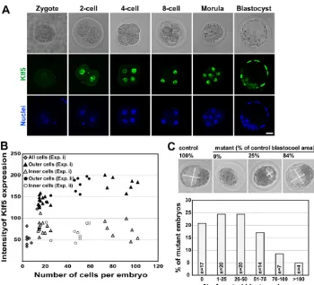
Nuclear Klf5 protein was first detected at the 2-cell stage and was found in all cells through the blastocyst stage (Fig. 1A). From morula to blastocyst stages, Klf5 protein significantly increased in the outer cells, whereas the inner cells had relatively low levels (Fig. 1B). As blastocysts developed, outer cells maturing into TE demonstrated further increases in Klf5 expression, whereas inner ICM cells maintained low levels (Fig. 1B). Klf5transcription was monitored with b-galactosidase activity in Klf5+/bgeo(KB) embryos
and coincided with Klf5 protein staining (see Fig. S2A in the supplementary material). Thus, Klf5 is expressed throughout pre-implantation development with differential levels between outer (TE) and inner (ICM) lineages. Consistent with this expression pattern, Klf5–/–embryos arrested at the blastocyst stage, as was
previously observed (Ema et al., 2008). Analysis of Klf5+/–
intercrosses revealed no null embryos between E6.5 and E8.5 (Table 2), confirming that Klf5 is required during pre-implantation development (Ema et al., 2008; Shindo et al., 2002). To identify the developmental basis for this arrest, we investigated the cellular and molecular roles of Klf5 during specification of the TE, EPI and PE lineages using two mutant alleles of Klf5(KDand KB; see Fig. S1 in the supplementary material), an inducible Klf5 transgenic system, aggregation chimeras, embryo culture and inducible ESCs. To investigate the role of Klf5 during pre-implantation development, E2.5 and E3.5 embryos were isolated from Klf5+/–
intercrosses and their morphology analyzed (Fig. 1C; see Fig. S2B in the supplementary material). E2.5 Klf5–/– morulae were
morphologically indistinguishable from controls. However, E3.5 Klf5–/–embryos had a variable phenotype in blastocoel formation
(Fig. 1C). The majority (70%) of E3.5 mutant embryos had either no cavity (n17) or a small cavity (n40) that was less than half the size of that of control embryos (control, n161; mutant, n82). Regardless of the size of the cavity, mutant embryos exhibited consistent defects in lineage marker expression (see below). To further investigate the phenotype, morulae were placed in culture and monitored over time (Fig. 2A). After 72 hours, control embryos (n18) formed fully expanded blastocysts and started to hatch from their zonae pellucidae. By contrast, Klf5–/–embryos
(n5) did not form expanded blastocoels, failed to hatch and eventually collapsed. A similar phenotype was observed when culturing E3.5 embryos (see Fig. S3 in the supplementary material). Thus, Klf5–/–embryos arrested prior to the expanded
blastocyst stage and failed to hatch. Klf5+/– embryos were
indistinguishable from Klf5+/+ embryos and were grouped as control embryos with the designation ‘+/’.
Early regulation of apical/basal cell polarity has been proposed to initiate segregation of the TE lineage to the outside and the ICM lineage to the inside of the embryo (Johnson and McConnell, 2004; Rossant and Tam, 2009; Yamanaka et al., 2006). Given the early arrest of Klf5–/–embryos, we investigated whether Klf5 affected early cell polarization. Examination of the apical epithelial marker protein kinase C (PKCz, Prkcz) (see Fig. S4A in the supplementary material), the basolateral markers E-cadherin (see Fig. S4B in the supplementary material) and b-catenin (see Fig. S4C in the supplementary material) and the tight junction protein ZO-1 (Tip1) (see Fig. S4D in the supplementary material) in the TE revealed no obvious changes in Klf5–/–embryos as compared
with controls. Since cell polarization occurred normally in Klf5–/–
embryos, we conclude that Klf5 regulates pre-implantation development downstream of cell polarization.
Klf5 acts cell-autonomously upstream of Cdx2 and downstream of Fgf4 to specify the TE lineage TE is the first lineage to be specified in the blastocyst and is required for hatching and implantation. Specification of the TE requires Cdx2 and its upstream regulators Tead4-Yap (Nishioka et al., 2009; Nishioka et al., 2008; Strumpf et al., 2005; Yagi et al., 2007) and Gata3 (Home et al., 2009; Ralston et al., 2010). In control embryos, Cdx2 was expressed throughout the morula and then restricted to the outer prospective TE cells (Fig. 2B) (Niwa et al., 2005; Ralston and Rossant, 2008; Strumpf et al., 2005). By
D
E
V
E
LO
P
M
E
N
contrast, even though morphologically indistinguishable from controls, Klf5–/–morulae had no detectable Cdx2 (Fig. 2B). Other TE markers downstream of Cdx2, Eomes and the intermediate filament protein Krt8, were also absent from E3.5 Klf5–/–embryos
(Fig. 2C,D), suggesting that Klf5 functions upstream of Cdx2 to specify the TE lineage.
Although the outer cells of Klf5–/– blastocysts were
morphologically distinct from the inner cells, it is possible that they have adopted an ICM fate, as was reported in Cdx2–/–cells that inappropriately express Oct4 and Nanog (Ralston and Rossant, 2008; Strumpf et al., 2005). We found no evidence for ectopic expression of Oct4 or Nanog in the outer cells of Klf5–/–embryos
(see below), suggesting that the TE lineage was arrested in an early state and did not adopt an alternative fate. Arrested TE development in Klf5mutants could be due to non-cell-autonomous defects of the ICM as previously reported (Chai et al., 1998; Nichols et al., 1998), where Fgf4 from the ICM acted as a paracrine signal for trophoblast development. However, adding recombinant FGF4 to E2.5 morulae in culture for 24 hours did not rescue TE defects (a lack of expression of Cdx2, Eomes and Krt8) in Klf5–/–embryos (control, n27; mutant, n5) (data not shown).
In addition, the staining pattern of phospho-ERK indicated that
MAP kinase activation downstream of FGF/receptor tyrosine kinase signaling (Lu et al., 2008) was also unaffected (Fig. 2E).
We investigated whether the arrested TE development in Klf5–/–
embryos was due to cell-autonomous defects by generating chimeric blastocysts using wild-type embryos and GFP-expressing embryos from Klf5heterozygote intercrosses (Fig. 2F). Cdx2 was not detected in Klf5mutant cells in mutant chimeras (n6), whereas Cdx2 was readily observed in Klf5non-mutant cells in non-mutant chimeras (n34) (Fig. 2G). Interestingly, whereas non-mutant cells contributed to both populations of TE and ICM, Klf5 mutant cells preferentially contributed to the ICM pole of the chimeric embryos (non-mutant, n34; mutant, n6) (see Fig. S5 in the supplementary material), indicating that Klf5–/– cells were impaired in TE
development and were excluded from the TE by competition from wild-type cells. Thus, Klf5 acts downstream or independently of FGF signaling but upstream of Cdx2 to cell-autonomously regulate TE development.
Low levels of Klf5 differentially regulate the EPI and PE lineages
[image:4.612.52.402.59.377.2]The ICM gives rise to the EPI and the PE, and we investigated the role of Klf5 in allocating these lineages. Oct4 and Nanog are required for establishing the EPI and for maintaining pluripotency in mouse embryos (Mitsui et al., 2003; Nichols et al., 1998) and ESCs (Niwa, 2007). Immunostaining of E2.5 and E3.5 embryos revealed that Oct4 protein was significantly reduced in 25% of mutant embryos (4 of 16) (Fig. 3A), whereas no gross change was observed in Nanog protein (Fig. 3B). However, qPCR analysis of single embryos showed that Oct4 and Nanog mRNAs were reduced by ~70% in mutant embryos (Fig. 3C). In addition,
Table 2. Genotypes of progeny from Klf5+/–matings
Klf5genotype
Stage +/+ +/– –/–
E8.5 5 11 0
E7.5 17 26 0
[image:4.612.49.298.682.741.2]E6.5 22 24 0
Fig. 1. Klf5 is required for normal mouse pre-implantation
development. (A)Confocal
cross-sections showing DIC images (top) and Klf5 (middle) and Draq5 nuclei (bottom) staining. Nuclear Klf5 protein was first detected in the 2-cell embryo and persisted in all cells throughout pre-implantation development. In the blastocyst, much lower levels of Klf5 protein were detected in the inner cell mass (ICM). Scale bar: 20mm. (B)Differential expression of Klf5 between outer and inner cells. Embryos at different developmental stages were stained for Klf5 in two experiments (i, n29; ii, n9). Klf5 expression was quantified from stacked confocal images. (C)E3.5 Klf5–/–embryos have
varying abilities to form blastocoels. Bright-field images show representative mutant embryos with different sizes of blastocoels relative to control
littermates. The length and width of the blastocoel was measured on confocal images and the area was calculated as length ⫻width. The blastocoel area of individual mutant embryos was compared with the average blastocoel size of control littermates. In total, 161 control and 82 mutant embryos from 34 litters were included.
D
E
V
E
LO
P
M
E
N
mutants had a significant reduction of Sox2and c-MycmRNAs (Fig. 3C). The reduction in expression of pluripotency genes, such as Oct4, is sufficient to cause loss of pluripotency (Niwa et al., 2000), and thus could be one cause for the arrest of Klf5–/–
embryos.
These data suggest that Klf5 maintains pluripotency gene expression in vivo, which is consistent with its role in maintaining ESC pluripotency (Ema et al., 2008; Parisi et al., 2008). To directly test whether Klf5 positively regulates pluripotency genes, we generated a mouse ESC line expressing a Dox-inducible v5-tagged Klf5 protein (Klf5/v5-iresGFP) (Kyba et al., 2002). Dox-induced Klf5 was sufficient to upregulate Oct4 and Nanog proteins after 12 hours and their levels remained elevated for up to 48 hours (see Fig. S6 in the supplementary material). Thus, Klf5 promotes the EPI/ESC lineage by regulating the expression of a network of pluripotency-related genes.
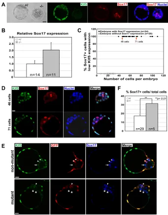
Contrary to its positive effect on the EPI, we found that Klf5 levels profoundly impact the PE lineage, as experimentally lowering Klf5 levels promotes a PE fate and increasing Klf5 levels suppresses the PE lineage. We used Sox17 as a marker of the presumptive PE (Morris et al., 2010) and found that the formation and segregation of the Sox17+/PE from the Nanog+/EPI cells was grossly normal in Klf5
mutants (Fig. 4A; see Fig. S7 in the supplementary material). However, Sox17mRNA levels in mutants increased 2-fold (Fig. 4B), suggesting that reducing Klf5 promotes Sox17 expression. This is consistent with the endogenous expression of Klf5 and Sox17 during pre-implantation development. Sox17 was never observed in cells with high Klf5 levels (Fig. 1B, Fig. 4C,D), suggesting that high Klf5 levels do not support a PE fate. We only observed Sox17 expression in inner cells with low Klf5 levels, suggesting that lowering the levels of Klf5 permits PE development (Fig. 4C,D), To test whether further reducing Klf5 levels can promote PE lineage commitment, we generated chimeric blastocysts to examine whether Klf5mutant cells preferentially contribute to the PE. Chimeric embryos were generated using GFP+wild-type embryos and GFP–embryos from Klf5heterozygote intercrosses (Fig. 4E,F). Klf5mutant cells were twice as likely to become Sox17+PE cells than were non-mutant cells (Fig. 4F), suggesting that a lack of Klf5 actively promotes PE development.
[image:5.612.51.391.60.485.2]To investigate whether upregulating Klf5 is sufficient to suppress the PE lineage, we generated transgenic embryos with a Dox-inducible Klf5 transgene (TRE-Klf5) (Sur et al., 2006) and a transgene ubiquitously expressing tetracycline transactivator ( R26-M2rtTA) (Hochedlinger et al., 2005). In double-transgenic embryos
Fig. 2. Klf5 is required for TE lineage
specification. (A)Cultured E2.5 Klf5–/–
mouse embryos do not form an expanded blastocoel or hatch. Morulae fromKlf5+/–
intercrosses were cultured and monitored for 72 hours. Klf5+/(Klf5+/–and Klf5+/+grouped as controls) and Klf5–/–morulae were
indistinguishable. After 72 hours,Klf5+/
embryos formed an expanded blastocoel (red dashed line) and began to hatch (arrowhead), whereas Klf5–/–embryos formed either no
cavity or a small cyst (red dashed line), remained enclosed within the zonae and degenerated. (B-D)Trophectoderm (TE) marker expression is largely absent inKlf5–/–
embryos. (B)E2.5 or E3.5 embryos were immunostained for Klf5, Cdx2 and b-catenin. Cdx2 was markedly reduced or undetectable in Klf5–/–embryos at E2.5 and absent at E3.5.
(C,D)E3.5 embryos were immunostained for Klf5/Eomes (C) or Cdx2/Krt8 (D). Klf5–/–
embryos showed no expression of Eomes (C) or Krt8 (D). (E)Normal FGF-RTK-MAP kinase signaling in Klf5–/–embryos. Phosphorylation
(P) of ERK (p44/42 MAPK, also known as Erk1/2 and Mapk3/1) was at similar levels in control and mutant embryos. No background staining was detected in the absence of pERK antibodies. (F)Strategy to examine the lineage potential of Klf5mutant cells in chimeric blastocysts. (G)Immunostaining of Klf5, GFP and Cdx2 in chimeric blastocysts. Cdx2 expression was not detected in Klf5mutant cells (arrows, Klf5–GFP+Cdx2–) in mutant chimeras but was present in non-mutant cells (arrowheads, Klf5+GFP+Cdx2+) in non-mutant chimeras. Images (B-E,G) are from confocal cross-sections. DNA was labeled with Draq5 (blue). Scale bars: 20mm.
D
E
V
E
LO
P
M
E
N
(TRE-Klf5; R26-M2rtTA), Dox-induced Klf5 overexpression resulted in blastocysts with half the number of Sox17+PE cells compared with single-transgenic controls (R26-M2rtTA) (Fig. 5A,B). We tested whether Klf5 could suppress the PE lineage by regulating Sox17promoter activity in vitro and found that Klf5 overexpression in HEK293 cells repressed a Sox17 promoter-luciferase reporter in a dose-dependent manner (Fig. 5C). Therefore, reducing the levels of Klf5 promotes the Sox17+/PE lineage, whereas experimentally elevating Klf5 levels suppresses it. Klf5 has the opposite effect on EPI lineage development, as low Klf5 levels are required to promote the EPI lineage and the expression of pluripotency genes, and increasing Klf5 levels further enhances the expression of pluripotency genes (see Fig. S6 in the supplementary material) (Ema et al., 2008; Parisi et al., 2008).
Impaired cell proliferation and increased cell death in Klf5–/–blastocysts
The failure of Klf5–/–embryos to expand at the late blastocyst stage
suggests that cell proliferation and/or cell death might be affected. We performed cell proliferation and TUNEL assays on control and Klf5–/–blastocysts. The percentage of BrdU+cells was significantly reduced in Klf5–/– embryos (45.67±5.75%, n7) compared with
controls (67.88±1.97%, n23) (see Fig. S8A,C in the supplementary material), and there were significantly more TUNEL+nuclei in Klf5–/– embryos (18.74±1.97%, n6) than in control littermates
(8.20±0.95%, n25) (see Fig. S8B,C in the supplementary material). However, Klf5deficiency did not differentially affect TE (outer cells) versus ICM (inner cells) regarding proliferation and cell death. The reduction in BrdU+cells and the 2-fold increase in TUNEL+cells were similar in the TE and the ICM ofKlf5–/–embryos relative to
control embryos (see Fig. S8C in the supplementary material). At the morula stage, no overt difference in the number of cells was
observed between control and mutants (control, 11.9±0.4 cells, n42; mutant, 13.6±0.5 cells, n15) and there was no significant difference in BrdU labeling or cell death (as assessed by cleaved caspase 3 staining) (data not shown). Therefore, in Klf5–/–embryos, both the
TE and ICM lineages failed to expand normally between the morula and blastocyst stages.
Outgrowths ofKlf5–/–blastocysts have reduced numbers of TE and EPI cells but an elevated percentage of PE cells
The arrest of Klf5–/–embryos within their zonae precluded further
examination of lineage development in the embryo. To investigate the impact of Klf5deficiency on later development of the TE, PE and EPI lineages, the zonae were removed, thereby alleviating the hatching defect, and blastocysts were cultured in vitro. Controls formed both trophoblast and ICM outgrowths after 48-72 hours (n43) (Fig. 6A). Klf5–/–blastocysts attached to the culture surface
and formed limited outgrowths without morphological evidence of ICM derivatives (n21) (Fig. 6A). ICM outgrowth was verified by alkaline phosphatase (AP) activity, a marker of undifferentiated pluripotent stem cells (Pease et al., 1990). AP staining was intense in control ICM outgrowths, whereas Klf5–/–outgrowths had largely
undetectable AP activity (Fig. 6A), demonstrating the requirement for Klf5 in generating pluripotent ICM-like cells in culture.
[image:6.612.53.390.60.377.2]The three-dimensional architecture of the outgrowths was further analyzed by performing z-series stacking using confocal microscopy with lineage markers (Fig. 6B; see Fig. S9 in the supplementary material). Control ICM outgrowths were highly organized three-dimensional structures, with an inner core of Oct4+ICM/EPI-like cells surrounded by Sox17+PE cells and then by Krt8+TE cells in the outermost layer. Control outgrowths also had cells that spread out on the plate and were a mix of all three lineages (Fig. 6B; see Fig.
Fig. 3. Klf5 is required to maintain normal transcriptional levels of the ICM
markers Oct4, Nanog and Sox2. (A,B)ICM
markers are present but reduced in Klf5–/–
mouse embryos. (A,B)E2.5 or E3.5 embryos were immunostained for Klf5/Oct4 (A) or Klf5/Nanog (B).Klf5–/–embryos showed
reduced Oct4 protein (A) and only a subset of cells from E3.5 Klf5–/–blastocysts
expressed Nanog (B). Images are from projected confocal z-series. DNA was labeled with Draq5 (blue). Scale bars: 20mm. (C)Klf5–/–blastocysts have reduced
expression of TE/epiblast (EPI) markers. qPCR analysis comparing the mRNA expression of early lineage markers between control and Klf5–/–blastocysts. Shown are the
mean±s.e.m. for the indicated numbers of embryos of each genotype. *P<0.05, **P<0.001.
D
E
V
E
LO
P
M
E
N
S9 in the supplementary material). By contrast, Klf5–/–outgrowths
had no ICM-derived cells and the remaining cells had spread out and contained scattered Oct4+cells mixed with abundant Sox17+PE cells (Fig. 6B; see Fig. S9 in the supplementary material). The Oct4+cells in Klf5–/– outgrowths had abnormally large nuclei, indicating a
significant phenotypic change (Fig. 6B). Cdx2+cells were almost absent in Klf5–/–outgrowths, in contrast to controls (Fig. 6B).
Quantification of cell numbers (control, 1682.9±306.1, n11; mutant, 155.9±27.9, n12) indicated that the expansion of the ICM/EPI, TE and PE lineages was reduced in Klf5–/–outgrowths
(Fig. 6Ca), consistent with the reduced proliferation and elevated apoptosis observed at the blastocyst stage (see Fig. S8 in the supplementary material). Oct4+cells were reduced by 89% (control, 106.4±50.1, n5; mutant, 11.8±6.8, n6) and Cdx2+TE cells by 98% (control, 331.5±60, n6; mutant, 4.2±1.8, n6) per embryo. The Sox17+PE lineage was least affected, with an 85% reduction per embryo (control, 224.7±55.1, n11; mutant, 33.1±7.7, n12).
To determine whether individual cell lineages were differentially affected, we normalized the number of TE/EPI/PE cells as a percentage of total cells per embryo (Fig. 6Cb). The proportion of Oct4+EPI cells relative to total cells was similar between control and
Klf5–/– outgrowths. By contrast, there was a 2-fold increase of
Sox17+PE cells in the mutant compared with control embryos (control, 12.4±1.4%, n11; mutant, 22.1±4.2%, n12), consistent with our findings that mutant embryos had increased Sox17 expression in vivo (Fig. 4B) and that Klf5 overexpression repressed Sox17expression in cultured cells and embryos (Fig. 5). There was a dramatic reduction in the percentage of Cdx2+TE cells in mutant outgrowths (control, 21.6±3.4%, n6; mutant, 1.6±0.8%, n6). The percentage of marker-negative cells was similar between control and mutant outgrowths (control, 60.4%; mutant, 67.3%). The outgrowth data indicate that Klf5 is required for the maintenance of the EPI/ESC and TE lineages and that Klf5 suppresses PE development, possibly by regulating the expression of factors involved in PE differentiation, such as Sox17.
DISCUSSION
Distinctive roles of Klf5in early embryonic lineage establishment
[image:7.612.48.389.57.493.2]Genetic loss-of-function studies in mice demonstrate that Klf2and Klf4are dispensable for early embryo development and ESC lineage establishment in vivo (Kuo et al., 1997; Segre et al., 1999). By
Fig. 4. Klf5 is not required for the initial segregation of EPI and PE lineages but represses subsequent PE development.
(A)E3.5 mouse embryos were
immunostained for Klf5 and Sox17. Sox17+ primitive endoderm (PE) cells are present in the Klf5–/–mutant embryos. (B)qPCR analysis
indicating that Klf5–/–blastocysts have
increased expression of the PE marker Sox17. Shown is the mean±s.e.m. (C)Sox17 was only observed in cells with low levels of Klf5 in the ICM and PE. (D)Immunostaining of Klf5 and Sox17 in representative embryos (arrows in C) showed colocalization of Sox17 and low Klf5 expression. (E)Immunostaining of Klf5, GFP and Sox17 in chimeric blastocysts generated from GFP+wild-type embryos and GFP–‘Klf5’ embryos from Klf5 heterozygote intercrosses. Sox17 expression in the ICM was detected in wild-type cells (arrowheads, Klf5+GFP+Sox17+), in non-mutant cells (arrows, Klf5+GFP–Sox17+) and in Klf5–/–cells (asterisks, Klf5–GFP–Sox17+). (F)Klf5mutant cells are more likely to become Sox17+PE cells in chimeras. The percentage of Sox17+cells (GFP–Sox17+) of total ‘Klf5’ cells (GFP–) in chimeras derived from non-mutant or mutant embryos is shown as mean±s.e.m. **P<0.01. Images are from projected confocal z-series (A,D) or from confocal cross-sections (E). DNA was labeled with Draq5 (blue). Scale bars: 20mm.
D
E
V
E
LO
P
M
E
N
contrast, Klf5–/–embryos arrest at the blastocyst stage (Ema et al.,
2008) and the cause is not known. We have found that Klf5 regulates multiple cell fate decisions during pre-implantation development. High levels of Klf5 act upstream of Cdx2 and downstream of, or in parallel to, Fgf4 to promote TE development. For the ICM lineages, a low amount of Klf5 is required for maintaining pluripotency factors to promote the EPI lineage. Having low Klf5 levels in the ICM is also important for the PE lineage, where cells with low or no Klf5 preferentially become Sox17+/PE cells and transgenic expression of Klf5 in pre-implantation embryos suppresses the PE fate. The requirement of Klf5 during early lineage development is illustrated in the model in Fig. 7.
Klf family members act redundantly to maintain ESC self-renewal (Jiang et al., 2008) and to induce pluripotency (Guo et al., 2009; Hall et al., 2009; Nakagawa et al., 2008). However, the presence of Klf2 and Klf4during pre-implantation development (Jiang et al., 2008) cannot rescue the Klf5mutant phenotype. The basis for the functional distinction of Klf5 is not known, but it could be due to the presence of Klf5-specific co-factors in the pre-implantation embryo. It is also plausible that Klf5 has unique DNA target sequences by which it activates or suppresses transcription. The identification of such Klf5-specific factors, binding sites and transcriptional targets will enable us to understand this functional distinction.
Phenotypic differences in the TE betweenKlf5–/– and Cdx2–/–embryos
It is likely that the TE defect in Klf5–/–embryos is responsible for the
failure to hatch. As Cdx2 and its downstream targets are essential for the specification and differentiation of TE (Ralston and Rossant, 2008; Strumpf et al., 2005), lack of Cdx2 could account for the TE defects seen in Klf5–/– embryos. However, there are phenotypic
differences between Klf5–/–and Cdx2–/–embryos. First, zona-free
Klf5–/–embryos attached and formed blastocyst outgrowths, whereas
Cdx2mutant embryos failed to attach (Ralston and Rossant, 2008; Strumpf et al., 2005). Second, Cdx2–/–embryos expressed Oct4 and
Nanog ectopically in the TE, but we found no such changes inKlf5–/–
embryos. Therefore, ectopic expression of Oct4 and Nanog in the TE of Cdx2–/–embryos requires Klf5. This is consistent with our finding
that Klf5 positively regulates the expression of pluripotency genes such as Oct4 and Nanogin the ICM (Fig. 3; see Fig. S6 in the supplementary material).
Because the outer cells of E3.5 Klf5–/–embryos did not express
any lineage markers, it is possible that these cells are trapped in an uncommitted state between the TE and ICM lineages. This phenotype is not due to defects in the Fgf4 signaling from the ICM
that is required for trophoblast proliferation and development (Chai et al., 1998; Nichols et al., 1998), as exogenous FGF4 did not rescue TE development. Moreover, pERK staining was unchanged in Klf5–/– embryos, suggesting that Klf5 affects neither FGF
receptor expression nor other FGF signaling components. Finally, TE defects that resulted from Klf5deficiency were not rescued by neighboring wild-type cells in chimeric embryos. These data show that Klf5 regulates TE development cell-autonomously and acts downstream or independently of FGF signaling. However, we do not know whether Klf5 directly regulates TE Cdx2 expression because no TE enhancer has been identified for Cdx2(Benahmed et al., 2008) and we failed to detect any effects of Klf5 on several Cdx2promoter-luciferase constructs (data not shown).
Klf5 maintains the ICM lineage cell-autonomously It is possible that the TE defect in Klf5–/–embryos in turn impacts
ICM development, causing reduced expression of pluripotency markers. However, we do not think that this is the mechanism underlying the ICM/EPI defects seen in Klf5–/–embryos. In Cdx2
(Strumpf et al., 2005) and Tead4(Nishioka et al., 2008; Yagi et al., 2007) mutants with defective TE development, delineation of the ICM lineage appeared normal. ESCs could be derived and there is ectopic, but not reduced, expression of ICM markers in those models. Thus, the defective ICM in Klf5–/–embryos is likely to be
due to a cell-autonomous role for Klf5 in regulating the pluripotency gene network required for ICM lineage development. Consistent with this interpretation, we have demonstrated that Klf5 overexpression is sufficient to upregulate key pluripotency genes (Nanogand Oct4) in ESCs.
A novel role of Klf5 in suppressing the PE lineage while promoting the EPI lineage
[image:8.612.52.363.59.228.2]Prospective EPI and PE cells in the ICM express the lineage-specific factors Nanog and Sox17/Gata6, respectively (Chazaud et al., 2006; Gerbe et al., 2008; Morris et al., 2010; Plusa et al., 2008). Nanog is required for maintaining pluripotency in the ICM (Chambers et al., 2003; Mitsui et al., 2003; Silva et al., 2009), and in the absence of Grb2, no Gata6+PE is formed and all ICM cells adopt Nanog+EPI characteristics (Chazaud et al., 2006; Cheng et al., 1998). Reduction of either Gata6 or Sox17 in ICM cells impairs their ability to contribute to the PE (Kanai-Azuma et al., 2002; Morris et al., 2010) and Nanog overexpression can inhibit PE development (Qu et al., 2008; Shimoda et al., 2007). Thus, the development of the EPI and PE lineages involves the mutual suppression of lineage-specific factors in the opposite lineage. Our
Fig. 5. Overexpression of Klf5 represses Sox17 expression in PE development.
(A)Immunostaining of Klf5, Nanog and Sox17 in single-transgenic control (Ctl) and double-transgenic (Tg) embryos. Double-transgenic embryos had reduced numbers of Sox17+cells. Images are from projected confocal z-series. Scale bar: 20mm. (B)Induced Klf5 expression led to a decrease in Sox17+PE cells. The percentage of Sox17+cells in cells positive for Sox17 or Nanog is shown. (C)Klf5 represses Sox17promoter activity. Mouse Sox17 promoter-luciferase plasmid (pSox17-5.6kb) was transfected into HEK293 cells together with the vector or different doses of the Klf5 expression plasmid. Error bars in B,C indicate mean±s.e.m.
D
E
V
E
LO
P
M
E
N
findings that low levels of Klf5 can both promote the EPI lineage and permit the PE lineage place it in a key position to modulate PE/EPI lineage allocation from the ICM. Moreover, Klf5deficiency did not significantly affect the expression of Gata6 (data not shown), indicating that Klf5 acts through the regulation of Sox17, which is known to be required for the formation of the PE lineage (Kanai-Azuma et al., 2002; Morris et al., 2010).
It is intriguing that one transcription factor, Klf5, differentially regulates three sets of lineage-determining factors. Klf5 is cell-autonomously required for TE development and acts upstream of Cdx2 and downstream of, or parallel to, Fgf4. In the ICM/EPI, Klf5 maintains normal levels of the key lineage factors Oct4, Nanog, Sox2 and c-Myc. Indeed, Klf5 also positively regulates these genes in ESCs, as shown in ChIP-on-chip analyses (Jiang et al., 2008). By contrast, we found that Klf5 can negatively regulate embryonic PE lineage development by repressing PE factors such as Sox17. Ultimately, the multiple defects in lineage gene expression brought about by Klf5deficiency result in blastocyst arrest. In conclusion, our data indicate that Klf5 is required at various stages to support the development of the earliest cell lineages in the mouse embryo. Klf5 is permissive in that it is required for the continuous expression of pluripotency genes in the ICM/EPI, and is instructive in promoting Cdx2 expression in the TE and repressing Sox17 expression in non-PE lineages. It was recently shown that another zinc-finger transcription factor, Sall4, can regulate distinct core circuits in two different blastocyst-derived stem cells lines: ESCs and extraembryonic endoderm (XEN) cells (Lim et al., 2008). An examination of whether Klf5 acts directly as an activator or repressor, and/or does so via interaction with other lineage-specific factors, is likely to further elucidate the complex biological activities of this molecule.
Acknowledgements
For their insight and help, we thank Drs. R. Nagai (Klf5 antibodies), M. Kyba (inducible Klf5/v5-ires-GFP ESCs), N. Brown (embryo manipulation), M. Kofron (microscopy) and A. Ralston (genotyping). We also thank P. Wakenight, S. K. Dey, A. Zorn, K. Campbell, J. Heasman and members of the J.M.W. and A. Zorn labs for their input. This work was supported by a Career Development Award from the Juvenile Diabetes Research Foundation (JDRF-2-2003-530) and grants from the NIH (R01GM072915 and R01DK080823). S.-C.J.L. was supported by a postdoctoral fellowship from the JDRF (3-2005-213). Deposited in PMC for release after 12 months.
Competing interests statement
The authors declare no competing financial interests.
Supplementary material
Supplementary material for this article is available at
[image:9.612.51.297.55.471.2]http://dev.biologists.org/lookup/suppl/doi:10.1242/dev.054775/-/DC1
Fig. 6. Outgrowth cultures from Klf5–/–embryos lack TE cells, fail
to form an ICM colony and have elevated PE contribution.
(A)Outgrowths of zona-free E3.5 mouse blastocysts. Klf5–/–embryos
attached but formed limited outgrowths that were devoid of ICM-like colonies. Alkaline phosphatase staining identified potential embryonic stem cell (ESC) colonies in control, but not in Klf5–/–, outgrowths.
(B)Outgrowths of zona-free E3.5 blastocysts were analyzed for lineage marker expression after 72 hours in culture. DNA was labeled with Draq5 (blue). Images are from projected confocal z-series. Klf5–/–
outgrowths failed to form an ICM-like structure and had cells that spread out and contained only a few Oct4+or Cdx2+cells alongside Sox17+PE cells. (C)The numbers of Oct4+, Cdx2+or Sox17+cells were counted from confocal optical sections of individual outgrowths. Data are shown either as the mean number of positive cells per embryo (a) or as a percentage of total cells per embryo (b) ± s.e.m. *P<0.05, **P<0.01, ***P<0.005. Scale bars: 20mm.
Fig. 7. A model for the role of Klf5 in regulating early embryonic
lineage decisions. Klf5 function is required at several stages of early
mouse embryonic lineage commitment. In the early blastocyst stage, Klf5 is upregulated and is crucial for the formation of the TE lineage (downstream or independently of FGF signaling). In the ICM lineages, low levels of Klf5 maintain normal expression of key ICM/EPI
transcription factors (Oct4, Nanog), while permitting Sox17 expression in the PE. Klf5 is not required for the initial specification of the EPI and PE lineages. However, the relative number of cells contributing to the EPI is controlled by Klf5 through its upregulation of Oct4 and Nanog (EPI fate) and suppression of Sox17 (PE fate) expression. In embryos and in outgrowths, Klf5deficiency leads to loss of EPI cells and to an increase in Sox17+PE cells.
D
E
V
E
LO
P
M
E
N
[image:9.612.315.564.60.147.2]References
Benahmed, F., Gross, I., Gaunt, S. J., Beck, F., Jehan, F., Domon-Dell, C., Martin, E., Kedinger, M., Freund, J. N. and Duluc, I.(2008). Multiple regulatory regions control the complex expression pattern of the mouse Cdx2 homeobox gene. Gastroenterology135, 1238-1247.
Chai, N., Patel, Y., Jacobson, K., McMahon, J., McMahon, A. and Rappolee, D. A.(1998). FGF is an essential regulator of the fifth cell division in preimplantation mouse embryos. Dev. Biol. 198, 105-115.
Chambers, I., Colby, D., Robertson, M., Nichols, J., Lee, S., Tweedie, S. and Smith, A.(2003). Functional expression cloning of Nanog, a pluripotency sustaining factor in embryonic stem cells. Cell113, 643-655.
Chazaud, C., Yamanaka, Y., Pawson, T. and Rossant, J.(2006). Early lineage segregation between epiblast and primitive endoderm in mouse blastocysts through the Grb2-MAPK pathway. Dev. Cell10, 615-624.
Cheng, A. M., Saxton, T. M., Sakai, R., Kulkarni, S., Mbamalu, G., Vogel, W., Tortorice, C. G., Cardiff, R. D., Cross, J. C., Muller, W. J. et al.(1998). Mammalian Grb2 regulates multiple steps in embryonic development and malignant transformation. Cell95, 793-803.
Cockburn, K. and Rossant, J.(2010). Making the blastocyst: lessons from the mouse. J. Clin. Invest.120, 995-1003.
Conkright, M. D., Wani, M. A., Anderson, K. P. and Lingrel, J. B.(1999). A gene encoding an intestinal-enriched member of the Kruppel-like factor family expressed in intestinal epithelial cells. Nucleic Acids Res. 27, 1263-1270.
Dang, D. T., Pevsner, J. and Yang, V. W.(2000). The biology of the mammalian Kruppel-like family of transcription factors. Int. J. Biochem. Cell Biol. 32, 1103-1121.
Ema, M., Mori, D., Niwa, H., Hasegawa, Y., Yamanaka, Y., Hitoshi, S., Mimura, J., Kawabe, Y., Hosoya, T., Morita, M. et al.(2008). Kruppel-like factor 5 is essential for blastocyst development and the normal self-renewal of mouse ESCs. Cell Stem Cell3, 555-567.
Evans, M. J. and Kaufman, M. H.(1981). Establishment in culture of pluripotential cells from mouse embryos. Nature292, 154-156.
Fujikura, J., Yamato, E., Yonemura, S., Hosoda, K., Masui, S., Nakao, K., Miyazaki Ji, J. and Niwa, H.(2002). Differentiation of embryonic stem cells is induced by GATA factors. Genes Dev.16, 784-789.
Gardner, R. L., Papaioannou, V. E. and Barton, S. C.(1973). Origin of the ectoplacental cone and secondary giant cells in mouse blastocysts reconstituted from isolated trophoblast and inner cell mass. J. Embryol. Exp. Morphol.30, 561-572.
Gerbe, F., Cox, B., Rossant, J. and Chazaud, C.(2008). Dynamic expression of Lrp2 pathway members reveals progressive epithelial differentiation of primitive endoderm in mouse blastocyst. Dev. Biol. 313, 594-602.
Guo, G., Yang, J., Nichols, J., Hall, J. S., Eyres, I., Mansfield, W. and Smith, A.
(2009). Klf4 reverts developmentally programmed restriction of ground state pluripotency. Development136, 1063-1069.
Hadjantonakis, A. K., Gertsenstein, M., Ikawa, M., Okabe, M. and Nagy, A.
(1998). Generating green fluorescent mice by germline transmission of green fluorescent ES cells. Mech. Dev.76, 79-90.
Hall, J., Guo, G., Wray, J., Eyres, I., Nichols, J., Grotewold, L., Morfopoulou, S., Humphreys, P., Mansfield, W., Walker, R. et al.(2009). Oct4 and LIF/Stat3 additively induce Kruppel factors to sustain embryonic stem cell self-renewal.
Cell Stem Cell5, 597-609.
Hochedlinger, K., Yamada, Y., Beard, C. and Jaenisch, R.(2005). Ectopic expression of Oct-4 blocks progenitor-cell differentiation and causes dysplasia in epithelial tissues. Cell121, 465-477.
Home, P., Ray, S., Dutta, D., Bronshteyn, I., Larson, M. and Paul, S.(2009). GATA3 is selectively expressed in the trophectoderm of peri-implantation embryo and directly regulates Cdx2 gene expression. J. Biol. Chem.284, 28729-28737.
Jiang, J., Chan, Y. S., Loh, Y. H., Cai, J., Tong, G. Q., Lim, C. A., Robson, P., Zhong, S. and Ng, H. H.(2008). A core Klf circuitry regulates self-renewal of embryonic stem cells. Nat. Cell Biol. 10, 353-360.
Johnson, M. H. and McConnell, J. M.(2004). Lineage allocation and cell polarity during mouse embryogenesis. Semin. Cell Dev. Biol. 15, 583-597.
Kanai-Azuma, M., Kanai, Y., Gad, J. M., Tajima, Y., Taya, C., Kurohmaru, M., Sanai, Y., Yonekawa, H., Yazaki, K., Tam, P. P. et al.(2002). Depletion of definitive gut endoderm in Sox17-null mutant mice. Development129, 2367-2379.
Kuo, C. T., Veselits, M. L., Barton, K. P., Lu, M. M., Clendenin, C. and Leiden, J. M.(1997). The LKLF transcription factor is required for normal tunica media formation and blood vessel stabilization during murine embryogenesis. Genes Dev.11, 2996-3006.
Kurimoto, K., Yabuta, Y., Ohinata, Y., Ono, Y., Uno, K. D., Yamada, R. G., Ueda, H. R. and Saitou, M.(2006). An improved single-cell cDNA amplification method for efficient high-density oligonucleotide microarray analysis. Nucleic Acids Res. 34, e42.
Kyba, M., Perlingeiro, R. C. and Daley, G. Q.(2002). HoxB4 confers definitive lymphoid-myeloid engraftment potential on embryonic stem cell and yolk sac hematopoietic progenitors. Cell109, 29-37.
Li, Y., McClintick, J., Zhong, L., Edenberg, H. J., Yoder, M. C. and Chan, R. J.
(2005). Murine embryonic stem cell differentiation is promoted by SOCS-3 and inhibited by the zinc finger transcription factor Klf4. Blood105, 635-637.
Lim, C. Y., Tam, W. L., Zhang, J., Ang, H. S., Jia, H., Lipovich, L., Ng, H. H., Wei, C. L., Sung, W. K., Robson, P. et al.(2008). Sall4 regulates distinct transcription circuitries in different blastocyst-derived stem cell lineages. Cell Stem Cell3, 543-554.
Lu, C. W., Yabuuchi, A., Chen, L., Viswanathan, S., Kim, K. and Daley, G. Q.
(2008). Ras-MAPK signaling promotes trophectoderm formation from embryonic stem cells and mouse embryos. Nat. Genet. 40, 921-926.
Martin, G. R.(1981). Isolation of a pluripotent cell line from early mouse embryos cultured in medium conditioned by teratocarcinoma stem cells. Proc. Natl. Acad.
Sci. USA78, 7634-7638.
McConnell, B. B., Ghaleb, A. M., Nandan, M. O. and Yang, V. W.(2007). The diverse functions of Kruppel-like factors 4 and 5 in epithelial biology and pathobiology. BioEssays 29, 549-557.
Meilhac, S. M., Adams, R. J., Morris, S. A., Danckaert, A., Le Garrec, J. F. and Zernicka-Goetz, M.(2009). Active cell movements coupled to positional induction are involved in lineage segregation in the mouse blastocyst. Dev. Biol.
331, 210-221.
Mitsui, K., Tokuzawa, Y., Itoh, H., Segawa, K., Murakami, M., Takahashi, K., Maruyama, M., Maeda, M. and Yamanaka, S.(2003). The homeoprotein Nanog is required for maintenance of pluripotency in mouse epiblast and ES cells. Cell113, 631-642.
Molkentin, J. D., Lin, Q., Duncan, S. A. and Olson, E. N.(1997). Requirement of the transcription factor GATA4 for heart tube formation and ventral morphogenesis. Genes Dev.11, 1061-1072.
Morris, S. A., Teo, R. T., Li, H., Robson, P., Glover, D. M. and Zernicka-Goetz, M.(2010). Origin and formation of the first two distinct cell types of the inner cell mass in the mouse embryo. Proc. Natl. Acad. Sci. USA 107, 6364-6369.
Morrisey, E. E., Tang, Z., Sigrist, K., Lu, M. M., Jiang, F., Ip, H. S. and Parmacek, M. S.(1998). GATA6 regulates HNF4 and is required for
differentiation of visceral endoderm in the mouse embryo. Genes Dev.12, 3579-3590.
Nagy, A., Gertsenstein, M., Vintersten, K. and Behringer, R.(2003).
Manipulating the Mouse Embryo: a Laboratory Manual. Cold Spring Harbor, NY:
Cold Spring Harbor Laboratory Press.
Nakagawa, M., Koyanagi, M., Tanabe, K., Takahashi, K., Ichisaka, T., Aoi, T., Okita, K., Mochiduki, Y., Takizawa, N. and Yamanaka, S.(2008). Generation of induced pluripotent stem cells without Myc from mouse and human fibroblasts.Nat. Biotechnol.26, 101-106.
Niakan, K. K., Ji, H., Maehr, R., Vokes, S. A., Rodolfa, K. T., Sherwood, R. I., Yamaki, M., Dimos, J. T., Chen, A. E., Melton, D. A. et al.(2010). Sox17 promotes differentiation in mouse embryonic stem cells by directly regulating extraembryonic gene expression and indirectly antagonizing self-renewal. Genes Dev.24, 312-326.
Nichols, J., Zevnik, B., Anastassiadis, K., Niwa, H., Klewe-Nebenius, D., Chambers, I., Scholer, H. and Smith, A.(1998). Formation of pluripotent stem cells in the mammalian embryo depends on the POU transcription factor Oct4.
Cell95, 379-391.
Nishioka, N., Yamamoto, S., Kiyonari, H., Sato, H., Sawada, A., Ota, M., Nakao, K. and Sasaki, H.(2008). Tead4 is required for specification of trophectoderm in pre-implantation mouse embryos. Mech. Dev.125, 270-283.
Nishioka, N., Inoue, K., Adachi, K., Kiyonari, H., Ota, M., Ralston, A., Yabuta, N., Hirahara, S., Stephenson, R. O., Ogonuki, N. et al.(2009). The Hippo signaling pathway components Lats and Yap pattern Tead4 activity to distinguish mouse trophectoderm from inner cell mass. Dev. Cell16, 398-410.
Niwa, H.(2007). How is pluripotency determined and maintained? Development
134, 635-646.
Niwa, H., Miyazaki, J. and Smith, A. G.(2000). Quantitative expression of Oct-3/4 defines differentiation, dedifferentiation or self-renewal of ES cells. Nat. Genet. 24, 372-376.
Niwa, H., Toyooka, Y., Shimosato, D., Strumpf, D., Takahashi, K., Yagi, R. and Rossant, J.(2005). Interaction between Oct3/4 and Cdx2 determines trophectoderm differentiation. Cell123, 917-929.
Nuez, B., Michalovich, D., Bygrave, A., Ploemacher, R. and Grosveld, F.
(1995). Defective haematopoiesis in fetal liver resulting from inactivation of the EKLF gene. Nature375, 316-318.
Parisi, S., Passaro, F., Aloia, L., Manabe, I., Nagai, R., Pastore, L. and Russo, T.
(2008). Klf5 is involved in self-renewal of mouse embryonic stem cells. J. Cell Sci.
121, 2629-2634.
Pease, S., Braghetta, P., Gearing, D., Grail, D. and Williams, R. L.(1990). Isolation of embryonic stem (ES) cells in media supplemented with recombinant leukemia inhibitory factor (LIF). Dev. Biol. 141, 344-352.
Perkins, A. C., Sharpe, A. H. and Orkin, S. H.(1995). Lethal beta-thalassaemia in mice lacking the erythroid CACCC-transcription factor EKLF. Nature375, 318-322.
Pfaffl, M. W.(2001). A new mathematical model for relative quantification in real-time RT-PCR. Nucleic Acids Res. 29, e45.
D
Plusa, B., Piliszek, A., Frankenberg, S., Artus, J. and Hadjantonakis, A. K.
(2008). Distinct sequential cell behaviours direct primitive endoderm formation in the mouse blastocyst. Development135, 3081-3091.
Qu, X. B., Pan, J., Zhang, C. and Huang, S. Y.(2008). Sox17 facilitates the differentiation of mouse embryonic stem cells into primitive and definitive endoderm in vitro. Dev. Growth Differ.50, 585-593.
Ralston, A. and Rossant, J.(2008). Cdx2 acts downstream of cell polarization to cell-autonomously promote trophectoderm fate in the early mouse embryo. Dev. Biol. 313, 614-629.
Ralston, A., Cox, B. J., Nishioka, N., Sasaki, H., Chea, E., Rugg-Gunn, P., Guo, G., Robson, P., Draper, J. S. and Rossant, J.(2010). Gata3 regulates trophoblast development downstream of Tead4 and in parallel to Cdx2.
Development137, 395-403.
Rossant, J.(2004). Lineage development and polar asymmetries in the peri-implantation mouse blastocyst. Semin. Cell Dev. Biol. 15, 573-581.
Rossant, J. and Cross, J. C.(2001). Placental development: lessons from mouse mutants.Nat. Rev. Genet.2, 538-548.
Rossant, J. and Tam, P. P.(2009). Blastocyst lineage formation, early embryonic asymmetries and axis patterning in the mouse. Development136, 701-713.
Segre, J. A., Bauer, C. and Fuchs, E.(1999). Klf4 is a transcription factor required for establishing the barrier function of the skin. Nat. Genet. 22, 356-360.
Seguin, C. A., Draper, J. S., Nagy, A. and Rossant, J.(2008). Establishment of endoderm progenitors by SOX transcription factor expression in human embryonic stem cells. Cell Stem Cell3, 182-195.
Shimoda, M., Kanai-Azuma, M., Hara, K., Miyazaki, S., Kanai, Y., Monden, M. and Miyazaki, J.(2007). Sox17 plays a substantial role in late-stage differentiation of the extraembryonic endoderm in vitro. J. Cell Sci. 120, 3859-3869.
Shindo, T., Manabe, I., Fukushima, Y., Tobe, K., Aizawa, K., Miyamoto, S., Kawai-Kowase, K., Moriyama, N., Imai, Y., Kawakami, H. et al.(2002). Kruppel-like zinc-finger transcription factor KLF5/BTEB2 is a target for angiotensin II signaling and an essential regulator of cardiovascular remodeling.
Nat. Med.8, 856-863.
Silva, J., Nichols, J., Theunissen, T. W., Guo, G., van Oosten, A. L.,
Barrandon, O., Wray, J., Yamanaka, S., Chambers, I. and Smith, A.(2009). Nanog is the gateway to the pluripotent ground state. Cell138, 722-737.
Sinner, D., Kirilenko, P., Rankin, S., Wei, E., Howard, L., Kofron, M., Heasman, J., Woodland, H. R. and Zorn, A. M.(2006). Global analysis of the transcriptional network controlling Xenopus endoderm formation. Development
133, 1955-1966.
Soudais, C., Bielinska, M., Heikinheimo, M., MacArthur, C. A., Narita, N., Saffitz, J. E., Simon, M. C., Leiden, J. M. and Wilson, D. B.(1995). Targeted mutagenesis of the transcription factor GATA-4 gene in mouse embryonic stem cells disrupts visceral endoderm differentiation in vitro. Development121, 3877-3888.
Strumpf, D., Mao, C. A., Yamanaka, Y., Ralston, A., Chawengsaksophak, K., Beck, F. and Rossant, J.(2005). Cdx2 is required for correct cell fate specification and differentiation of trophectoderm in the mouse blastocyst.
Development132, 2093-2102.
Sur, I., Rozell, B., Jaks, V., Bergstrom, A. and Toftgard, R.(2006). Epidermal and craniofacial defects in mice overexpressing Klf5 in the basal layer of the epidermis. J. Cell Sci. 119, 3593-3601.
Takahashi, K. and Yamanaka, S.(2006). Induction of pluripotent stem cells from mouse embryonic and adult fibroblast cultures by defined factors. Cell126, 663-676.
Wang, H. and Dey, S. K.(2006). Roadmap to embryo implantation: clues from mouse models.Nat. Rev. Genet.7, 185-199.
Wan, H., Luo, F., Wert, S. E., Zhang, L., Xu, Y., Ikegami, M., Maeda, Y., Bell, S. M. and Whitsett, J. A.(2008). Kruppel-like factor 5 is required for perinatal lung morphogenesis and function. Development135, 2563-2572.
Wani, M. A., Means, R. T., Jr and Lingrel, J. B.(1998). Loss of LKLF function results in embryonic lethality in mice. Transgenic Res.7, 229-238.
Yagi, R., Kohn, M. J., Karavanova, I., Kaneko, K. J., Vullhorst, D., DePamphilis, M. L. and Buonanno, A.(2007). Transcription factor TEAD4 specifies the trophectoderm lineage at the beginning of mammalian development. Development134, 3827-3836.
Yamanaka, Y., Ralston, A., Stephenson, R. O. and Rossant, J.(2006). Cell and molecular regulation of the mouse blastocyst. Dev. Dyn.235, 2301-2314.
Zernicka-Goetz, M., Morris, S. A. and Bruce, A. W.(2009). Making a firm decision: multifaceted regulation of cell fate in the early mouse embryo.Nat. Rev. Genet.10, 467-477.