Epigenetic reprogramming in somatic cells induced by extract from germinal vesicle stage
pig oocytes
Hong-Thuy Bui*, Deug-Nam Kwon, Min-Hui Kang, Mi-Hye Oh, Mi-Ryung Park, Woo-Jin Park, Seung-Sam Paik, Nguyen Van Thuan and Jin-Hoi Kim*
There was an error published in Development 139, 4330-4340.
Nguyen Van Thuan was incorrectly listed as an author for correspondence. The correct authors for correspondence are shown below:
*Authors for correspondence (bthuy@konkuk.ac.kr; jhkim541@konkuk.ac.kr).
The authors apologise to Nguyen Van Thuan and to readers for this mistake. Development 140, 687 (2013) doi:10.1242/dev.092239
© 2013. Published by The Company of Biologists Ltd
D
E
V
E
LO
P
M
E
N
Development 139, 4330-4340 (2012) doi:10.1242/dev.086116 © 2012. Published by The Company of Biologists Ltd
INTRODUCTION
Epigenetic reprogramming is severely deficient in both somatic cell nuclear transfer (SCNT)-generated cloned embryos and induced pluripotent stem (iPS) cells: most exhibit DNA and histone H3-K9 hypermethylation (Kang et al., 2002; Santos et al., 2003; Kim et al., 2010). Extracts of Xenopusoocytes or embryonic cells can remodel mammalian somatic cell genomes (Hansis et al., 2004; Alberio et al., 2005; Freberg et al., 2007). Our first report showed that a cytoplasmic extract from germinal vesicle (GV) stage oocytes improved the efficiency of producing cloned mouse offspring by the demethylation of histones in the donor somatic cell nucleus (Bui et al., 2008). Although extracts of whole GV stage oocytes and mature metaphase II (MII) oocytes induced site-specific demethylation in the gene region upstream of that encoding the transcription factor Nanog (Miyamoto et al., 2009), MII oocyte extracts had no effect on DNA methylation in the pig (Tong et al., 2006). In addition, methylation of histone H3 at lysine 9 (Me-H3-K9) in somatic cell nuclei is stable after being injected into MII stage oocytes in the pig and mouse (Bui et al., 2006; Bui et al., 2008). We demonstrated in mice that only the GV cytoplasm can induce histone demethylation, whereas the GV nucleus cannot (Bui et al., 2008). Treatment with a cytoplasmic extract from XenopusGV oocytes was also reported to result in the birth of live offspring and increased ovine cloning efficiency (Rathbone et al., 2010).
We believe that the factors in the GV oocyte cytoplasm are necessary to induce some reprogramming event (such as histone
demethylation), but that they are not sufficient to completely reprogram transferred nuclei toward pluripotency. We have developed a simple extract of GV pig oocytes (GVcyto-extract) for establishing stem-like cells by two-step reprogramming. First, oocyte cytoplasmic factors in GVcyto-extract induce chromatin remodeling with histone modifications in treated somatic cell nuclei, and then oocyte nuclear factors continue to reprogram somatic cells to a pluripotent state.
The pig has long been used as a valid model in many branches of medicine because of its morphological and functional similarities to human systems (Prather et al., 2003). Nevertheless, the isolation of fully competent and validated embryonic stem (ES) cells has not been achieved in the pig. Furthermore, progress in establishing iPS cells from pigs lags far behind that for humans and mice, for which many successful isolations have been reported. One reason might be a lack of knowledge regarding the regulatory mechanisms of differentiation and genomic reprogramming in pig cells and embryos. Such reprogramming in pig cells might be more complex than in mice and human models and more investigations should be conducted to overcome these challenges to successful applications.
Methylation of histone H3 at lysine 9 is associated with the appearance of heterochromatin in the nucleus and transcriptional repression (Fischle et al., 2003). Functional differences between the various methylation states have been reported; however, the trimethylated state is often regarded as being the most significant. In pig, dimethylated H3-K9 was found to be present at equivalent levels in both parental sets of chromatin, and trimethylated H3-K9 was established gradually in the paternal chromatin during the pronucleus stage in the pig (Jeong et al., 2007). Both of these situations are in contrast to sperm-derived chromatin in the mouse, which is not modified until the 4-cell stage (Liu et al., 2004). Because there is a specific epigenetic interplay between DNA methylation and TriMe-H3-K9 in the pig paternal pronucleus, as in other mammals (Jeong et al., 2007), we examined TriMe-H3-K9 levels in SCNT-generated cloned pig embryos, along with the
1Department of Animal Biotechnology, College of Animal Bioscience and
Biotechnology/Animal Resources Research Center, Konkuk University, 1 Hwayang-dong, Gwangjin-gu, Seoul 143-701, Korea. 2Hanyang University Hospital,
Department of Histopathology, Molecular pathology, 17 Haengdang-dong, Seondong-gu, Seoul 133-792, Korea.
*Authors for correspondence (bthuy@konkuk.ac.kr; vanthuan@konkuk.ac.kr;
jhkim541@konkuk.ac.kr)
Accepted 18 September 2012
SUMMARY
Genomic reprogramming factors in the cytoplasm of germinal vesicle (GV) stage oocytes have been shown to improve the efficiency of producing cloned mouse offspring through the exposure of nuclei to a GV cytoplasmic extract prior to somatic cell nuclear transfer (SCNT) to enucleated oocytes. Here, we developed an extract of GV stage pig oocytes (GVcyto-extract) to investigate epigenetic reprogramming events in treated somatic cell nuclei. This extract induced differentiation-associated changes in fibroblasts, resulting in cells that exhibit pluripotent stem cell-like characteristics and that redifferentiate into three primary germ cell layers both in vivo and in vitro. The GVcyto-extract treatment induced large numbers of high-quality SCNT-generated blastocysts, with methylation and acetylation of H3-K9 and expression of Oct4 and Nanog at levels similar to in vitro fertilized embryos. Thus, GVcyto-extract could elicit differentiation plasticity in treated fibroblasts, and SCNT-mediated reprogramming reset the epigenetic state in treated cells more efficiently than in untreated cells. In summary, we provide evidence for the generation of stem-like cells from differentiated somatic cells by treatment with porcine GVcyto-extract.
KEY WORDS: Epigenetic reprogramming, Histone methylation, Nuclear transfer, Reprogrammed cells, Oocyte extract
Epigenetic reprogramming in somatic cells induced by
extract from germinal vesicle stage pig oocytes
Hong-Thuy Bui1,*, Deug-Nam Kwon1, Min-Hui Kang1, Mi-Hye Oh1, Mi-Ryung Park1, Woo-Jin Park2, Seung-Sam Paik2, Nguyen Van Thuan1,* and Jin-Hoi Kim1,*
D
E
V
E
LO
P
M
E
N
acetylation of histone H3-K9, an epigenetic mark that is typically associated with active genes and is mutually exclusive with TriMe-H3-K9.
We demonstrated that fibroblasts treated with GVcyto-extract partially and transiently dedifferentiate after culture, with the formation of colonies and expression of specific stem cell-associated genes. The GVcyto-extract treatment induced continuous expression of Oct4in blastocysts developed from embryos reconstructed with Oct4-EGFP-expressing fibroblasts. The use of donor nuclei treated with GVcyto-extract and used for SCNT significantly increased the number of high-quality blastocysts that exhibited Me-H3-K9 and Ac-H3-K9 and expressed Oct4 and Nanog, similar to in vitro fertilized (IVF) embryos. Fibroblasts treated with GVcyto-extract could differentiate in vivo and in vitro into neuronal, pancreatic, cardiac and endothelial cell lineages under specific culture conditions. Our data provide evidence for the generation of functional stem-like cells from somatic cells without the introduction of retrovirally mediated transgenes or ES cell fusion. These results suggest that a combination of reprogramming techniques could improve the efficiency or frequency of normal development in SCNT-generated clones.
MATERIALS AND METHODS
Ethics statement
The treatment of the pigs used in this research followed the guidelines of the National Institute of Animal Science’s Institutional Animal Care and Use Committee, Suwon, South Korea (approval no. 2009-004, D-grade).
Collection of GV oocytes and culture for maturation
Ovaries were collected from prepubertal gilts at a local slaughterhouse. Oocyte-cumulus complexes (OCCs) were aspirated from antral follicles (2-6 mm diameter) using a 1(2-6 g needle. Groups of 50-100 OCCs were cultured in maturation medium for 38-40 hours (Bui et al., 2007).
Preparation of ear skin fibroblasts
Fibroblasts were cultured from ear skin biopsies taken from adult male miniature pigs. Briefly, small pieces of ear skin tissue were washed in Dulbecco’s phosphate-buffered saline (DPBS; Invitrogen, Carlsbad, CA, USA) and minced with a surgical blade on a 100 mm Petri dish. Cells were then dissociated from the tissues in 0.25% trypsin-EDTA (Invitrogen) for 10 minutes at 39°C. After being washed three times, cells were cultured
for 6-8 days in Dulbecco’s modified Eagle’s medium (DMEM; Invitrogen) supplemented with 10% (v/v) fetal bovine serum (FBS; Hyclone, Logan, UT, USA). After removal of unattached clumps of cells by washing the culture plates with DMEM, attached cells were further cultured until confluent and subcultured at intervals of 5-7 days by trypsinization for 5 minutes using 0.25% trypsin-EDTA. Cultured cells were used for experiments after fewer than ten passages.
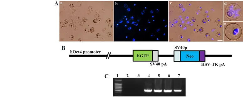
Fibroblast permeabilization and preparation of oocyte extracts Cell permeabilization and oocyte extract preparation were conducted as described (Bui et al., 2008), with modifications. Briefly, fibroblasts were washed in Ca2+- and Mg2+-free Hank’s balanced salt solution (Gibco Life Technologies, Carlsbad, CA, USA) and incubated in 300 ng/ml streptolysin O in the same solution for 50 minutes at 38.5°C with occasional agitation. The oocyte extract was prepared as follows: ~1000 GV oocytes had the cumulus cells stripped and the zonae pellucidae were dissolved using acidic Tyrode’s solution. Zona-free oocytes were transferred to a 1.5 ml tube containing 1 ml extraction buffer (Bui et al., 2008). After centrifugation at 800 rpm (50 g) for 1-2 minutes, 990 l of the supernatant was discarded, leaving 10 l of extraction buffer with the oocytes. The oocytes were lysed by mouth pipetting through a fine glass pipette (internal diameter of ~70 m). Because pig GV oocytes (including growing and fully grown oocytes) are 100-120 m in diameter and the nucleus is 35-40 m in diameter (Bui et al., 2007), this pipetting did not disrupt the nuclei (Fig. 1A). The lysed GV oocyte extract in 10 l extraction buffer (GVcyto-extract) was kept on ice until use.
Permeabilized fibroblasts (5⫻103cells) were suspended in 10 l GVcyto-extract and incubated for 1 hour at 38.5°C in a water bath with occasional agitation. Then, 40 l ES culture medium (Wakayama et al., 2005) was added to the treated cells and a narrow pipette (internal diameter of ~20 mm) was used to disrupt the nuclei of the GV stage oocytes in the extract, and continuously incubated for 4 hours at at 38°C in a 5% CO2atmosphere with occasional agitation. To reseal the plasma membrane, ES medium containing 2 mM CaCl2was added to treated cells and cultured in a 4-well dish for 2 hours. The medium was changed to fresh ES culture medium and cells were cultured until use.
[image:3.612.69.457.489.644.2]Extracts from whole GV oocytes (GVoocyte extract) were prepared in the same medium, but zona-free oocytes and their nuclei were ruptured by high-speed centrifugation at 4°C as described (Miyamoto et al., 2009). Control permeabilized fibroblasts were incubated in the same medium without any oocyte extract.
Fig. 1. Details of preparation of oocyte extract and plasmid used for transfection.(A) Appearance of oocyte nuclei in pig germinal vesicle (GV) stage oocyte cytoplasmic extract (GVcyto-extract; preparation described in Materials and methods) after 1 hour of incubation with
permeabilized fibroblasts. Shown are brightfield (a), DNA (DAPI, blue) (b) and merge (c). Because the isolated GV oocytes (from follicles of 2-6 mm in diameter) contain growing and fully grown oocytes, the remaining nuclei have patterns typical of stringy chromatin (d) and heterochromatin ring (e), respectively, as described previously (Bui et al., 2007). Scale bar: 40 m. (B) The plasmid used for miniature pig ear fibroblast (MPEF)
transfection. The plasmid consists of two expression units: a human OCT4promoter (hOct4)-driven EGFPgene and a neomycin resistance gene (Neo). pA, polyadenylation signal. (C) PCR analysis of genomic DNA to detect the EGFPinsert in G418-resistant MPEF clones stably transfected with hOct4-EGFP plasmid. A primer set was used corresponding to the central region of the EGFPsequence in the plasmid transgene. Lane 1, 100 bp
marker; lane 2, control with water; lane 3, control non-transfected MPEF cells; lane 4, hOct4-EGFP vector; lanes 5-7, cell lines.
D
E
V
E
LO
P
M
E
N
Reprogramming of fibroblasts by GVcyto-extract treatment GVcyto-extract-treated fibroblasts were cultured in ES medium for 1, 2, 3, 4, 5 and 6 weeks. After culture, cells were collected to examine the expression of stem cell markers (Oct4, Rex1, Nanog and Sox2) and the repression of somatic cell markers (Ckap2, Npr3, LmnA and Col5a2) at the mRNA level by reverse transcription PCR (RT-PCR) and at the protein level by immunostaining.
The reprogrammed cells were prepared as follows. GVcyto-extract-treated cells were cultured for 3 weeks and colonies were picked and dispersed to single cells by treatment for 5 minutes in 0.25% trypsin-EDTA. Cells were used for SCNT or 8-cell embryo injection and another group of cells was cultured on mitomycin C-treated mouse embryonic fibroblasts as feeder cells. The medium was changed every 2 days and cells were subcultured using trypsinization every 5-7 days.
Construction of hOct4-EGFP plasmids
To generate hOct4-EGFP, the human (h) OCT4promoter region spanning 2.6 kb was amplified by PCR with the following primers (XhoI and HindIII sites underlined): hOct4-F, 5⬘CTCGAGGGATGGCAAGCTGAGA -AACA-3⬘; and hOct4-R, 5⬘ -AAGCTTGGGGAAGGAAGGCGCCCCAA-3⬘. The amplified DNA was inserted upstream of EGFP cDNA in a pEGFP-N1 vector (Clontech, Palo Alto, CA, USA) (Fig. 1B). The cytomegalovirus (CMV) promoter region was then removed and the construct was confirmed by sequencing. The plasmid was purified using the EndoFree Plasmid Maxi Kit (Qiagen, Valencia, CA, USA) and linearized by digestion with ApaI for stable transfection.
Establishment of Oct4-EGFP transgenic miniature pig ear fibroblast (MPEF) cell lines
MPEF cells were transfected with linearized hOct4-EGFP using a nucleofector-mediated transfection system according to the manufacturer’s instructions (Invitrogen). Stable lines carrying hOct4-EGFP were selected in the presence of 400 g/ml G418 (Geneticin, Invitrogen). The presence of hOct4-EGFPin these lines was confirmed by PCR (Fig. 1C) and three lines were used for SCNT to check the expression of Oct4-EGFP in reconstructed early blastocysts (5 days), of which cell line 2, which showed strong EGFP expression, was chosen. The cells were cultured in DMEM supplemented with 10% FBS, 400 g/ml G418, 100 U/ml penicillin and 100 g/ml streptomycin under 5% CO2in air at 38.5°C.
RT-PCR
mRNA was extracted using the Dynabeads mRNA Direct Kit (Invitrogen Dynal, Oslo, Norway) according to the manufacturer’s instructions. Reverse transcription was performed with 5 l mRNA using the QuantiTect Reverse Transcription Kit (Qiagen). cDNAs were subjected to PCR using AccuPower PCR PreMix (Bioneer, Daejon, Korea) and the specific primers listed in supplementary material Table S1. PCR products were visualized on agarose gels stained with ethidium bromide under u.v. light and relative band intensities were determined using ImageJ software (NIH).
Nuclear transfer to MII oocytes and activation and culture of embryos
For enucleation, mature oocytes were transferred to a droplet of North Carolina State University-23 (NCSU23) medium with 0.4% (w/v) bovine serum albumin (BSA) containing 5 g/ml cytochalasin B and overlaid with sterile mineral oil. A slit in the zona pellucida was introduced using the XYClone laser system with 80% pulse strength and a pulse length of 500 seconds (Hamilton Thorne Biosciences, Beverly, MA, USA), and a small amount of cytoplasm containing metaphase chromosomes was extruded by squeezing the oocyte with the holding pipette and a glass needle. A treated or control fibroblast was introduced into the enucleated oocyte by direct injection by piezo-driven micromanipulators as described (Wakayama et al., 1998). Oocytes subjected to SCNT were cultured in NCSU23 medium for 3 hours and then subjected to parthenogenetic activation (Van Thuan et al., 2002). Control IVF-derived embryos were produced as described (Gupta et al., 2009). After parthenogenetic activation or sperm penetration, all embryos were cultured in NCSU23 medium with an osmolarity of 290-310 mOsM and supplementary amino acids (Van Thuan et al., 2002; Nguyen et al., 2003). In our experimental conditions, embryos reached the
2-cell, morula, early blastocyst, late blastocyst and hatched blastocyst stages at 2, 5, 6, 7 and 8 days, respectively.
Induction of differentiation of reprogrammed cells
Neuronal ectoderm
Reprogrammed cells were seeded in complete DMEM at 5⫻105cells in 90 mm sterile culture dishes. Suspension cultures were maintained for 24 hours before adding 10 M all-trans retinoic acid (Sigma-Aldrich, St Louis, MO, USA). Cells were cultured for 2 weeks in retinoic acid, with the medium replaced every 2 days. Cell aggregates were washed in complete DMEM and plated onto poly-L-lysine (10 g/ml; Sigma-Aldrich)-coated
plates in complete DMEM containing the mitotic inhibitors fluorodeoxyuridine (10 M), cytosine arabinoside (1 M) and uridine (10 M) (all Sigma-Aldrich). Anti-synaptophysin, anti-neuron-specific enolase (NSE), anti-neurofilament, anti-nestin and anti-b-tubulin III antibodies were used to detect neuronal markers.
Pancreatic endoderm
Reprogrammed cells were cultured in RPMI 1640 medium (Invitrogen) containing 11.1 mM glucose supplemented with B27 (Invitrogen) (RPMI 1640/B27 medium), 4 nM activin A (R&D Systems, Minneapolis, MN, USA) and 1 mM sodium butyrate (Sigma-Aldrich) for 1 day. After 24 hours, the medium was replaced with fresh RPMI 1640/B27 supplemented with 4 nM activin A and 0.5 mM sodium butyrate and the cells cultured for 6 days. Cells were dissociated with 200 U/ml collagenase IV (Invitrogen) at 37°C for 3-5 minutes, then transferred to ultralow-attachment 6-well plates (Corning, St Louis, MO, USA) and scraped off the plate in RPMI 1640/B27 supplemented with 20 ng/ml epidermal growth factor (EGF; R&D Systems), 2 ng/ml basic fibroblast growth factor (bFGF; Invitrogen) and 100 ng/ml noggin (R&D Systems) at a ratio of 1:1. The cells were fed with fresh medium every 2 days for 1 week. Anti-Foxa2 and anti-Gata4 antibodies were used to identify pancreatic endoderm markers.
Cardiomyocyte and endothelial cell mesoderm
For cardiomyocyte lineage differentiation, reprogrammed cells were cultured in complete DMEM containing 5 ng/ml leukocyte inhibitory factor (LIF; Millipore, Temecula, CA, USA) and 3 ng/ml bone morphogenetic protein 2 (BMP2; Sigma-Aldrich) in 6-well culture plates (1⫻106cells per well) and 4-well chamber slides (1⫻104cells per well) coated with 0.5% gelatin for 7 days. These cells were immunostained for cardiomyocyte markers using Anti-cardiac troponin T and anti-connexin 43 antibodies. For endothelial lineage differentiation, cells were cultured in endothelial differentiation medium (10% FBS/EBM-2; Clonetics, San Diego, CA, USA) containing supplements (SingleQuot Kit; Clonetics) for 7 days. Cells were stained for endothelial cell markers using anti-von Willebrand factor and anti-CD31 antibodies.
Differential nuclear staining
Zonae pellucidae were removed from blastocysts using acidic Tyrode’s solution. Zona-free embryos were exposed to a 1:7 dilution of rabbit anti-pig whole serum (Sigma-Aldrich) for 60 minutes. Embryos were then washed three times for 5 minutes each in DPBS. Finally, embryos were incubated in a 1:10 dilution of guinea pig complement (Sigma-Aldrich) containing 10 g/ml propidium iodide and 10 g/ml bisbenzimide (Hoechst 33342; Sigma-Aldrich) for 35 minutes. After being washed in DPBS five times, the stained embryos were mounted onto slides and observed under u.v. light with an epifluorescence microscope. Cells stained blue and pink were considered to be inner cell mass and trophectoderm cells, respectively. The ratio of inner cell mass cells was calculated as a percentage of total cell number.
Immunohistochemical staining
Oocytes and embryos were fixed and then immunostained as described (Bui et al., 2007). Quantitative analysis was conducted as described (Bui et al., 2008). Antibodies used are summarized in supplementary material Table S2.
Flow cytometry
Cells were fixed in 2% neutral-buffered paraformaldehyde and permeabilized with 0.1% Triton X-100 for 15 minutes. Then, cells were
D
E
washed twice in DPBS supplemented with 1% BSA and incubated at 4°C overnight with primary antibodies (supplementary material Table S2). After washing, cells were incubated in Alexa Fluor 488 anti-rabbit or anti-mouse IgG for 1 hour. Labeled cells were subjected to FACS (BD Bioscience, Franklin Lakes, NJ, USA). Data were analyzed using WinMDI 2.8 software. Differences in intensity were quantified by dividing the intensity of signal fluorescence by that of the counterstain used. Arbitrary units were used for graphic representation.
Karyotype analysis
Cells were incubated in culture with colchicine (20 ng/ml; Sigma) for 3 hours, followed by hypotonic treatment with 75 mM KCl, fixed with methanol:acetic acid (3:1) and spread onto clean glass slides. Metaphase chromosomes were prepared and karyotypes evaluated using an Applied Spectral Imaging Band View digital imaging system (Malvern Instruments, Malvern, UK).
Teratoma formation
Approximately 1⫻106reprogrammed cells were injected subcutaneously or into the testis of 5-week-old immunodeficient nude mice to evaluate teratoma formation. For controls, mouse ES cells were injected into some of the mice. After 5-6 weeks, teratomas were fixed in paraformaldehyde, embedded in paraffin wax and sectioned for histology.
Statistical analysis
Each experiment was repeated at least four times. More than 50 immunostained oocytes were examined in each group. All data were subjected to one-way analysis of variance (F1 test) followed by the Tukey multiple range test to determine differences between experimental groups. Data on embryo development rates were analyzed using the 2test and P<0.05 was considered statistically significant. For quantitative analyses, fluorescent images were subjected to densitometric analysis using ImageJ.
RESULTS
Induction of dedifferentiation in GVcyto-extract-treated fibroblasts
[image:5.612.54.370.301.742.2]Fibroblasts treated with GVcyto-extract showed changes in cell morphology as early as day 7 post-treatment and started forming small colonies. The colonies enlarged, resembling ES colony morphology, after 3 weeks in culture (Fig. 2A). Colony formation was concomitant with weak expression of Oct4 (also known as Pou5f1) protein in treated cells after 2 weeks in culture (supplementary material Fig. S1Aa,b), whereas the permeabilized control cells that were not treated with GVcyto-extract did not show any morphological changes or marker expression during this
Fig. 2. GVcyto-extract induces stem cell-like characteristics.(A) Colony formation in GVcyto-extract-treated fibroblasts after culture for 0-3 weeks. Arrow indicates appearance of small colony after culture for 1 week. (B) The colonies expressed high levels of Oct4 protein after 3 weeks of culture, whereas the co-cultured cells did not form colonies and did not express any stem cell markers. (C-F) Expression of stem cell-specific genes (Oct4, Rex1, Nanogand Sox2) and repression of somatic cell-specific genes (Ckap2,
Npr3, Lmnaand Col5a2) in treated cells. Pig fibroblasts were treated with GVcyto-extract, cultured for 0-6 weeks, isolated and mRNA expression quantified. (C,D) PCR analysis of the indicated genes. b-actin was used as a normalization control. BL, blastocyst; (–), water control; pF, pig ear fibroblast. (E,F) Relative band intensities of Oct4and Ckap2. Lanes are ordered as in C,D, i.e. BL, –, pF and 0-6 weeks in culture. Scale bars: 50 m.
D
E
V
E
LO
P
M
E
N
time in culture (supplementary material Fig. S1Ac,d). High levels of Oct4 and Nanog protein were detected in colonies after 3 weeks in culture (Fig. 2B; supplementary material Fig. S1B). The levels of Oct4and Nanogtranscripts were significantly upregulated after culture (Fig. 2C,E). Other pluripotent markers, such as Rex1(also known as Zfp42) and Sox2, were also activated, concomitant with the repression of somatic markers Ckap2and Npr3during this time (Fig. 2C-F). These results show that reprogramming of somatic nuclei by GVcyto-extract is a gradual process that requires the expression of pluripotent factors before cells begin to enter a stable self-sustaining pluripotent state.
To date, a cell type that both proliferates continuously in culture without differentiation and demonstrates full pluripotency potential has not been produced in the pig (Telugu et al., 2010). LmnA/C, which is not expressed in undifferentiated mouse and human ES cells (Constantinescu et al., 2006), has been detected in porcine ES-like cells (Vackova et al., 2011). Similarly, in the present study, LmnA and Col5a2 expression levels were maintained in fibroblasts treated with GVcyto-extract during culture (Fig. 2D).
GVcyto-extract induces demethylation of H3-K9 in somatic donor cells and reconstructed embryos We demonstrated in mice that the GV cytoplasm could induce histone demethylation but that the GV nucleus could not (Bui et al.,
2008). To examine whether GVcyto-extract could demethylate H3-K9 in somatic donor nuclei, histone H3-H3-K9 methylation was examined in reprogrammed fibroblasts after extract treatment and this ‘epigenetic memory’ was then examined in pig SCNT-generated embryos. In addition, we used extracts of whole GV stage pig oocytes (GVoocyte extract; see Materials and methods) to compare with GVcyto-extract for its effects on histone modification in treated donor fibroblasts and reconstructed embryos.
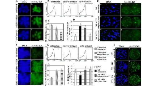
[image:6.612.55.510.336.613.2]Fibroblasts treated with GVcyto-extract had low levels of Me-H3-K9 (Fig. 3Af) compared with untreated cells (Fig. 3Ab). However, GVoocyte extract could not induce significant histone demethylation in the treated fibroblasts, such that the cells exhibited high levels of Me-H3-K9, similar to control untreated cells (Fig. 3Ab,d). Flow cytometry analysis revealed that the methylation of H3-K9 was reduced significantly in GVcyto-extract-treated cells compared with untreated controls (Fig. 3B,C). Thus, GVcyto-extract clearly contained demethylation activity. The low level of Me-H3-K9 in SCNT-generated embryos derived from the treated fibroblasts was maintained until the late blastocyst stage and showed a level similar to IVF-derived blastocysts (Fig. 3D,Ef). By contrast, embryos derived from GVoocyte extract-treated and untreated donor cells showed high methylation levels at all blastocyst stages (Fig. 3Eb,d). In addition, both extracts increased the acetylation of H3-K9
Fig. 3. Effect of GVcyto-extract and whole pig GV stage oocyte extract (GVoocyte-extract) on histone modification in treated cells and reconstructed embryos.(A) Histone H3 methylation (Me-H3-K9) in fibroblast nuclei with no treatment (b, control), GVoocyte-extract treated (d) or GVcyto-extract treated (f). (a-c) DNA staining (DAPI). (B,C) FACS and quantification of FACS result showing a significant reduction in the intensity of Me-H3-K9 in GVcyto-extract-treated compared with untreated cells (D,E) Quantification of Me-H3-K9 in reconstructed blastocysts with
untreated fibroblasts (NT untreated) or GVoocyte-extract-treated fibroblasts (NT+GVoocyte-extract) or GVcyto-extract-treated fibroblasts (NT+GVcyto-extract). (F) Histone H3 acetylation (Ac-H3-K9) in fibroblast nuclei with untreated (b, control) or GVoocyte-extract treatment (d) or GVcyto-extract treatment (f). (G,H) FACS and quantification of FACS result showing significantly increased intensity of Ac-H3-K9 in both the GVoocyte-extract-treated and GVcyto-extract-treated groups. (I,J) Quantification of Ac-H3-K9 in reconstructed blastocysts with untreated fibroblasts (NT untreated) or GVoocyte-extract-treated fibroblasts (NT+GVoocyte-extract) or GVcyto-extract-treated fibroblasts (NT+GVcyto-extract). In vitro fertilization (IVF)-derived embryos were used as controls. Each column represents the normalized mean value of the intensity of Me-H3-K9 or Ac-H3-K9 per group. The data are presented as mean ± s.e.m. In bar charts, values marked with different lowercase letters were significantly different (P<0.05). NT or SCNT, somatic cell nuclear transfer. Scale bars: 20 m in A,F; 30 m in E,J.
D
E
V
E
LO
P
M
E
N
significantly in treated cells and reconstructed embryos compared with the untreated group (Fig. 3F-J).
The duration of pig oocyte maturation is longer than in any other mammalian species, such that the process of germinal vesicle breakdown (GVBD) takes ~18 hours in vitro (Bui et al., 2007), whereas it takes only 3 hours in the mouse (Bui et al., 2008). Thus, the nuclei of GV stage pig oocytes could remain intact in GVcyto-extract because GVBD was not induced during the 1-hour incubation of permeabilized fibroblasts (Fig. 1A). In addition, based on the successes of embryonic stem cell nuclear transfer (ESNT) in selectively enucleated or metaphase zygotes, it appears likely that some components of the pronuclei are essential to nuclear reprogramming activity in zygotes (Greda et al., 2006; Egli et al., 2007). However, the capacity to completely reprogram and support preimplantation development after SCNT is likely to require multiple factors located in the nucleus as well as the cytoplasm, as we demonstrated that cytoplasmic extracts of mouse GV oocytes enhanced the cloning efficiency when treated nuclei were transferred into mature oocytes (Bui et al., 2008). Therefore, we developed a simple method for establishing stem-like cells by two-step reprogramming (see Materials and methods).
EGFP expression in GVcyto-extract-treated Oct4-EGFP cells and pig chimeric blastocysts
A non-invasive monitoring system using Oct4promoter-driven EGFP expression would be very helpful to study differentiation and reprogramming mechanisms in pig embryos and cells (Miyoshi et al., 2009; Huang et al., 2011). We established a human OCT4 promoter-driven EGFP reporter system for monitoring the reprogramming of GVcyto-extract-treated cells (Fig. 1B). We established three Oct4-EGFP cell lines from ear skin fibroblasts (Fig. 1C). Differential immunostaining revealed that embryos derived from the treated fibroblasts by SCNT had higher inner cell mass (ICM), trophectoderm (TE) and total cell numbers than in the untreated group (Fig. 4A,B) and numerous high-quality blastocysts
were found in the SCNT+GVcyto-extract group (Fig. 4C). Because the GVoocyte extract treatment did not improve the quality of SCNT-derived embryos, we used GVcyto-extract treatment of fibroblasts for all subsequent experiments.
The Oct4-EGFP cells were treated with GVcyto-extract and cultured to examine EGFP expression. Reactivation of the OCT4 promoter in treated cells was detected weakly by 10 days post-treatment (Fig. 4Da,b). After 3 weeks, colonies were larger and EGFP fluorescence was clearly detected (Fig. 4Dc,d). These reprogrammed cells were injected into 8-cell parthenogenetically activated host embryos and examined at later developmental stages. EGFP expression was found in all host morulae, indicating that Oct4-EGFP cells were distributed throughout the embryos. Most of the reprogrammed cells became localized in the ICM of the host blastocysts (Fig. 4Eb). These data strengthened our hypothesis that reprogrammed fibroblasts would have stem cell-like characteristics.
GVcyto-extract promotes Oct4-EGFP expression in SCNT-derived embryos
Jaenisch et al. (Jaenisch et al., 2002) examined the potency of blastocysts derived from ESNT and SCNT and showed that ESNT-derived embryos developed to term at a 10- to 20-fold higher efficiency than SCNT-derived embryos generated using cumulus or fibroblast donor cell nuclei (Wakayama et al., 1998; Rideout et al., 2000). We performed SCNT using reprogrammed cells collected from GVcyto-extract-treated Oct4-EGFP cells (SCNT+GVcyto-extract) or Oct4-EGFP cells that were not treated with extract (SCNT untreated). Embryos were cultured to the hatched blastocyst stage. EGFP expression was not found in any embryos from the 1-cell to the 16-cell stage (data not shown). EGFP expression was first weakly detectable in the morula stage of SCNT-derived embryos (Table 1; Fig. 4Fb,Gb). EGFP expression levels in the SCNT+GVcyto-extract blastocysts were significantly higher than in SCNT untreated blastocysts (91% and 22%, respectively; Table 1; Fig. 4Fd,Gd). Interestingly, EGFP expression was found in the
SCNT+GVcyto-Fig. 4. Effect of pig GVcyto-extract on development of SCNT-derived embryos reconstructed with treated cells.(A,B) Effect of pig GV extracts on the total number of blastomeres, trophectoderm (TE) cells, inner cell mass (ICM) cells and ICM formation rates in reconstructed blastocysts was examined by differential staining of SCNT untreated (NT untreated), SCNT+GVoocyte-extract and SCNT+GVcyto-extract embryos. (A) TE nuclei are stained pink and ICM nuclei are stained blue. (B) Values marked with different lowercase letters were significantly different in the same item (total cells or ICM or TE) between groups (P<0.05). Error bars indicate s.e.m. (C) High-quality SCNT+GVcyto-extract blastocysts at 7 days. (D-G) EGFP expression (green) in pig GVcyto-extract-treated Oct4-EGFP fibroblasts. (D) EGFP expression in treated cells after 10 days (a,b) and 3 weeks (c,d) in culture.
(E) GVcyto-extract-treated Oct4-EGFP fibroblasts after 3 weeks in culture (reprogrammed cells) were injected into parthenogenetically activated 8-cell host embryos and cultured to the blastocyst stage. EGFP expression was detected in all morulae (5 days) and localized to the ICM cells of blastocysts (6 days). (F,G) EGFP expression in SCNT-derived embryos reconstructed from Oct4-EGFP fibroblasts (SCNT untreated, F) or Oct4-EGFP reprogrammed cells
(SCNT+GVcyto-extract, G). Scale bars: 30 m.
D
E
V
E
LO
P
M
E
N
[image:7.612.53.365.460.731.2]extract group in hatched blastocysts, whereas the untreated cells did not show any EGFP expression at this stage (18% and 0%, respectively; Table 1; Fig. 4Ff,Gf). Control parthenogenetic embryos did not show any EGFP fluorescence (Table 1). There were no significant differences in the rates of morula formation in the SCNT untreated and SCNT+GVcyto-extract groups. However, the rates of formation of late blastocysts (27% and 10%, respectively) and hatched blastocysts (25% and 7%) were significantly higher for the SCNT+GVcyto-extract embryos than for the SCNT untreated group.
Embryos reconstructed by SCNT using GVcyto-extract-treated somatic cells have increased cell numbers and higher-quality blastocysts
In early blastocysts, Oct4 and Cdx2 were colocalized in nearly all nuclei (Fig. 5A). There was a decrease in Cdx2 expression in some
[image:8.612.54.569.83.154.2]nuclei at the late blastocyst stage, indicating that Cdx2 expression was starting to be downregulated in the ICM (Fig. 5Bj, arrows). The expression of endogenous Oct4 in blastocysts was significantly lower in the SCNT untreated group than in SCNT+GVcyto-extract-derived embryos (Fig. 5A-C). Interestingly, GVcyto-extract increased the Oct4 level in SCNT-derived embryos to a level similar to IVF-derived embryos (Fig. 5C). Nanog expression was not detected in any embryos (supplementary material Fig. S2). In hatched blastocysts, Cdx2 expression was downregulated in some of the ICM cells (Fig. 5Dc,f,i, circles), whereas Nanog expression was more concentrated in these nuclei (Fig. 5Db,e,h). This result indicated that Nanog tends to be expressed in ICM cells of the hatched blastocyst, perhaps in preparation for localization into the epiblast in later development. IVF-derived embryos contained more Nanog-positive nuclei than SCNT-derived embryos (Fig.
Table 1. Effect of pig GV stage oocyte cytoplasmic extract on the expression of EGFP and development of SCNT-derived embryos reconstructed with Oct4-EGFP cells
2-cell (2 days) Morula (5 days) Late BL (7 days) Hatched BL (8 days)
No (%) of EGFP/total No (%) of EGFP/total No (%) of EGFP/total No (%) of EGFP/total Groups 2-cell 2-cell morulae morulae BL BL hatched BL hatched BL
Parthenogenetic 93 0 (0) 50 (54)a 0 (0) 35 (38)a 0 (0)a 29 (31)a 0 (0)a SCNT untreated 90 0 (0) 38 (42)a 38 (100)a 9 (10)b 2 (22)b 6 (7)b 0 (0)a SCNT+GVcyto-extract 87 0 (0) 44 (51)a 44 (100)a 23 (27)a 20 (91)c 22 (25)a 4 (18)b
Values with different superscripts (a-c) in the same column differ significantly (P<0.05). BL, blastocyst.
Fig. 5. Effects of pig GVcyto-extract on Oct4 and Nanog expression levels.(A,B) Expression of Oct4 or Cdx2 in SCNT-derived embryos reconstructed from fibroblasts (SCNT untreated) or GVcyto-extract-treated fibroblasts (SCNT+GVcyto-extract) in early blastocysts (6 days) (A) and late blastocysts (7 days) (B). IVF-derived embryos were used as controls. (C) Quantification of Oct4 in blastocysts. Each column represents the normalized mean value (± s.e.m.) of the intensities at each developmental stage. (D) Typical expression pattern of Nanog and Cdx2 in hatched blastocysts from IVF-derived, SCNT untreated and SCNT+GVcyto-extract embryos. Circles indicate inner cell mass of embryos. (E) Effect of GVcyto-extract on the proportions of hatched blastocysts containing different numbers of nuclei expressing Nanog as assessed by immunoreactivity: Type I, more than 30 positive cells; Type II, fewer than 30 positive cells. Scale bars: 30 m.
D
E
V
E
LO
P
M
E
N
[image:8.612.55.533.369.661.2]5Db). Of note, hatched blastocysts containing more than 30 Nanog-positive blastomeres (designated Type I good embryos) appeared significantly more frequently in the SCNT+GVcyto-extract group than in the SCNT untreated group (Fig. 5Dh,E).
These results demonstrated that Oct4 expression was severely deficient in SCNT-derived embryos, with a low concentration in the ICM at the late blastocyst stage, concomitant with low expression of Nanog in hatched blastocysts. GVcyto-extract promoted somatic cell reprogramming and cloned embryo development by the production of increased numbers of high-quality blastocysts that exhibited Me-H3-K9 and expressed Oct4 and Nanog at levels similar to IVF-derived embryos.
Cells reprogrammed with GVcyto-extract can redifferentiate into multiple cell lineages
Although the reprogrammed cells showed reduced colony formation after 4 months of culture (corresponding to 20 passages), the forming colonies contained cells of normal karyotype, a normal ability to form teratomas when transferred to immunodeficient mice, and rapidly differentiated into several cell types in culture (Fig. 6). Under specific culture conditions (see Materials and
[image:9.612.53.386.315.710.2]methods), reprogrammed cells acquired the morphology and expression of markers for each of the three germ layers. For redifferentiation into ectoderm, the cell morphology changed in all-trans retinoic acid after 2 weeks in culture (Fig. 6Fa) and cells became neuron-like after culture in differentiation medium for an additional week (Fig. 6Fb,c). These neuron-like cells showed expression of specific markers after retinol induction by 2 weeks, including synaptophysin, neuron-specific enolase, neurofilament and, after full redifferentiation at 3 weeks, nestin and b-tubulin III (Fig. 6Fd-i). For redifferentiation into endoderm, the majority of reprogrammed cells expressed the pancreatic cell marker Foxa2 after 1 week of definitive endoderm induction (Fig. 6Ga). After being transferred to low-attachment plates for aggregate formation, various sizes of aggregates were produced in culture for an additional week and these strongly expressed pancreatic cell markers such as Foxa2 and Gata4 (Fig. 6Gb). Similarly, for redifferentiation into mesoderm lineages, cardiomyocyte markers such as cardiac troponin T and connexin 43 (Fig. 6H) and the endothelial cell markers CD31 and von Willebrand factor (Fig. 6H) were expressed. Control permeabilized fibroblasts cultured under the same conditions did not show expression of any of these
Fig. 6. Characteristics of reprogrammed cells.After 20 passages, the stem-like cells showed normal karyotypes, formed teratomas when transplanted into
immunodeficient mice and differentiated into several cell types in culture. (A) Cytogenetic analysis showed a normal karyotype (38, XY). (B) Representative images of teratomas formed subcutaneously (a) or in the testis (b,c) by transplantation of reprogrammed cells or control ES cell (asterisks). (C) Flow cytometric characterization of three primary germ layers in teratoma cells. Specific antibodies staining for ectoderm: Beta-tubulin and neuron-specific enolase (NSE); for endoderm: Gata-4 and Foxa2; and for mesoderm: connexin 43 and cardiac troponin T. (D) Histochemistry of teratomas in control ES cells (a-c) and reprogrammed cells (d-f). (E) Immunostaining of tissues indicates expression of S100 (a), p63 (b) and muscle creatine kinase (MCK, c) (green). (F) In vitro differentiation of neurons after 2 weeks (a). They became neuron-like cells after culture for an additional week in differentiation medium (b,c). Expression of neuronal markers in neuron-like cells after culture for 2 weeks: synaptophysin (d), NSE (e), neurofilament (f); and by 3 weeks: nestin (g) and b-tubulin III (h), with merge (i). (G) Expression of pancreatic markers in redifferentiated cells after culture for 1 week: (a) Foxa2 (red); and for 2 weeks: (b) Foxa2 (green) and Gata4 (red). (H) Expression of cardiomyocyte markers in redifferentiated cells, such as cardiac troponin T (a, red) and connexin 43 (b, green); and endothelial cell markers, such as CD31 (c, red) and von Willebrand factor (d, green), with DNA (e) and merge (f). Scale bars: 20 m.
D
E
V
E
LO
P
M
E
N
markers. Further evidence of the stem cell-like characteristics of the GVcyto-extract-treated cells included their ability to undergo cell passaging at least ten times without changes in their morphology or Oct4 and Nanog expression levels.
DISCUSSION
Cloned embryos have higher levels of histone methylation and DNA methylation than naturally formed embryos (Kang et al., 2002; Santos et al., 2003). This might result from the incomplete erasure of pre-existing methylation in the donor cells. We reported previously that genomic reprogramming factors present in the cytoplasm of the mouse GV stage oocyte induced histone demethylation in donor somatic cell chromosomes and in reconstructed embryos (Bui et al., 2008). Similarly, in the pig, the low methylation level of H3-K9 in embryos derived from fibroblasts treated with GVcyto-extract was maintained until the hatching blastocyst stage. Importantly, GVcyto-extract could induce histone demethylation in treated fibroblasts whereas GVoocyte-extract could not. Thus, GVcyto-extract caused partial erasure of pre-existing epigenetic marks of donor cells and SCNT-mediated reprogramming reset the treated cell states to conform to the environment of the recipient cytoplasm. This enhanced the development of cloned embryos. It was reported that GV oocyte extracts activated pluripotent marker genes more effectively than did MII stage oocyte extracts (Miyamoto et al., 2009). However, GV stage oocyte extracts could not improve the efficiency of producing cloned blastocysts in the cow (Tang et al., 2009) or in the pig (this study). Therefore, GVcyto-extract is better for improving the quality of cloned embryos.
In contrast to the situation in the mouse, IVF-derived pig embryos at the blastocyst stage appear not to downregulate the expression of Oct4 in the TE (Kirchhof et al., 2000). In the present study, localization of Oct4-EGFP expression in early blastocysts from SCNT-derived embryos was not confined to the ICM, but was also seen in the TE (Fig. 4D). This result confirmed that Oct4 is expressed in both the ICM and TE of blastocysts. The lack of Oct4-EGFP expression in SCNT-derived embryos might inhibit their development because late blastocysts developed in vivo express Oct4 at high levels in the pig (Lee et al., 2006). The present study demonstrated that GVcyto-extract treatment induced continuous expression of Oct4 in late blastocysts developed from SCNT-derived embryos and that this treatment might be important for improving cloning efficiency. In addition, Nanog protein expression is undetectable in pig oocytes and embryos (Kuijk et al., 2008) but appears in the epiblast at a later stage of development (Hall et al., 2009). In the present study, expression of Nanog was detected in some nuclei in hatched blastocysts, perhaps in preparation for later localization into the epiblast. However, the SCNT-derived embryos derived from donor cells without GVcyto-extract treatment had only a few nuclei expressing Nanog at this stage. Interestingly, GVcyto-extract resulted in a significant increase in Nanog expression relative to the untreated group (Fig. 5D). Therefore, high expression of Oct4 and Nanog in the SCNT+GVcyto-extract-derived embryos might result from successful reactivation of the Oct4and Nanoggenes in treated donor cells.
Despite two decades of effort, the establishment of ES cells from pigs has remained elusive, as putative ES cell-like colonies can be initially established from pig embryos but they cannot be sustained in long-term culture (Telugu et al., 2010). Establishment of pig ES cells from zona-enclosed blastocyst and ICM indicated that Oct4 is expressed during the first passage for a maximum of 7 passages, when its expression becomes completely downregulated (Brevini
et al., 2007). The culture of pig ES cells using human ES cell culture conditions, however, does not sustain undifferentiated growth of the pig ICM and epiblast, as they differentiate within four passages (du Puy et al., 2011). Pig ES or NT-ES cells cultured in the presence of -MEM medium with EGF and activin, or treated with trichostatin A, can show an increase in the number of passages, although it is unclear whether these cells can be cultured beyond 14 or 15 passages, respectively (Vassiliev et al., 2010; Vassiliev et al., 2011). In addition, it has been shown that epigenetic memory is often retained in mouse iPS cells, but not in cells reprogrammed by mouse oocytes (Kim et al., 2010), and clinical application of iPS cells is limited by the fact that most protocols modify the genomes.
Here, we developed a simple method for establishing stem-like cells with the aim of overcoming the difficulties in establishing ES-like cells in the pig. The GVcyto-extract-treated fibroblasts were shown to dedifferentiate partially and transiently after culture, with the formation of colonies and expression of specific stem-like genes. Our results indicate that GVcyto-extract mediated the dedifferentiation of terminally differentiated pig fibroblasts and enabled the reprogrammed cells to undergo multilineage redifferentiation. These findings not only support the feasibility of such an approach, but also provide evidence that the stem-like cells obtained by this method can be used to study tissue repair. In addition, the application of transgenic pigs to research on gene function and the regulation of human diseases is widely sought and could become a source of organs or cells that would not be rejected by human patients. However, success in producing transgenic pigs is still inefficient. In the present study, GVcyto-extract significantly improved the number and quality of SCNT-derived blastocysts and might be an important source for establishing SCNT-generated ES cell lineages for future studies of therapeutic cloning and transgenic pig production. However, the stem-like cells in the present study showed reduced colony formation after several months of culture. This suggests that other factors are necessary to maintain the self-renewal of these cells in an undifferentiated state. We intend to standardize and optimize the derivation and culture protocols or treatments for these stem-like cells to enable their further development.
Generally, it is well documented that the GV stage oocyte must undergo an intensive growth phase during which the cell accumulates all the necessary material that will be used subsequently during early embryo development (Sorensen and Wassarman, 1976). This is accompanied by very high levels of RNA polymerase I and II transcription that are indispensable for the production of ribosomes and specific mRNAs (Picton et al., 1998). In the mouse, immature oocytes can reprogram the immature nucleus, whereas MII stage oocytes cannot (Bao et al., 2000; Obata et al., 2002), and pups can be produced by transferring the nuclei of growing oocytes into the cytoplasm of enucleated fully grown oocytes following IVF (Obata et al., 2011). Therefore, there must be several reprogramming factors from the cytoplasm of growing and fully grown oocytes in our GVcyto-extract. Histone deacetylases in the cytoplasm of GV oocytes are important for the regulation of histone acetylation status throughout the genome (Endo et al., 2008). In addition, the protein DJ-1 (also known as Park7) is translocated from the cytoplasm to the nucleus during the cell cycle after mitogen stimulation (Nagakubo et al., 1997). This protein is also present at high concentration in oocytes from the GV stage until the 4-cell embryo stage and it has been suggested that DJ-1 is required for the development of SCNT-derived embryos (Miyamoto et al., 2011). It was also reported that mature human oocytes can reprogram somatic cells to a pluripotent
D
E
V
E
LO
P
M
E
N
state (Noggle et al., 2011). In the present study, we carried out reprogramming procedures using a combination of factors from the cytoplasm and nucleus of oocytes via two steps. The first reprogramming step occurs through oocyte cytoplasmic factors that initialize the genomic memory of the differentiated somatic cells, and then oocyte nuclear factors continue to reprogram somatic cells to a pluripotent state.
Taken together, these data suggest that GVcyto-extract could provide the necessary regulatory components required for inducing somatic cell nuclear reprogramming and for altering the differentiation status of non-embryonic cells. Furthermore, SCNT was able to reset the reprogrammed cells to improve cloned embryo development. Much remains to be learned about the mechanisms involved in the genomic reprogramming of somatic cell nuclei. Studies on factors in the GV oocyte that might be involved in the remodeling of somatic cell nuclei during cloning by SCNT are now ongoing in our laboratory.
Acknowledgements
We thank Prof. Takashi Miyano and Prof. Teruhiko Wakayama for valuable discussions.
Funding
Financial support for this research was provided by a Woo Jang-Choon project grant [PJ007849] from the Research and Development Agency (RDA) and Institute of Planning & Evaluation for Technology (IPET) [111047-5] of the Republic of Korea.
Competing interests statement
The authors declare no competing financial interests.
Supplementary material
Supplementary material available online at
http://dev.biologists.org/lookup/suppl/doi:10.1242/dev.086116/-/DC1
References
Alberio, R., Johnson, A. D., Stick, R. and Campbell, K. H.(2005). Differential nuclear remodeling of mammalian somatic cells by Xenopus laevis oocyte and egg cytoplasm. Exp. Cell Res.307, 131-141.
Bao, S., Obata, Y., Carroll, J., Domeki, I. and Kono, T.(2000). Epigenetic modifications necessary for normal development are established during oocyte growth in mice. Biol. Reprod.62, 616-621.
Brevini, T. A., Tosetti, V., Crestan, M., Antonini, S. and Gandolfi, F.(2007). Derivation and characterization of pluripotent cell lines from pig embryos of different origins. Theriogenology67, 54-63.
Bui, H. T., Van Thuan, N., Wakayama, T. and Miyano, T.(2006). Chromatin remodeling in somatic cells injected into mature pig oocytes. Reproduction131, 1037-1049.
Bui, H. T., Van Thuan, N., Kishigami, S., Wakayama, S., Hikichi, T., Ohta, H., Mizutani, E., Yamaoka, E., Wakayama, T. and Miyano, T.(2007). Regulation of chromatin and chromosome morphology by histone H3 modifications in pig oocytes. Reproduction133, 371-382.
Bui, H. T., Wakayama, S., Kishigami, S., Kim, J. H., Van Thuan, N. and Wakayama, T.(2008). The cytoplasm of mouse germinal vesicle stage oocytes can enhance somatic cell nuclear reprogramming. Development135, 3935-3945.
Constantinescu, D., Gray, H. L., Sammak, P. J., Schatten, G. P. and Csoka, A. B.(2006). Lamin A/C expression is a marker of mouse and human embryonic stem cell differentiation. Stem Cells24, 177-185.
du Puy, L., Lopes, S. M., Haagsman, H. P. and Roelen, B. A.(2011). Analysis of co-expression of OCT4, NANOG and SOX2 in pluripotent cells of the porcine embryo, in vivo and in vitro. Theriogenology75, 513-526.
Egli, D., Rosains, J., Birkhoff, G. and Eggan, K.(2007). Developmental reprogramming after chromosome transfer into mitotic mouse zygotes. Nature 447, 679-685.
Endo, T., Kano, K. and Naito, K.(2008). Nuclear histone deacetylases are not required for global histone deacetylation during meiotic maturation in porcine oocytes. Biol. Reprod.78, 1073-1080.
Fischle, W., Wang, Y. and Allis, C. D.(2003). Histone and chromatin cross-talk.
Curr. Opin. Cell Biol.15, 172-183.
Freberg, C. T., Dahl, J. A., Timoskainen, S. and Collas, P.(2007). Epigenetic reprogramming of OCT4 and NANOG regulatory regions by embryonal carcinoma cell extract. Mol. Biol. Cell18, 1543-1553.
Greda, P., Karasiewicz, J. and Modlinski, J. A.(2006). Mouse zygotes as recipients in embryo cloning. Reproduction132, 741-748.
Gupta, M. K., Jang, J. M., Jung, J. W., Uhm, S. J., Kim, K. P. and Lee, H. T.
(2009). Proteomic analysis of parthenogenetic and in vitro fertilized porcine embryos. Proteomics9, 2846-2860.
Hall, V. J., Christensen, J., Gao, Y., Schmidt, M. H. and Hyttel, P.(2009). Porcine pluripotency cell signaling develops from the inner cell mass to the epiblast during early development. Dev. Dyn.238, 2014-2024.
Hansis, C., Barreto, G., Maltry, N. and Niehrs, C.(2004). Nuclear
reprogramming of human somatic cells by Xenopus egg extract requires BRG1.
Curr. Biol.14, 1475-1480.
Huang, L., Fan, N., Cai, J., Yang, D., Zhao, B., Ouyang, Z., Gu, W. and Lai, L.
(2011). Establishment of a porcine Oct-4 promoter-driven EGFP reporter system for monitoring pluripotency of porcine stem cells. Cell. Reprogram.13, 93-98.
Jaenisch, R., Eggan, K., Humpherys, D., Rideout, W. and Hochedlinger, K.
(2002). Nuclear cloning, stem cells, and genomic reprogramming. Cloning Stem Cells4, 389-396.
Jeong, Y. S., Yeo, S., Park, J. S., Lee, K. K. and Kang, Y. K.(2007). Gradual development of a genome-wide H3-K9 trimethylation pattern in paternally derived pig pronucleus. Dev. Dyn.236, 1509-1516.
Kang, Y. K., Park, J. S., Koo, D. B., Choi, Y. H., Kim, S. U., Lee, K. K. and Han, Y. M.(2002). Limited demethylation leaves mosaic-type methylation states in cloned bovine pre-implantation embryos. EMBO J.21, 1092-1100.
Kim, K., Doi, A., Wen, B., Ng, K., Zhao, R., Cahan, P., Kim, J., Aryee, M. J., Ji, H., Ehrlich, L. I. et al.(2010). Epigenetic memory in induced pluripotent stem cells. Nature467, 285-290.
Kirchhof, N., Carnwath, J. W., Lemme, E., Anastassiadis, K., Schöler, H. and Niemann, H.(2000). Expression pattern of Oct-4 in preimplantation embryos of different species. Biol. Reprod.63, 1698-1705.
Kuijk, E. W., Du Puy, L., Van Tol, H. T., Oei, C. H., Haagsman, H. P., Colenbrander, B. and Roelen, B. A.(2008). Differences in early lineage segregation between mammals. Dev. Dyn.237, 918-927.
Lee, E., Lee, S. H., Kim, S., Jeong, Y. W., Kim, J. H., Koo, O. J., Park, S. M., Hashem, M. A., Hossein, M. S., Son, H. Y. et al.(2006). Analysis of nuclear reprogramming in cloned miniature pig embryos by expression of Oct-4 and Oct-4 related genes. Biochem. Biophys. Res. Commun.348, 1419-1428.
Liu, H., Kim, J. M. and Aoki, F.(2004). Regulation of histone H3 lysine 9 methylation in oocytes and early pre-implantation embryos. Development131, 2269-2280.
Miyamoto, K., Tsukiyama, T., Yang, Y., Li, N., Minami, N., Yamada, M. and Imai, H.(2009). Cell-free extracts from mammalian oocytes partially induce nuclear reprogramming in somatic cells. Biol. Reprod.80, 935-943.
Miyamoto, K., Nagai, K., Kitamura, N., Nishikawa, T., Ikegami, H., Binh, N. T., Tsukamoto, S., Matsumoto, M., Tsukiyama, T., Minami, N. et al.(2011). Identification and characterization of an oocyte factor required for development of porcine nuclear transfer embryos. Proc. Natl. Acad. Sci. USA108, 7040-7045.
Miyoshi, K., Mori, H., Mizobe, Y., Akasaka, E., Ozawa, A., Yoshida, M. and Sato, M.(2009). Development of a noninvasive monitoring system for evaluation of Oct-3/4 promoter status in miniature pig somatic cell nuclear transfer embryos. J. Reprod. Dev.55, 661-669.
Nagakubo, D., Taira, T., Kitaura, H., Ikeda, M., Tamai, K., Iguchi-Ariga, S. M. and Ariga, H.(1997). DJ-1, a novel oncogene which transforms mouse NIH3T3 cells in cooperation with ras. Biochem. Biophys. Res. Commun.231, 509-513.
Nguyen, V. T., Kure-bayashi, S., Harayama, H., Nagai, T. and Miyake, M.
(2003). Stage-specific effects of the osmolarity of a culture medium on the development of parthenogenetic diploids in the pig. Theriogenology59, 719-734.
Noggle, S., Fung, H. L., Gore, A., Martinez, H., Satriani, K. C., Prosser, R., Oum, K., Paull, D., Druckenmiller, S., Freeby, M. et al.(2011). Human oocytes reprogram somatic cells to a pluripotent state. Nature478, 70-75.
Obata, Y., Kono, T. and Hatada, I.(2002). Gene silencing: maturation of mouse fetal germ cells in vitro. Nature418, 497.
Obata, Y., Hiura, H., Fukuda, A., Komiyama, J., Hatada, I. and Kono, T.
(2011). Epigenetically immature oocytes lead to loss of imprinting during embryogenesis. J. Reprod. Dev.57, 327-334.
Picton, H., Briggs, D. and Gosden, R.(1998). The molecular basis of oocyte growth and development. Mol. Cell. Endocrinol.145, 27-37.
Prather, R. S., Hawley, R. J., Carter, D. B., Lai, L. and Greenstein, J. L.(2003). Transgenic swine for biomedicine and agriculture. Theriogenology59, 115-123.
Rathbone, A. J., Fisher, P. A., Lee, J. H., Craigon, J. and Campbell, K. H.
(2010). Reprogramming of ovine somatic cells with Xenopus laevis oocyte extract prior to SCNT improves live birth rate. Cell. Reprogram.12, 609-616.
Rideout, W. M., 3rd, Wakayama, T., Wutz, A., Eggan, K., Jackson-Grusby, L., Dausman, J., Yanagimachi, R. and Jaenisch, R.(2000). Generation of mice from wild-type and targeted ES cells by nuclear cloning. Nat. Genet.24, 109-110.
Santos, F., Zakhartchenko, V., Stojkovic, M., Peters, A., Jenuwein, T., Wolf, E., Reik, W. and Dean, W.(2003). Epigenetic marking correlates with developmental potential in cloned bovine preimplantation embryos. Curr. Biol. 13, 1116-1121.
Sorensen, R. A. and Wassarman, P. M.(1976). Relationship between growth
and meiotic maturation of the mouse oocyte. Dev. Biol.50, 531-536.
D
Tang, S., Wang, Y., Zhang, D., Gao, Y., Ma, Y., Yin, B., Sun, J., Liu, J. and Zhang, Y.(2009). Reprogramming donor cells with oocyte extracts improves in vitro development of nuclear transfer embryos. Anim. Reprod. Sci.115, 1-9.
Telugu, B. P., Ezashi, T. and Roberts, R. M.(2010). The promise of stem cell research in pigs and other ungulate species. Stem Cell Rev.6, 31-41.
Tong, G. Q., Heng, B. C. and Ng, S. C.(2006). Exposure of mouse cumulus cell nuclei to porcine ooplasmic extract eliminates TATA box protein binding to chromatin, but has no effect on DNA methylation. J. Assist. Reprod. Genet.23, 413-419.
Vackova, I., Novakova, Z., Krylov, V., Okada, K., Kott, T., Fulka, H. and Motlik, J.(2011). Analysis of marker expression in porcine cell lines derived from blastocysts produced in vitro and in vivo. J. Reprod. Dev.57, 594-603.
Van Thuan, N., Harayama, H. and Miyake, M.(2002). Characteristics of preimplantational development of porcine parthenogenetic diploids relative to the existence of amino acids in vitro. Biol. Reprod.67, 1688-1698.
Vassiliev, I., Vassilieva, S., Beebe, L. F., Harrison, S. J., McIlfatrick, S. M. and Nottle, M. B.(2010). In vitro and in vivo characterization of putative porcine embryonic stem cells. Cell. Reprogram.12, 223-230.
Vassiliev, I., Vassilieva, S., Truong, K. P., Beebe, L. F., McIlfatrick, S. M., Harrison, S. J. and Nottle, M. B.(2011). Isolation and in vitro characterization of putative porcine embryonic stem cells from cloned embryos treated with trichostatin A. Cell. Reprogram.13, 205-213.
Wakayama, T., Perry, A. C., Zuccotti, M., Johnson, K. R. and Yanagimachi, R.
(1998). Full-term development of mice from enucleated oocytes injected with cumulus cell nuclei. Nature394, 369-374.
Wakayama, S., Kishigami, S., Van Thuan, N., Ohta, H., Hikichi, T., Mizutani, E., Yanagimachi, R. and Wakayama, T.(2005). Propagation of an infertile hermaphrodite mouse lacking germ cells by using nuclear transfer and embryonic stem cell technology. Proc. Natl. Acad. Sci. USA102, 29-33.