Copyright © 2002, American Society for Microbiology. All Rights Reserved.
Pulmonary Infiltrates in Immunosuppressed Patients: Analysis of
a Diagnostic Protocol
Cristina Danés,
1Julián González-Martín,
1* Tomàs Pumarola,
1Ana Rañó,
2Natividad Benito,
3Antoni Torres,
2Asunción Moreno,
3Montserrat Rovira,
4and Jorge Puig de la Bellacasa
1Servei de Microbiologia, Institut Clínic d’Infeccions i Immunologia,1Servei de Pneumologia i Al.lèrgia Respiratòria, Institut Clínic de Pneumologia i Cirurgia Toràcica,2Servei de Malalties Infeccioses, Institut Clínic d’Infeccions i Immunologia,3
and Servei d’Hematologia, Institut Clínic d’Hematologia i Oncologia,4Institut d’Investigacions Biomèdiques Agustí Pi i Sunyer, Hospital Clínic de Barcelona, Barcelona, Spain
Received 30 April 2001/Returned for modification 20 September 2001/Accepted 6 March 2002
A diagnostic protocol was started to study the etiology of pulmonary infiltrates in immunosuppressed patients. The diagnostic yields of the different techniques were analyzed, with special emphasis on the importance of the sample quality and the role of rapid techniques in the diagnostic strategy. In total, 241 patients with newly developed pulmonary infiltrates within a period of 19 months were included. Noninvasive or invasive evaluation was performed according to the characteristics of the infiltrates. Diagnosis was achieved in 202 patients (84%); 173 patients (72%) had pneumonia, and specific etiologic agents were found in 114 (66%). Bronchoaspirate and bronchoalveolar lavage showed the highest yields, either on global analysis (23 of 35 specimens [66%] and 70 of 134 specimens [52%], respectively) or on analysis of each type of pneumonia. A tendency toward better results with optimal-quality samples was observed, and a statistically significant difference was found in sputum bacterial culture. Rapid diagnostic tests yielded results in 71 of 114 (62.2%) diagnoses of etiological pneumonia.
Both infectious and noninfectious pulmonary complications are a major cause of morbidity and mortality in the immuno-suppressed host (1, 6, 29). The marked increase in the number and types of transplantations together with the advances in immunosuppressive treatment have created a subset of pa-tients with increased risk for pulmonary complications (8). In addition, almost 70% of the individuals with human immuno-deficiency virus (HIV) infection have at least one respiratory episode during the course of their disease (23). Even though a wide variety of etiologic agents are responsible for pneumonia in these patients, the type and timing of immunosuppression predispose the patient to infections by certain pathogens (1). Accordingly, in solid-organ transplant recipients, first bacterial
pneumonia, mainly due to gram-negative bacilli and
Staphylo-coccus aureus, and then opportunistic infections by
cytomega-lovirus (CMV) and Pneumocystis carinii are most commonly
found (1). In neutropenic patients, mainly patients with hema-tological malignancies and bone marrow transplant recipients, bacterial and fungal pneumonia is the most important chal-lenge (1, 9). On the other hand, in HIV-infected patients
P. carinii remains a common respiratory pathogen together withStreptococcus pneumoniaeandMycobacterium tuberculosis
(22–24).
Prompt and accurate diagnosis of the cause of pulmonary infiltrates is essential in order to begin specific antimicrobial treatment. Various procedures have been proposed for this purpose, but the optimal management strategy remains con-troversial (6). Besides fiber optic bronchoscopy techniques, other new procedures have been introduced. For example,
induced sputum for the detection ofP. carinii(13) and
naso-pharyngeal washing (NPW) for the detection of respiratory viruses (32) are noninvasive techniques that play a role in the diagnostic strategy. In addition, recent advances in
microbio-logical techniques such asLegionella pneumophila(28) andS.
pneumoniaeantigen detection in urine,Aspergillussp. antigen detection in serum (20), and molecular biology methods (11, 14, 27) allow rapid diagnostic tests and therefore prompt ini-tiation of specific therapy if pertinent.
We report the results of a prospective study on the etiology of pulmonary infiltrates in immunosuppressed patients. Sev-eral groups have been included: patients with bone marrow transplantation, solid-organ transplantation, hematologic ma-lignancies, and HIV infection. The diagnostic yields of the different methods used were analyzed in order to optimize, if possible, the diagnostic strategy in these patients.
MATERIALS AND METHODS
Patient population.We prospectively studied immunosuppressed patients ad-mitted to the Hospital Clínic of Barcelona with a new pulmonary infiltrate from March 1998 to October 1999. The following groups were included: patients with bone marrow transplantation, solid-organ transplantation, hematologic malig-nancies, and HIV infection and patients undergoing continued immunosuppres-sive treatment for other reasons. A total of 241 patients were considered for evaluation.
Demographic and clinical data. In all cases, the following variables were recorded at admission: age, sex, type of immunosuppression, temperature, white blood cell count, type (alveolar, interstitial, or mixed) and distribution (unilateral or bilateral) of radiographical infiltrates, and antibiotic status at the time of respiratory sampling (less or more than 48 h of antibiotic treatment).
Sampling.Noninvasive or invasive evaluation was performed according to the characteristics of the pulmonary infiltrates. Patients with a single alveolar pul-monary infiltrate underwent noninvasive evaluation consisting of two sets of blood cultures, a sputum specimen (induced sputum if no expectoration), bron-choaspiration (BAS) in intubated patients, detection ofL. pneumophila sero-group 1 antigen in urine, NPW for detection of respiratory viruses, and serolog-* Corresponding author. Mailing address: Servei de Microbiologia,
Hospital Clínic de Barcelona, Villarroel 170, 08036 Barcelona, Spain. Phone: 34.3.2275522. Fax: 34.3.2279372. E-mail: [email protected].
2134
on May 15, 2020 by guest
http://jcm.asm.org/
ical tests for atypical pneumonia due toL. pneumophila serogroups 1 to 6 (immunoglobulin G [IgG]),Chlamydia pneumoniae (IgM, IgG),Mycoplasma
pneumoniae(IgM, IgG), andCoxiella burnetii(IgM, IgG) and viral respiratory
infections (adenovirus, respiratory syncytial virus, parainfluenza viruses 1, 2, and 3, and influenza viruses A and B).
Patients with multiple pulmonary infiltrates or interstitial infiltrate underwent invasive evaluation. Patients presenting with a single alveolar pulmonary infil-trate and remaining unresponsive after 3 days of empirical treatment also un-derwent invasive evaluation. This consisted of fiber optic bronchoscopy exami-nation including a protected specimen brush (PSB) and bronchoalveolar lavage (BAL). These techniques were performed as previously reported (9, 33). Briefly, fiber optic bronchoscopy was performed transnasally or transorally after topical nasal anesthesia was instilled. Once PSB was performed, the brush was asepti-cally severed and introduced into 1 ml of thioglycolate broth for transport. BAL sampling was carried out by instilling three aliquots of 50 ml of sterile saline solution. The first aspirated portion of the fluid lavage was discarded, and the remainder was processed for stain and culture.
Patients without diagnosis after bronchoscopic evaluation and patients with persistent pulmonary infiltrates were subjected to transbronchial biopsy if pos-sible.
Once the protocol had been started and due to recent advances in microbio-logical methods, several new diagnostic procedures were performed for selected patients: detection ofS. pneumoniaeantigen in urine, detection ofAspergillussp. antigen in serum, detection by PCR enzyme immunoassay (PCR-EIA) ofC.
pneumoniaein nasopharyngeal swabs, and detection by ligase chain reaction
(LCR) ofM. tuberculosisin clinical specimens.
Laboratory processing of specimens. (i) Culture procedures.Sputum speci-mens were cultured for bacterial pathogens, fungi, and mycobacteria. Fiber optic bronchoscopy specimens were first homogenized by vortexing and then subjected to quantitative bacterial culture and qualitative culture for fungi andLegionella. For quantitative culture, 0.1 ml of PSB fluid and 0.1 ml and a 10-fold dilution of BAL fluid were plated onto each medium. BAL and BAS specimens were also cultured for mycobacteria. Media and incubation conditions were as follows. For bacterial culture, secretions were plated on sheep blood agar and chocolate agar and incubated at 37°C in a CO2-enriched atmosphere for 2 days. For fungal culture, secretions were inoculated on Sabouraud’s agar and incubated at 30°C in aerobic conditions for 4 weeks. Buffered charcoal-yeast extract agar incubated at 37°C in a CO2-enriched atmosphere for 10 days was used for isolation of
Legio-nella. For mycobacteria, Löwenstein-Jensen medium and radiometric BACTEC
12B broth culture bottles (Becton Dickinson) were incubated at 37°C in aerobic conditions for 6 weeks.
NPW and BAL specimens were examined for viruses by cell culture. After decontamination treatment, 0.3 ml of each sample was inoculated onto mono-layers of HEp-2, A-549, MRC-5, and MDCK cell lines (Vircell, Granada, Spain). Cultures were maintained for 4 weeks and examined regularly for cytopathic effect. Confirmation of viral growth was done by immunofluorescence staining with monoclonal antibodies (Respiratory Panel 1 viral screening and identifica-tion kit; Light Diagnostics, Temecula, Calif.).
(ii) Direct microscopy.Sputum and BAS specimens were stained with the Gram and Ziehl-Neelsen methods. PSB and BAL smears were obtained by cytocentrifuge preparation in accordance with a modification of a described protocol (15) (0.25 and 0.5 ml of fluid for PSB and BAL, respectively, at 1,500 rpm for 15 min; Cytospin 3 centrifuge; Shandon Instruments, Cheshire, England) and subsequently Gram stained for detection of bacteria and fungi and deter-mination of percentage of intracellular organisms. The May-Grünwald Giemsa technique was used for fungal detection and differential cell count of macro-phages, squamous epithelial cells, ciliated bronchial cells, neutrophils, lympho-cytes, eosinophils, and erythrocytes for assessment of specimen quality. Addi-tional stains were used for BAL smears; these include Ziehl-Neelsen stain for detection of acid-fast organisms, Gomori methenamine silver (GMS) forP.
cariniiand fungi; periodic acid-Schiff stain, hematoxylin-eosin, and Papanicolau
stain for detection of malignant cells; and Pearl’s stain for detection of sideroph-ages. Viral antigens were detected in NPW and BAL specimens by direct fluo-rescence employing a pool of monoclonal antibodies against a panel of respira-tory viruses (influenza virus A and B, parainfluenza virus 1, 2, and 3, respirarespira-tory syncytial virus, and adenovirus). In addition, CMV detection was done by means of an immunocytochemistry method (alkaline phosphatase anti-alkaline phos-phatase) using a monoclonal antibody against the pp65-CMV protein (Dako, Glostrup, Denmark).
(iii) Non-culture-dependent methods.Urinary antigen tests were performed for detection ofL. pneumophilaserogroup 1 by EIA (BiotestLegionellaUrin Antigen EIA; Biotest) and for detection ofS. pneumoniaeby immunochromatog-raphy (Binax NowS. pneumoniae urinary antigen test; Binax). Detection in
serum ofAspergillussp. galactomannan antigen was by EIA (PlateliaAspergillus; Sanofi Diagnostics Pasteur).M. tuberculosiswas determined in clinical samples by LCR (LCxMycobacterium tuberculosisassay; Abbott Laboratories, Abbot Park, Ill.), and detection ofC. pneumoniaein nasopharyngeal swabs was per-formed by PCR-EIA (Diasorin).
Diagnostic criteria.The diagnosis of pneumonia was based on the develop-ment of new or progressive pulmonary infiltrates together with at least two of the following: fever, cough productive of sputum, leukocytosis, or leukopenia. The diagnosis of pneumonia was definite if the above criteria were associated with one of the following: (i) histopathologic demonstration of pneumonia, with or without isolation of specific etiologic agent, (ii) positive culture of pleural fluid, (iii) positive blood cultures (two positive sets, taken within a period of 48 h), (iv) histopathologic demonstration of tissue invasion by fungal hyphae in lung biopsy specimens, (v) isolation ofL. pneumophilaorM. tuberculosisfrom respiratory samples, (vi) positive urinary antigen test forL. pneumophilaserogroup 1, (vii) serologic diagnosis (demonstration of seroconversion), (viii) cytologic evidence of CMV inclusion bodies in cytocentrifuge smears, (ix) presence ofP. cariniiin the GMS-stained induced sputum or BAL specimens. If the criteria for definite infection were not met, a diagnosis of a probable case of pneumonia was made if one of these criteria were met: (i) bacterial pathogens yieldingⱖ103CFU/ml in cultures of PSB,ⱖ104CFU/ml in cultures of BAL, andⱖ106CFU/ml in cultures of BAS; (ii) bacterial pathogen isolated from sputum only if there was heavy growth of the pathogen, sputum was included in group 4 or 5 of Murray and Washington’s grading system (10 to 25 epithelial cells and⬎25 leukocytes per low-power field [group 4] and⬍10 epithelial cells and⬎25 leukocytes per low-power field [group 5]) (25), and Gram stain findings correlated with culture results; (iii) positive culture for fungal pathogen in at least two bronchoscopy specimens; (iv) positive culture for fungal pathogen in at least two sputum samples and presence of fungal hyphae in the Gram stain (pseudohyphae and yeasts were excluded); (v) positive cell culture for respiratory viruses from NPW or BAL specimens, (vi) positive antigen detection for respiratory viruses from NPW or BAL specimens.
Absence of pneumonia was indicated by failure to meet the criteria for diag-nosis of pneumonia. Patients to whom this applied included those with an alternative diagnosis and those with an undetermined diagnosis.
Criteria for respiratory specimen rejection.Rejection of specimens was based on the following criteria: (i) sputum, induced sputum, and BAS specimens in group 1, 2, or 3 of Murray and Washington’s grading system (⬎25 epithelial cells and⬍10 leukocytes per low-power field [group 1],⬎25 epithelial cells and 10 to 25 leukocytes per low-power field [group 2], and⬎25 epithelial cells and⬎25 leukocytes per low-power field [group 3]) (25); and (ii) BAL and PSB specimens with⬎1% squamous epithelial cells. When BAL specimens were processed for detection ofP. cariniior respiratory viruses, these criteria were not applied.
Criteria for optimum specimen quality.Optimum conditions associated with the highest diagnostic yield were determined for each specimen as follows: (i) sputum and BAS specimens included in group 4 or 5 of Murray and Washing-ton’s grading system (25) when considering bacterial pneumonia or in group 4, 5, or 6 (⬍10 epithelial cells and⬍10 leukocytes per low-power field) when consid-ering fungal pneumonia; (ii) BAL and PSB specimens with⬎25% inflammatory cells (neutrophils plus lymphocytes) for bacterial pneumonia or with⬎10% inflammatory cells for fungal andP. cariniipneumonia. For viral pneumonia these criteria were not applied.
Data analysis.Before the data were analyzed, patients were divided into those with and without pneumonia and subsequently pneumonias were divided into definite and probable and into those with an established microbial etiology and those without one. The criteria used to define these groups are the ones shown above (see “Diagnostic criteria”).
(i) Diagnostic yield.The diagnostic yields of the different techniques were evaluated according to different points of view. First, a global diagnostic yield was evaluated, including yields for pneumonias with both infectious- and noninfec-tious-agent etiologies. For this analysis, specimens collected from all patients included in the study were considered. The diagnostic yield was calculated (a) by including all the respiratory specimens collected, irrespective of their quality, and (b) by excluding those specimens fulfilling criteria for respiratory specimen rejection.
Second, the diagnostic yield was evaluated according to the specific etiologic agent, bacterial, fungal, or viral agent. Therefore, only patients with a diagnosis of pneumonia etiology were considered. The diagnostic yield was calculated (a) by including all the specimens collected from patients with a diagnosis of bac-terial pneumonia and/or fungal pneumonia and/or viral pneumonia and (b) by excluding specimens not fulfilling the criteria for optimum specimen quality.
Third, the diagnostic yields of the rapid diagnostic techniques were evaluated. The following methods were considered. (a) Staining methods included the
on May 15, 2020 by guest
http://jcm.asm.org/
Gram, Ziehl-Neelsen, and GMS methods. The Gram stain was defined as pos-itive if microorganisms were seen predominantly (⬎10 bacteria per high-power field) and correlated with what grew in significant concentration in culture (bacterial pathogens yieldingⱖ103CFU/ml in cultures of PSB orⱖ104CFU/ml in cultures of BAL orⱖ106CFU/ml in cultures of BAS; for sputum specimens, a heavy growth was considered significant). (b) Antigen detection methods in-cluded detection ofL. pneumophilaserogroup 1 antigen in urine, detection ofS.
pneumoniaeantigen in urine, detection ofAspergillussp. antigen in serum, and
detection of respiratory virus antigen from NPW and BAL specimens. (c) Mo-lecular biology-based methods included an LCR-based assay for detection ofM.
tuberculosisand PCR-EIA for detection ofC. pneumoniae. Analysis of the
diag-nostic yield was performed by grouping the different methods according to the type of pneumonia; within each type, the rates of diagnoses obtained by rapid methods were calculated.
(ii) Cytologic patterns of BAL specimens according to type of pneumonia.To analyze whether specific cytologic patterns were associated with each type of pneumonia, three patterns were defined depending on the percentage of inflam-matory cells (neutrophils plus lymphocytes): (a)⬎50%; (b) 25 to 50%; (c) ⬍25%.
Statistical analysis.Epi Info software, version 6.02, (Epidemiology Program Office, Centers for Disease Control and Prevention, Atlanta, Ga.) was used in conjunction with the two-tailed Fisher exact test to compare pairs of proportions. Values ofP⬍0.05 were considered significant.
RESULTS
Study population. Overall, 241 patients (160 males, 81 fe-males; age range, 22 to 73 years, mean age, 43 years) were included in the study. Of these, 135 were HIV-infected pa-tients, 25 had undergone bone marrow transplantation, 26 had hematological malignancies, 40 were solid-organ transplant re-cipients (26 kidney, 10 liver, 4 heart), and 15 had received continued immunosuppressive treatment for other reasons.
The type and distribution of the pulmonary infiltrates were as follows: 103 patients (43%) had an alveolar infiltrate, 89 (37%) had an interstitial infiltrate, and 49 (20%) had a mixed pattern. These were unilateral in 119 (49%) and bilateral in 122 (51%).
As for the number of diagnostic procedures performed, blood cultures were obtained for 208 (86%) patients, sputum was obtained from 187 (78%), induced sputum was obtained from 56 (23%), and BAS was obtained from 35 (15%).
Detec-tion of antigen forL. pneumophilain urine was performed for
151 patients (63%), serologic tests for atypical pneumonias and respiratory viruses were performed for 35 (15%) and 20 (8%) patients, respectively, and serologic tests of NPW were per-formed for 42 patients (17%). Bronchoscopy examination was performed for 146 patients (61%). Among patients with a single alveolar pulmonary infiltrate, 25% underwent bronchos-copy, whereas, among the rest, bronchoscopy was performed for 79%. On the whole, 134 BALs and 110 PSBs were per-formed. Ten patients underwent transbronchial biopsy.
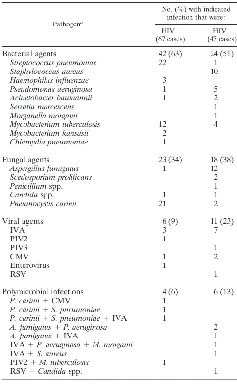
Etiology of pulmonary infiltrates.According to the diagnos-tic criteria, a diagnosis of either infectious or noninfectious was achieved in 202 out of 241 patients included in the study (84%). One hundred seventy-three patients (72%) had pneu-monia. Among them, 108 (63%) were classified as having a probable pneumonia and the remaining 65 (37%) were classi-fied as definitely having pneumonia. A specific etiologic agent was found in 114 (66%). Bacterial pneumonia was the most frequent (65 cases, including 7 mixed infections), followed by fungal pneumonia (41 cases, including 7 mixed infections) and viral pneumonia (17 cases, including 6 mixed infections). The etiological agents are summarized in Table 1. HIV-infected
patients had a significantly (P⬍0.0005) higher incidence ofS.
pneumoniae and P. carinii infection than non-HIV-infected patients, and non-HIV-infected patients had a significantly
higher incidence ofS. aureusand Aspergillus fumigatus
infec-tion than HIV-infected patients.
The association of the radiographic pattern with etiology was analyzed, and a significant relationship between bacterial pneumonia and unilateral alveolar infiltrate was found (24 of
35 patients versus 23 of 88 patients;P⫽0.0002). All patients
withP. cariniipneumonia had a diffuse interstitial infiltrate (P
⫽0.002). Mycobacterial, fungal, and viral pneumonias mainly
presented a diffuse interstitial pulmonary infiltrate, but there was no statistical significance.
The relationship between CD4 counts of HIV-infected pa-tients and the type of pneumonia was also analyzed. Even though only 70% of patients’ CD4 counts were available, the following was determined: 85% of the patients with fungal
pneumonias had available CD4 counts of ⬍200 cells/mm3,
[image:3.587.299.541.91.483.2]whereas 67% of the patients with bacterial pneumonias had
TABLE 1. Microbial agents in 114 patients with diagnoses of pneumonia etiology
Pathogena
No. (%) with indicated infection that were:
HIV⫹
(67 cases) HIV
⫺
(47 cases)
Bacterial agents 42 (63) 24 (51)
Streptococcus pneumoniae 22 1 Staphylococcus aureus 10 Haemophilus influenzae 3
Pseudomonas aeruginosa 1 5 Acinetobacter baumannii 1 2 Serratia marcescens 1 Morganella morganii 1 Mycobacterium tuberculosis 12 4 Mycobacterium kansasii 2
Chlamydia pneumoniae 1
Fungal agents 23 (34) 18 (38)
Aspergillus fumigatus 1 12 Scedosporium prolificans 2
Penicilliumspp. 1
Candidaspp. 1 1
Pneumocystis carinii 21 2
Viral agents 6 (9) 11 (23)
IVA 3 7
PIV2 1
PIV3 1
CMV 1 2
Enterovirus 1
RSV 1
Polymicrobial infections 4 (6) 6 (13)
P. carinii⫹CMV 1 P. carinii⫹S. pneumoniae 1 P. carinii⫹S. pneumoniae⫹IVA 1
A. fumigatus⫹P. aeruginosa 2
A. fumigatus⫹IVA 1
IVA⫹P. aeruginosa⫹M. morganii 1
IVA⫹S. aureus 1
PIV2⫹M. tuberculosis 1
RSV⫹Candidaspp. 1
aIVA, influenza A virus; PIV2, parainfluenza 2 virus; RSV, respiratory syn-cytial virus.
on May 15, 2020 by guest
http://jcm.asm.org/
counts of ⬎200 cells/mm3 (P ⬍ 0.05). As for patients with
mycobacteriosis and viral pneumonias, their CD4 counts were evenly distributed among both categories.
Among patients not fulfilling the criteria for pneumonia, an alternative diagnosis was found for 29 (12%) and 39 (16%) remained with an undetermined diagnosis. Noninfectious
causes included pulmonary edema (n ⫽ 8), diffuse alveolar
hemorrhage (n⫽4), endocarditis (n⫽4), pulmonary
malig-nancies (n⫽4), bronchiolitis obliterans with organizing
pneu-monia (n ⫽ 3), pulmonary chronic diseases (n ⫽ 4), adult
respiratory distress syndrome (n ⫽ 1), and pulmonary drug
toxicity by bleomycin (n⫽1).
Diagnostic yield of diagnostic procedures. (i) Global diag-nostic yield.On the whole, BAS and BAL proved to be the techniques with the highest diagnostic yields. Diagnosis was obtained in 23 of 35 (66%) of the BASs and in 70 of 134 (52%) of the BALs performed (Table 2).
If respiratory specimens fulfilling criteria for respiratory specimen rejection are excluded, the diagnostic yield obtained is higher but the differences are not significant (data not shown).
The diagnostic yields of the newest diagnostic procedures
were not evaluated because they were performed only in se-lected patients. However, they contributed to the global
diag-nostic rate as follows: five patients had detectable S.
pneu-moniaeantigen in urine, three patients had detectable serum
Aspergillusantigen, LCR forM. tuberculosiswas positive in two
cases, and PCR-EIA forC. pneumoniae was positive in one
case.
(ii) Diagnostic yield according to the type of pneumonia.
With regard to bacterial pneumonias, the diagnostic procedure with the highest yield was BAS (12 of 15 specimens [80%]), followed by BAL (11 of 18 specimens [61%]) and PSB (8 of 13 specimens [61%]). The Gram stains of BAS, BAL, and PSB were positive in 73, 44, and 38% of the cases, respectively. A comparison of these results to those obtained if only specimens fulfilling criteria for optimum specimen quality were consid-ered indicated significant differences for sputum culture (20 of
35 [57%] compared to 20 of 21 [95%];P⬍0.005). The effect
of previous antibiotic administration on culture results was analyzed for each procedure, but no significant differences were observed.
[image:4.587.43.541.83.413.2]As for fungal pneumonias, BAS culture and GMS-stained
TABLE 2. Diagnostic yields of the different techniques according the type of pneumonia
Specimen
Yieldafor indicated type of:
Global diagnostic
yieldd Bacterial pneumonia Fungal pneumonia Mycobacteriosis Viral pneumonia,
ASb
ASb OSc ASb OSc ASb OSc
BAS 23/35 (66)
Culture 12/15 (80) 11/12 (92) 10/11 (91)e 10/11 (91) 1/1 1/1
Gram stain 11/15 (73) 11/12 (92) 9/11 (82)e 9/11 (82)
Ziehl-Neelsen stain 1/1 1/1
BAL 70/134 (52)
Culture 11/18 (61) 7/7 (100) 7/12 (58)e 6/8 (75) 7/8 (87) 2/2 (100) 14/15 (93)
Gram stain 8/18 (44) 5/7 (71) 2/12 (17)e 2/8 (25)
Ziehl-Neelsen stain 3/8 (37.5) 2/2 (100)
GMS stain 22/22 (100)f 10/10 (100)
Viral Ag detection 9/17 (53)
Sputum 38/187 (20)
Culture 20/35 (57) 20/21 (95)g 5/14 (36)e 5/13 (38) 13/15 (87) 13/14 (93)
Ziehl-Neelsen stain 9/15 (60) 9/14 (64)
PSB 14/110 (12)
Culture 8/13 (61) 4/4 (100) 6/12 (50)e 3/4 (75)
Gram stain 5/13 (38) 4/4 (100) 1/12 (8)e 1/4 (25)
Pleural fluid culture 0/3 0/3 0/11e 0/11 1/11 (9) 1/11 (9) 1/11 (9)
Blood culture 15/42 (36) 15/42 (36) 2/16 (13)e 2/16 (13) 17/208 (8)
NPW 3/47 (6)
Culture 3/10 (30)
Viral Ag detection 1/10 (10)
Bacterial serology 0/5 0/5 0/35
Viral serology 1/2 (50) 1/20 (5)
Induced-sputum GMS stain 1/8 (12)f 1/4 (25) 1/56 (2)
L. pneumophilaAg urine 0/28 0/28 0/151
aTotal number of diagnoses (diagnoses of pneumonia etiology)/total number of specimens fulfilling the criteria for AS or OS (the specimens included in each classification are described in footnotesbandc, respectively. Percentages are in parentheses.
bAll specimens (AS) from patients with the indicated diagnoses are included. cSpecimens not fulfilling the criteria for optimum specimen (OS) quality are excluded.
dTotal number of diagnoses (including both infectious- and noninfectious-agent etiologies)/total number of specimens collected. eCases of filamentous fungi and yeasts.
fCases ofP. carinii. gP⬍0.05.
on May 15, 2020 by guest
http://jcm.asm.org/
BAL specimens showed the highest diagnostic yield (10 of 11 specimens [91%] and 22 of 22 [100%], respectively).
The diagnostic yield of the Gram stain in sputum samples was not analyzed because a positive Gram stain was a criterion needed to consider sputum diagnostic of either bacterial or fungal pneumonia.
Mycobacteriosis were analyzed apart from bacterial pneu-monias, and BAS was excluded from the analysis because it was performed for only one patient in this group. BAL and sputum cultures proved to be the techniques with the highest yields (7 of 8 specimens [87%] and 13 of 15 specimens [87%], respectively).
Finally, among viral pneumonias, cell culture of BAL and NPW yielded more diagnoses than antigen detection methods. In addition, BAL seems to be more sensitive than NPW (14 of 15 specimens [93%] and 3 of 10 specimens [30%], respective-ly).
(iii) Diagnostic yields of rapid diagnostic techniques. Gram stain of BAS specimens was the rapid technique with the high-est yield in both bacterial and fungal pneumonias (11 of 15 specimens [73%] and 9 of 11 specimens [82%], respectively) (Table 3). As for mycobacteriosis, the best technique proved to be Ziehl-Neelsen staining of sputum (9 of 15 specimens [60%]), and antigen detection of BAL specimens was best for viral pneumonias (9 of 17 specimens [53%]). Considering all cases with a diagnosis of pneumonia, rapid methods yielded results in 71 of 114 (62.2%).
Cytologic patterns of BAL specimens according to type of pneumonia.With cases of polymicrobial infections and cases for which no cytology could be evaluated having been ex-cluded, cytologic patterns of BAL specimens were analyzed (Table 4). Among bacterial pneumonias, six out of the seven
(86%) with BAL available had ⬎25% inflammatory cells
(three of them had ⬎50% inflammatory cells). For fungal
pneumonias, the cytologic patterns for 6 BAL specimens were
evenly distributed among patterns 1 to 3, while 4 out of 8
patients (50%) with viral pneumonia had⬍25% inflammatory
cells and 16 out of 18 patients (89%) withP. cariniipneumonia
had⬍25% inflammatory cells.
DISCUSSION
A diagnostic protocol was started at our hospital in order to study the etiology of pulmonary infiltrates in immunosup-pressed patients and to analyze the diagnostic yields of the different techniques used. The aim of the study was to optimize the diagnostic strategy in this type of patients, both in terms of number of techniques performed and in terms of time. Almost 2 years after initiation of the protocol, the results have been analyzed.
Among the 241 episodes included, diagnosis was achieved in 202 (84%), which represents an important improvement in relation to previous studies performed at our hospital (9). The etiology was an infectious agent in 72% of the cases and a noninfectious agent in 12% of the cases. The most frequent infectious agent was bacterial, in both HIV- and
non-HIV-infected patients.S. pneumoniaewas the most frequent
bacte-rial pathogen in HIV-infected patients (22 cases out of 28),
whereasS. aureus was the leading pathogen in
non-HIV-in-fected patients (10 cases out of 24). These results are in ac-cordance with several recently published studies (8, 12). The long-known increased risk for mycobacterial infections among immunosuppressed patients, specially among individuals with HIV infection (4, 21, 30), was also confirmed in our study. Accordingly, 14 cases were diagnosed among HIV-infected patients and 4 cases were diagnosed among non-HIV-infected patients.
Fungal agents constituted the second-most-frequent etiol-ogy, followed by viral agents. Twenty-one out of 23 fungal
infections in HIV-infected patients were due toP. carinii. The
incidence ofP. carinii pneumonia in these patients remains
high although effective prophylaxis has been widely introduced (24). In the present study most cases can be explained by the fact that HIV infection status was diagnosed as a result of the
P. cariniipneumonia and therefore there had been no
prophy-laxis. Interestingly, in three casesP. cariniicoexisted with other
pathogens. On the other hand, A. fumigatus was the most
[image:5.587.43.283.84.311.2]common fungal agent among non-HIV-infected patients. Over the last few years in our institution there has been an increase in the number of cases of invasive aspergillosis. Numerous reports have documented this increased incidence, and it has
TABLE 3. Diagnostic yield of rapid diagnostic techniques Type of pneumonia
(rate of diagnosesb[%]) Technique Diagnosticyielda
Bacterial (37/48 [77]) Gram stain of sputum 20/35 (57)
Gram stain of BAS 11/15 (73)
Gram stain of PSB 5/13 (38)
Gram stain of BAL 8/18 (44)
Assay ofL. pneumophilaAg in urine 0 Assay ofS. pneumoniaeAg in urine 5
PCR-EIA ofC. pneumoniae 1
Mycobacteriosis
(12/18 [67]) Sputum Ziehl-Neelsen stainBAS Ziehl-Neelsen stain 9/15 (60)1/1 BAL Ziehl-Neelsen stain 3/8 (37.5)
LCR forM. tuberculosis 2
Fungal (31/41 [76]) Gram stain of sputum 5/14 (36)
Gram stain of BAS 9/11 (82)
Gram stain of PSB 1/12 (8)
Gram stain of BAL 2/12 (17)
GMS stain of BAL 22/34 (65)
GMS stain of induced sputum 1/56 (2) Assay ofAspergillusspp. in serum 3
Viral (10/17 [59]) BAL Ag detection 9/17 (53)
NPW Ag detection 1/10 (10)
aTotal number of diagnoses/total number of tests performed (percentage). bTotal number of diagnoses/total number of cases of pneumonia.
TABLE 4. Cytologic patterns of BAL according to type of pneumonia
Type of pneumonia
No. of cases with cytologic patterna:
Totalb
1 2 3 4
Bacterial 3 3 1 3 10
Fungal 2 2 2 0 6
Viral 1 3 4 2 10
P. carinii 1 1 16 1 19
aDefinition of cytologic patterns: 1,⬎50% neutrophils plus lymphocytes; 2, 25 to 50% neutrophils plus lymphocytes; 3,⬍25% neutrophils plus lymphocytes; 4, evaluation not possible.
bPolymicrobial infections have been excluded.
on May 15, 2020 by guest
http://jcm.asm.org/
[image:5.587.301.540.622.694.2]been related to the growing population of immunosuppressed patients, mainly those with prolonged and intense neutropenia and those undergoing high-dose corticosteroid therapy (10). In addition, outbreaks of nosocomial aspergillosis have been as-sociated with contaminated air due to nearby construction work (10, 18, 26). In our study, the hospital renovation work that is currently taking place may have played a role in the increased number of invasive-aspergillosis cases. This specula-tion is based on the fact that over the last year there has been
an increased isolation of fungi, among themAspergillusspp., in
air samples obtained within the hospital (data not shown). Viral agents are the third-leading cause of pneumonia in both groups, with 17 cases diagnosed. Among them, the influ-enza A virus is the most frequent viral pathogen followed by CMV. This is specially interesting for HIV-infected patients, in whom the role of respiratory viruses in pneumonia has recently been described (16). Besides upper respiratory illness, respira-tory viruses may cause pneumonia, which, due to the special characteristics of the immunosuppressed population, may have serious consequences. Accordingly, there have been several reports of moderate-to-severe pneumonia caused by these vi-ruses (5, 31). However, their real importance in pneumonias when coinfection with bacteria or fungi occurs remains to be assessed. In our study, 6 out of 17 respiratory virus pneumonias were associated with other microbial agents.
Besides the etiology of pulmonary infiltrates, the diagnostic yields of the different procedures were analyzed. Overall, BAS and BAL proved to be the best techniques, with yields of 66 and 52%, respectively. BAS was performed in only 35 cases, 31 being for non-HIV-infected patients. This procedure was done mainly when there was a high suspicion of bacterial infection, and this may partially explain the high yield observed. In con-trast, BAL was performed in patients with less-definite radio-logical patterns, including patients with a worse clinical re-sponse. This fact probably influenced the diagnostic yield. The
agents most frequently found by BAL were viruses and P.
carinii, mainly in HIV-infected patients, and the diagnostic yield was high in this group of patients. Seven cases caused by noninfectious agents were retrieved by BAL, all in non-HIV-infected patients. The wide range of infections and etiologies involving noninfectious agents which can be diagnosed by BAL, renders this technique very useful in immunosuppressed patients, in whom the great variety of pulmonary complications is precisely what makes their diagnosis difficult (15).
When the diagnostic yield for each type of pneumonia was considered, special emphasis was put on evaluating the impor-tance of the quality of the samples in the final yield. Thus, although a statistically significant difference was found only in sputum culture, there was a clear tendency toward obtaining better results when the quality of the samples in all categories was optimal. This was evident in bacterial pneumonias, where the rate of diagnosis was highly influenced by the quality of the specimens; the diagnostic yield of sputum bacterial culture
increased from 56 to 95% (P⬍0.05) and the BAL and PSB
culture yields increased from 61 to 100% when only optimal-quality specimens were used. Among bacterial pneumonias, all BAL and PSB specimens exhibiting inflammatory cytology
(⬎25% inflammatory cells) showed a diagnosis of bacterial
etiology. The lack of statistical significance may be explained by the small number of cases. BAS specimens were not as
strongly influenced due to the fact that most fulfilled the cri-teria for optimum quality.
On comparison of the diagnostic yields of the various tech-niques used for each type of pneumonia, it is clear that again BAS and BAL were the techniques with the highest yields, regardless of the type of pneumonia. It is interesting to point
out that in the diagnosis of P. carinii pneumonia, induced
sputum had a very poor yield (12%). These results are not in accordance with previous reports (2, 19) showing that induced
sputum was a reliable noninvasive means of diagnosing P.
cariniipneumonia. In the present study, only 1 out of 23 cases was diagnosed by this technique. Moreover, in 7 cases in which
BAL had diagnosedP. carinii, induced sputum was negative.
Obtaining good-quality induced-sputum specimens requires cooperation from the patients and special training of the ther-apists (3). In our institution, the technique has recently been introduced, and thus more experience is needed to reassess its usefulness.
For viral pneumonia, the lower diagnostic yield of NPW viral isolation (3 of 10 specimens) compared to that of BAL (14 of 15 specimens) may be explained by a delay in sample collec-tion. Seven out of 10 NPWs performed were collected more than 48 h after the onset of symptoms. Furthermore, among the three NPWs collected in the first 24 h, two yielded a positive result. Poor sample quality accounts for the low yield of antigen detection methods in NPW (1 of 10 specimens [10%]). It has been previously reported (7, 17) that the sensi-tivity of these methods can be enhanced by optimizing sample collection to obtain an adequate amount of material for testing without mucus contamination. In our study, 8 out of 42 (18%) NPWs performed did not yield an adequate number of cells for direct fluorescent-antibody assay (DFA) examination. Conse-quently, if poor quality samples were excluded from the analysis, the diagnostic yield of NPW DFA in viral pneumonias would be more correctly calculated as one of six specimens (17%).
The role of diagnostic techniques with rapid turnaround times was analyzed. Rapid reporting of results increases the clinical relevance of the information provided by the labora-tory and is essential for instituting specific therapy for the immunosuppressed host. Over the last few years, technologic advances have provided new rapid diagnostic procedures such as antigen detection (20, 28) and molecular biology (11, 14, 27) methods. In the present study, new rapid methods have been added to conventional ones. Accordingly, among antigen
de-tection methods, urine antigen tests for detectingL.
pneumo-philaserogroup 1 andS. pneumoniaehave been introduced as
have serological tests for detectingAspergillussp. antigen. As
for molecular biology methods, nucleic acid amplification
meth-ods for detectingM. tuberculosisandC. pneumoniaewere
per-formed. Except for the detection ofL. pneumophilain urine, the
remaining new procedures were performed in selected cases and therefore more studies are needed to evaluate their usefulness. Analysis of the diagnostic yield of the rapid diagnostic pro-cedures showed that the Gram stain of BAS and sputum spec-imens had a high yield in both bacterial and fungal pneumo-nias. Consequently, even though the information provided is not definitive, appropriate antimicrobial therapy can be se-lected on the basis of Gram stain results. Neither detection of
L. pneumophilaantigen in urine nor the conventional methods (serology and culture) achieved diagnosis, thereby possibly
on May 15, 2020 by guest
http://jcm.asm.org/
plaining the absence of L. pneumophila pneumonia in our study. Overall, nearly 80% of bacterial and fungal pneumonias were diagnosed by rapid methods. Regarding mycobacteriosis, the rate of rapid diagnosis was 67% and Ziehl-Neelsen sputum staining gave the best diagnostic yield. For viral pneumonias, DFA is a reliable technique for the rapid diagnosis of viral infections. In the present study, DFA allowed the diagnosis of 59% viral pneumonias.
Finally, the cytologic patterns of BAL specimens from pa-tients with a diagnosis of pneumonia etiology were also ana-lyzed in order to see if the type of inflammatory response could be useful in predicting the type of pneumonia. The clearest
association was withP. cariniipneumonia: in 89% of the cases
there were⬍25% inflammatory cells. For most of the bacterial
pneumonias, even though most of the patients were immuno-suppressed, an increased percentage of inflammatory cells was
noted. Among viral pneumonias a cytologic pattern of⬍25%
inflammatory cells was the most frequent, whereas in fungal pneumonias no specific pattern was found.
In conclusion, after this protocol was implemented, an in-crease in the number of diagnoses of the etiology of new pulmonary infiltrates in the immunosuppressed population was observed. However, in view of the analysis of the diagnostic yields of the different methods, new approaches should be considered to optimize the diagnostic strategy. Thus, in the presence of a unilateral alveolar pulmonary infiltrate, bacterial pneumonia should first be excluded by collecting a good-qual-ity sputum sample and empirical treatment could be started immediately based on Gram stain results. For bilateral or in-terstitial pulmonary infiltrates unresponsive to empirical
anti-biotics, BAL should be performed; considering the fact thatP.
cariniipneumonia in HIV-infected patients and viral or fungal pneumonia in non-HIV-infected patients are more likely to be found, appropriate laboratory tests should be part of the diag-nostic algorithm. Finally, emphasis should be placed on per-forming rapid diagnostic tests. The availability of results within a few hours makes rapid tests essential for the immunosup-pressed population. However, more studies to evaluate the newest procedures are needed in order to improve, if possible, rapid diagnosis in these patients.
ACKNOWLEDGMENT
This study was supported in part by the grant FIS 00/0966.
REFERENCES
1.Baughman, R. P.1999. The lung in the immunocompromised patient. In-fectious complications part 1. Respiration66:95–109.
2.Bigby, T. D., D. Margolskee, J. L. Curtis, P. F. Michael, D. Sheppard, W. K. Hadley, and P. C. Hopewell.1986. The usefulness of induced sputum in the diagnosis ofPneumocystis cariniipneumonia in patients with the acquired immunodeficiency syndrome. Am. Rev. Respir. Dis.133:515–518. 3.Bustamante, E. A., and H. Levy.1994. Sputum induction compared with
bronchoalveolar lavage by Ballard catheter to diagnosePneumocystis carinii
pneumonia. Chest105:816–822.
4.Caylà, J. A., H. Galdós-Tangüis, J. M. Jansà, P. García de Olalla, T. Brugal, and H. Panella.1998. Tuberculosis in Barcelona, Spain (1987–1995). Influ-ence of human immunodeficiency virus and control measures. Med. Clin. (Barcelona)111:608–615.
5.Couch, R. B., J. A. Englund, and E. Whimbey.1997. Respiratory viral infec-tions in immunocompetent and immunocompromised persons. Am. J. Med.
102:2–9.
6.Crawford, S.1999. Non-infectious lung diseases in the immunocompromised host. Respiration66:385–395.
7.Doing, K. M., M. A. Jerkofsky, E. G. Dow, and J. A. Jellison.1998. Use of
fluorescent-antibody staining of cytocentrifuge-prepared smears in combina-tion with cell culture for direct deteccombina-tion of respiratory viruses. J. Clin. Microbiol.36:2112–2114.
8.Dunagan, D. P., A. M. Baker, D. D. Hurd, and E. F. Haponik.1997. Bron-choscopic evaluation of pulmonary infiltrates following bone marrow trans-plantation. Chest111:135–141.
9.Ewig, S., A. Torres, R. Riquelme, M. El-Ebiary, M. Rovira, E. Carreras, A. Raño, and A. Xaubet.1998. Pulmonary complications in patients with hemato-logical malignancies treated at a respiratory ICU. Eur. Respir. J.12:116–122. 10.Fridkin, S. K., and W. R. Jarvis.1996. Epidemiology of nosocomial fungal
infections. Clin. Microbiol. Rev.9:499–511.
11.Gaydos, C. A., C. L. Fowler, V. J. Gill, J. J. Eiden, and T. C. Quinn.1993. Detection ofChlamydia pneumoniaeby PCR-EIA in an immunocompro-mised population. Clin. Infect. Dis.17:718–723.
12.Hirschtick, R. E., J. Glassroth, M. C. Jordan, T. C. Wilcosky, J. M. Wallace, P. A. Kvale, N. Markowitz, M. J. Rosen, B. T. Mangura, P. C. Hopewell, et al.1995. Bacterial pneumonia in persons infected with the human immuno-deficiency virus. N. Engl. J. Med.333:845–851.
13.Hopewell, P. C.1988.Pneumocystis cariniipneumonia. J. Infect. Dis.157:
1115–1119.
14.Ieven, M., and H. Goossens.1997. Relevance of nucleic acid amplification techniques for diagnosis of respiratory tract infections in the clinical labo-ratory. Clin. Microbiol. Rev.10:242–256.
15.Kahn, F. W., and J. M. Jones.1988. Analysis of bronchoalveolar lavage specimens from immunocompromised patients with a protocol applicable in the microbiology laboratory. J. Clin. Microbiol.26:1150–1155.
16.King, J. C.1997. Community respiratory viruses in individuals with human immunodeficiency virus infection. Am. J. Med.102:19–24.
17.Landry, M. L., S. Cohen, and D. Ferguson.2000. Impact of sample type on rapid detection of influenza virus A by cytospin-enhanced immunofluores-cence and membrane enzyme-linked immunosorbent assay. J. Clin. Micro-biol.38:429–430.
18.Leenders, A. C. A., A. Belkum, M. Behrendt, A. Luijendijk, and H. A. Ver-brgh.1999. Density and molecular epidemiology ofAspergillusin air and rela-tionship to outbreaks ofAspergillusinfection. J. Clin. Microbiol.37:1752–1757. 19.Leigh, T. R., P. Parsons, C. Hume, O. A. N. Husain, B. Gazzard, and J. V. Collins.1989. Sputum induction for diagnosis ofPneumocystis carinii pneu-monia. Lancetii:205–206.
20.Maertens, J., J. Verhaegen, H. Demuynck, P. Brock, G. Verhoef, P. Vanden-berghe, J. Van Eldere, L. Verbist, and M. Boogaerts.1999. Autopsy con-trolled evaluation of serial screening for circulating galactomannan by a sandwich enzyme-linked immunosorbent assay for hematological patients at risk for invasive aspergillosis. J. Clin. Microbiol.37:3223–3228.
21.Markowitz, N., N. I. Hansen, P. C. Hopewell, J. Glassroth, P. A. Kvale, B. T. Mangura, T. C. Wilcosky, J. M. Wallace, M. J. Rosen, L. B. Reichman, and The Pulmonary Complications of HIV Infection Study Group.1997. Inci-dence of tuberculosis in the United States among HIV-infected persons. Ann. Intern. Med.126:123–132.
22.Meduri, G. U., and D. S. Stein.1992. Pulmonary manifestations of acquired immunodeficiency syndrome. Clin. Infect. Dis.14:98–113.
23.Miller, R.1996. HIV-associated respiratory diseases. Lancet348:307–312. 24.Mitchell, D. M., and R. F. Miller.1995. AIDS and the lung: update 1995. 2.
New developments in the pulmonary diseases affecting HIV-infected indi-viduals. Thorax50:294–302.
25.Murray, P. R., and J. A. Washington II.1975. Microscopic and bacteriologic analysis of expectorated sputum. Mayo Clin. Proc.50:339–344.
26.Radford, S. A., E. M. Johnson, J. P. Leeming, M. R. Millar, J. M. Cornish, A. B. M. Foot, and D. W. Warnock.1998. Molecular epidemiological study of
Aspergillus fumigatusin a bone marrow transplantation unit by PCR
ampli-fication of ribosomal intergenic spacer sequences. J. Clin. Microbiol.36:
1294–1299.
27.Rohner, P., E. I. M. Jahn, B. Ninet, C. Ionati, R. Weber, R. Auckenthaler, and G. E. Pfyffer.1998. Rapid diagnosis of pulmonary tuberculosis with the
LCxMycobacterium tuberculosisassay and comparison with conventional
diagnostic techniques. J. Clin. Microbiol.36:3046–3047.
28.Stout, J. E., and V. L. Yu.1997. Legionellosis. N. Engl. J. Med.337:682–687. 29.Tamm, M.1999. The lung in the immunocompromised patient. Infectious
complications part 1. Respiration66:199–207.
30.Theuer, C. P., P. C. Hopewell, D. Elias, G. F. Schecter, G. W. Rutherford, and R. E. Chaisson.1990. Human immunodeficiency virus infection in tu-berculosis patients. J. Infect. Dis.162:8–12.
31.Whimbey, E., R. E. Champlin, R. B. Couch, J. A. Englund, J. M. Goodrich, I. Raad, D. Przepiorka, V. A. Lewis, N. Mirza, H. Yousuf, J. J. Tarrand, and G. P. Bodey.1995. Community respiratory virus infections among hospital-ized adult bone marrow transplant recipients. Clin. Infect. Dis.22:778–782. 32.Wiedbrauk, D. L., and S. L. G. Johnston.1993. Specimen collection and processing, p. 22–32.InD. L. Wiedbrauk and S. L. G. Johnston (ed.), Manual of clinical virology. Raven Press, Ltd., New York, N.Y.
33.Wimberly, N. W., L. J. Faling, and J. G. Bartlett.1979. A fiberoptic bron-choscopy technique to obtain uncontaminated lower airway secretions for bacterial culture. Am. Rev. Respir. Dis.119:337–343.